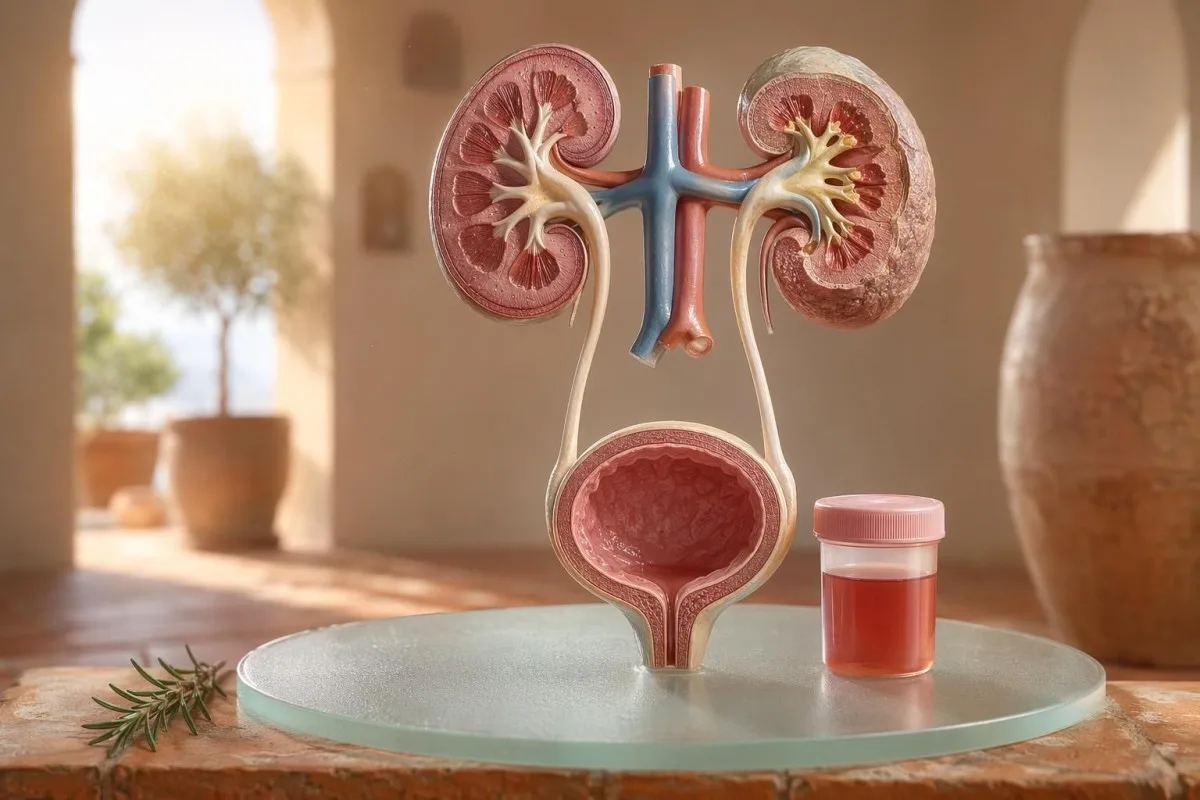
د ناروغ-لومړی لارښود د ښکاره او مایکروسکوپي هیماتوریا لپاره، په ګډون د دې چې ولې د ډیپسټیک وینه د مایکروسکوپ لاندې د سره حجرو (RBC) له وینې سره نه برابره ده.


- په ادرار کې وینه مانا لري ښکاره سره رنګه، د چای رنګه یا د کولاکولا رنګه ادرار، یا مایکروسکوپي هیماتوریا چې د ازموینې پر مهال وموندل شي؛ ښکاره وینه تل طبي تعقیب ته اړتیا لري.
- Microscopic hematuria عموماً د مناسب ډول راټول شوي ادرار مایکروسکوپي نمونې کې په هر لوړ ځواک میدان (high-power field) کې ۳ یا زیات سره د وینې حجرې (RBC) تعریفېږي.
- د ډیپسټیک وینه د هیم (heme) فعالیت کشفوي، نو کېدای شي مثبت وي د سالمو سره حجرو (RBC)، وړیا هیموګلوبین یا میوګلوبین له امله وروسته له عضلاتي ټپ څخه.
- د UTI نښې پکې سوځښت، پرلهپسې ادرار، leukocyte esterase، nitrites او سپینې حجرې شاملې دي؛ هیماتوریا باید وروسته له دې چې انتان (infection) پاک شي بیا وکتل شي.
- د ادرار رسوب موندنې لکه dysmorphic سره حجرې، د سره حجرو کاسټونه یا د پام وړ پروټین د پښتورګو د فلټر کولو واحد (kidney filtering-unit) د ناروغۍ لور ته اشاره کوي.
- د ادرار کاسټونه د پښتورګو د نلکو (tubules) څخه جوړ شوی مواد دی؛ د سره حجرو کاسټونه (red cell casts) د یوې اونۍ په موده کې د نیفرولوژي (nephrology) لپاره یو مهم خبرداری دی، په ځانګړي ډول که د وینې لوړ فشار وي یا eGFR کمېږي.
- د سرطان خطر نښې پکې بې درده ښکاره هیماتوریا (visible hematuria)، لخته (clots)، عمر له ۴۵ کلونو پورته، د سګرټ څکولو سابقه، د کار ځای کیمیاوي تماس او د بې سبب تکراري پېښې شاملې دي.
- د تمرین پر مهال هیماتوریا عموماً تر ۴۸–۷۲ ساعتونو پورې ارامېږي؛ که له استراحت وروسته هم وینه دوام وکړي، باید دا په منډه یا بایسکل چلولو ونه تړل شي.
- عاجله ارزونه ته اړتیا ده د ادرار په وینه کې چې تبه، د غاړې/بغل درد (flank pain)، لخته چې د ادرار د بندېدو سبب شي، امیندوارۍ، ټروما (trauma) یا د شدید انیمیا نښې وي.
په ادرار کې وینه څه معنا لري مخکې له دې چې وېرېږئ
په ادرار کې وینه کېدای شي د انتان (infection)، تیږو (stones)، د پښتورګو د فلټر کولو ناروغۍ، د پروسټېټ یا مثانې (bladder) شرایطو، درملو، تمرین، یا د حیض (periods) له امله ککړتیا څخه راشي. عملي قاعده ساده ده: ښکاره وینه، تکراري مایکروسکوپیک هیماتوریا، یا هیماتوریا د درد، تبه، لخته، لوړ وینې فشار، پروټین، یا د پښتورګو د کار کمېدو سره—باید ژر ارزونه وشي، نه دا چې یوازې انتظار وشي.
د چوغندر (beetroot) یا د نوي مکمل (supplement) وروسته په تشناب کې یو ځل ګلابي رنګ ښکاره کېدل د ستوني له درد (sore throat) وروسته د کولا رنګ ادرار په شان هماغه کلینیکي ستونزه نه ده. زما په عمل کې، د رنګ کیسه ډېری وخت راته وایي چې لومړی چیرته وګورو: د لخته سره روښانه سور رنګ عموماً د ټیټې ادراري لارې (lower urinary tract) لوري ته اشاره کوي، خو نسواري یا د چای رنګ ادرار ښایي د پښتورګو له فلټرونو څخه زاړه هیم (heme) رنګیزې (pigments) په ګوته کړي.
هیماتوریا تشخیص نه دی؛ دا یوه نښه (sign) ده. ښکاره هیماتوریا مانا دا ده چې تاسو د رنګ بدلون وینئ، پداسې حال کې چې مایکروسکوپیک هیماتوریا مانا دا ده چې ادرار عادي ښکاري خو مایکروسکوپي سره سره حجرې (red cells) موندل کېږي، معمولاً په هر لوړ ځواک میدان (high-power field) کې ۳ یا زیات سره د وینې حجرې.
کانټیسټي یو دی د AI د وینې معاینې شنونکی دا مرسته کوي ناروغان په یوه مهالویش (timeline) کې د ادرار موندنې د پښتورګو د نښو لکه کریټینین (creatinine)، eGFR، CRP، هیموګلوبین (hemoglobin) او پلیټلیټونو (platelets) سره ونښلوي. که رنګ مغشوشوونکی وي، زموږ د ادرار د رنګ لارښود (urine color guide) تشریح کوي چې ولې سور، نسواري، نارنجي او تیاره ژېړ ادرار ټول یو شان معنا نه لري.
د ډیپسټیک وینه د مایکروسکوپي (RBC) سره نه برابره ده
د ادرار ډیپ سټک (dipstick) چې نښه شوې وي وینه مثبت (blood positive) د هیم په شان کیمیاوي فعالیت (chemical activity) کشف کوي، حتمي نه چې بشپړې (intact) سره د وینې حجرې وي. مایکروسکوپي راتلونکی پوښتنه ځوابوي: ایا په ادرار کې د رسوب (sediment) دننه رښتینې RBCs شته، او که وي، نو څه ډول ښکاري؟
د ډیپ سټک ازموینه چټکه ده ځکه د ری ایجنټ پډ (reagent pad) د هیم د پېر اکسېډېز (peroxidase) فعالیت سره غبرګون کوي. دا مانا لري چې مثبت نتیجه کېدای شي د بشپړو RBCs سره، د سره حجرې له ماتېدو وروسته ازاد هیموګلوبین (free hemoglobin)، یا د پام وړ عضلاتي ټپ (significant muscle injury) وروسته مایوګلوبین (myoglobin) سره هم رامنځته شي.
مایکروسکوپي ورو ده خو ډېر مشخصه ده. یو دقیق لابراتوار به ادرار سنټریفیوژ (centrifuge) کړي، د د ادرار رسوب (urine sediment), به معاینه کړي، او راپور به ورکړي چې په هر لوړ ځواک میدان کې څو سره حجرې (red cells) دي؛ ډېر کلینیسینان په لویانو کې ۰–۲ RBC/hpf عادي ګڼي او ۳ یا زیات RBC/hpf غیرعادي.
دا ناسمون مهم دی. که ډیپ سټک ۳+ وینه وښيي او ۰–۲ RBC/hpf وي، زه له مثانې ناروغۍ سره سمدستي کود نه وهم؛ بلکې د rhabdomyolysis، hemolysis، اکسایډ کوونکو ککړوونکو (oxidizing contaminants)، او د نمونې (specimen) د سمبالولو (handling) په اړه پوښتنه کوم؛ توپیر زموږ د د ادرار معاینه او د کلتور پرتله.
کله د UTI نښې هیماتوریا تشریح کوي، او کله نه
د ادراري لارې انتان (UTI) کولی شي په ادرار کې وینه, ، په ځانګړي ډول د سوځښت، بیړنۍ اړتیا، پرلهپسې ادرار، د حوض ناراحتي، leukocyte esterase، نایټریټس او سپینو حجرو (white cells) سره. خو هیماتوریا باید د درملنې وروسته پاکه شي؛ د UTI وروسته هم دوامداره سره حجرې (red cells) د دویم ځل کتنې اړتیا لري.
په ساده ټیټ UTI کې، زه تمه لرم یوه ډله وي: ډیسوریا، پرلهپسې ادرار، leukocyte esterase مثبت، پیوریا اکثره له ۵-۱۰ سپینو حجرو/hpf څخه پورته وي، او کله ناکله نایټریټس هم وي. دودیز کلتور مثبتوالی ۱۰۰،۰۰۰ CFU/mL دی، خو په نښو لرونکو ښځو کې ټیټې شمېرې لکه ۱،۰۰۰-۱۰،۰۰۰ CFU/mL هم کولای شي په کلینیکي لحاظ مهم وي.
Leukocyte esterase د سپینو حجرو انزایم دی، د باکتریاوو ثبوت نه دی. د هیماتوریا سره مثبت نتیجه د انتان، تیږو، ککړتیا، یا د پښتورګو التهاب له امله رامنځته کېدای شي؛ له همدې امله زموږ leukocyte esterase لارښود عامو غلط مثبتو (false positives) څخه ګټورې نښې جلا کوي.
نایټریټس ډېر مشخص دي خو لږ حساسیت لري، ځکه ټول باکتریاوې نایټریټ نایټریټ ته نه بدلوي. که نښې کلاسیکې وي خو نایټریټس منفي وي، کلتور بیا هم مثبت کېدای شي؛ عملي جزئیات زموږ په د نایټریټ پایلې تشریح (explainer).
ښکاره هیماتوریا: د رنګ، لختهګانو او وخت نښې
ښکاره هیماتوریا هېڅکله هغه څه نه دي چې زه یې بېپامه پرېږدم، حتی که یوازې یو ځل هم پېښ شي. روښانه سره ادرار، لخته (clots)، یا بېدرد وینه بهیدل وختي ارزونې ته اړتیا لري، ځکه د ټیټې ادرارې لارې لاملونه د عمر او د سګرټ څکولو له زیاتېدو سره ډېر احتمال پیدا کوي.
وختبندي یې په حیرانوونکي ډول ګټور وي. یوازې د ادرار په پیل کې وینه راتلل ښايي د یوریترا (پیشاب نل) وي؛ د جریان په اوږدو کې وینه راتلل کېدای شي د مثانې یا پښتورګو له سرچینو وي، او د پای ته نږدې وینه کله ناکله د مثانې د غاړې سیمې ته اشاره کوي.
ټوټې (clots) عموماً مانا لري چې وینه د پښتورګو د فلټرونو لاندې ځای څخه راځي، ځکه ټوټې د ګلومیرولي او ټیوبولونو له لارې له تېرېدو وروسته په اسانۍ نه جوړېږي. هغه ټوټه چې د ادرار جریان بند کړي بیړنۍ ده: د مثانې پړسوب (distension) کېدای شي په څو ساعتونو کې دردناک شي، او د کیتیټر پاملرنه ممکنه وي.
نارینه ځینې وختونه ګومان کوي چې د ادراري نښو وروسته ښکاره هیماتوریا یوازې د پروسټېټ ځورښت دی. دا کېدای شي رښتیا وي، خو که PSA د عفونت پر مهال یا لږ وروسته وکتل شي، تفسیر یې ګډوډ کېدای شي؛ زموږ د UTI وروسته PSA لارښود تشریح کوي چې ولې ډېر کلینیشینان د PSA بیا تکرارولو مخکې ۶–۸ اونۍ انتظار کوي.
مایکروسکوپي هیماتوریا حدونه چې ډاکټران یې کاروي
مایکروسکوپیک هیماتوریا عموماً داسې تعریفېږي لکه په هر high-power field کې ۳ یا زیات RBC په سمه توګه راټول شوي د ادرار نمونه کې. د AUA/SUFU لارښود دا حد کاروي او سپارښتنه کوي چې د هر ناروغ لپاره یو شان درملنه نه وي، بلکې د خطر پر بنسټ ارزونه وشي (Barocas et al., 2020).
یوازې ډیپسټک (dipstick) باید د مایکروسکوپیک هیماتوریا د تشخیص لپاره ونه کارول شي. د Barocas et al. 2020 AUA/SUFU لارښود کې تعریف د مایکروسکوپي پر بنسټ دی، ځکه ډیپسټکونه د heme pigments له امله مثبت کېدای شي، حتی که بشپړ/صحتمند RBC موجود نه وي.
بیا ازموینه کمزوري نه ده؛ دا ښه طب دی. که یو څوک د سخت تمرین، تبه، جنسي اړیکې، منسټرېشن، یا ککړې راټولې شوې نمونې وروسته ۳–۱۰ RBC/hpf ولري، زه عموماً غواړم مخکې له دې چې سکینونه امر کړم، یوه پاکه تکراري نمونه ولرم؛ لکه څنګه چې موږ د د بیا غیرنورمالو لابراتواري ازموینو لارښود.
خطر (Risk) مسیر بدلوي. یو ۲۴ کلن کس چې د نیمه ماراتون وروسته ۴ RBC/hpf لري، د ۶۸ کلن پخواني سګرټ څښونکي په شان نه دی چې په دوو نمونو کې ۲۵ RBC/hpf لري، که څه هم دواړه تخنیکي ډول د هیماتوریا تعریف پوره کوي.
د ادرار رسوب، پروټین او د ادرار کاسټونه چې د ارزونې (workup) کار بدلوي
د ادرار رسوب ښايي څرګنده کړي چې هیماتوریا احتمالاً د پښتورګو د فلټرونو له لارې راځي. ډیسمورفیک سره حجرې (dysmorphic red cells)، د سره حجرو کاسټونه (red cell casts)، او د پام وړ پروټین بدلون اندېښنه د ګلومیرولر ناروغۍ لور ته اړوي، نه د ساده مثانې د عفونت.
هغه سره حجرې چې ناهموارې ښکاري، blebbed وي یا د حلقې په شان شکل ولري، dysmorphic RBCs بلل کېږي، او دا د التهاب لرونکي ګلومیرولر فلټر له لارې تېرېدو ته اشاره کوي. Acanthocytes چې د ادرار د سره حجرو شاوخوا له ۵۱TP54T څخه پورته وي، ډېری وخت د قوي ګلومیرولر نښې په توګه درملنه کېږي، که څه هم لابراتوارونه توپیر لري چې څومره باوري یې راپور ورکوي.
پروټین دویمه نښه ده چې زه یې له پامه نه غورځوم. د ادرار albumin-creatinine ratio له ۳۰ mg/g څخه پورته، یا شاوخوا ۳ mg/mmol، غیرعادي دی، او ښکاره هیماتوریا د پروټین سره د چټک پښتورګي-تمرکز بیاکتنې مستحقه ده؛ زموږ د ادرار پروټین لارښود د ناروغ په کچه حدونه (thresholds) وړاندې کوي.
د ادرار کاسټونه کوچني مولډونه (tiny molds) دي چې د پښتورګو په ټیوبولونو کې جوړېږي. Hyaline casts کېدای شي د ډیهایډریشن یا تمرین وروسته ښکاره شي، خو د سره حجرو کاسټونه غیرعادي دي او عموماً د ګلومیرولونفریت لور ته اشاره کوي؛ د پراخ urinalysis لپاره زموږ د ادرار د تحلیل بشپړ لارښود.
کاڼي، ورزش او لنډمهاله لاملونه د هیماتوریا
کاڼي (Stones) او سخت تمرین کولی شي هیماتوریا رامنځته کړي، خو د وختبڼې (timelines) یې بېلابېلې دي. د کاڼي اړوند هیماتوریا ډېری وخت د غاړې/بغل درد (flank pain) یا کریسټالونو سره وي، پداسې حال کې چې د تمرین هیماتوریا باید عموماً د استراحت په ۴۸–۷۲ ساعتونو کې آرامه شي.
د پښتورګو کاڼي په کلاسیک ډول د بغل درد څپې (waves)، نسیه (nausea)، او مایکروسکوپیک یا ښکاره هیماتوریا سبب کېږي. د کلسیم اکسالېټ کریسټالونه (calcium oxalate crystals) کاڼی ثابت نه کوي، خو د colicky درد سره یو ځای کېدل شک زیاتوي؛ زموږ د کلسیم اکسالېټ کریسټالونو لارښود تشریح کوي چې د ادرار مایکروسکوپي نښه څه ده.
د تمرین هیماتوریا رښتیا ده. ما د ماراتون منډهوهونکو لیدلي چې د ریس له ورځې وروسته په سبا کې ۱۰–۲۰ RBC/hpf لري، عادي کلتور (culture) لري، پروټین نه لري، او درې ورځې وروسته یې تکراري urinalysis په بشپړ ډول عادي وي.
خطر دا دی چې هر څه د تمرین/روزنې (training) پر غاړه واچوو. که هیماتوریا له ۷۲ ساعتونو زیات دوام وکړي، د تیاره کولا په شان ادرار سره ښکاره شي، یا د لوړ CK، د کریټینین لوړوالی، یا د عضلاتو درد سره راشي، نو د مثانې تر شا هم وګورئ؛ زموږ د کرېټینین د تمرین لارښود د پښتورګو او عضلې ترمنځ ګډ اغېز پوښي.
د پښتورګو خطر نښې چې ډاکټران یې له پامه نه غورځوي
د پروټین سره هیماتوریا، لوړ د وینې فشار، پړسوب، کم شوی eGFR، د سره حجرو سلنډرونه یا په چټکۍ سره د کرېټینین لوړوالی د پښتورګو لپاره سور بیرغ دی. دا موندنې ښيي چې د فلټر کولو واحدونه ښايي پړسیدلي وي، او د میاشتو انتظار د پښتورګو د کارکردګۍ لګښت تمامولی شي.
KDIGO 2024 مزمنه د پښتورګو ناروغي د پښتورګو د زیان د نښو له مخې تعریفوي، لکه البومینوریا، یا eGFR له 60 mL/min/1.73 m² څخه کم د لږ تر لږه 3 میاشتو لپاره. خو په حاد حالتونو کې، هیماتوریا د کرېټینین له څو ورځو یا څو اونیو لوړوالي سره، مخکې له دې چې د 3 میاشتو تعریف پوره شي، د زیاتولو لپاره کافي ده.
البومینوریا ډېر وخت د لومړني خبرداري نښه وي. د ACR 30-300 mg/g په منځنۍ کچه زیاتوالی دی، له 300 mg/g څخه پورته سخت زیاتوالی دی، او د دې هر یو سره د هیماتوریا یوځای کېدل خبرې بدلوي؛ زموږ د پښتورګو ACR لارښود تشریح کوي چې ولې د ادرار البومین د نښو له څرګندېدو مخکې زیان موندلی شي.
د وینې فشار دلته مهم دی. د 160/100 mmHg نوی BP د کولـا په شان ادرار او د ستوني له انتان وروسته د پښې په پړسوب سره ما ته د ګلومېرولونېفریت فکر راکوي، نه د سیسټایټس؛ که eGFR ناڅرګند وي، زموږ د GFR بیاکتنې لارښود تشریح کوي چې کله سیسټاټین C کولی شي د پښتورګو د کارکردګۍ روښانه کولو کې مرسته وکړي.
د سرطان خطر نښې پرته له وېرې
بې درده ښکاره هیماتوریا، په ځانګړي ډول د 45 کلنۍ وروسته، عاجله ارزونه غواړي ځکه د مثانې او پورتني ادراري لارې سرطانونه همداسې وړاندې کېدای شي. NICE NG12 د لویانو لپاره (45 کلن او تر دې پورته) د نه تشریح کېدونکي ښکاره هیماتوریا په صورت کې د شکمن-سرطان راجع کول سپارښتنه کوي، یا هغه ښکاره هیماتوریا چې د UTI درملنې وروسته دوام وکړي.
ډېره هیماتوریا سرطان نه وي. خو بیا هم هغه پېښې چې زه یې یادوم هغه خاموشې دي: درد نه وي، تبه نه وي، سوځښت نه وي—یوازې دوه ځلې د سره ادرار پېښې په 56 کلن سګرټ څښونکي کې چې نږدې یې له پامه غورځولې وې.
خطر جمع کېدونکی دی، نه دوه حالته. د 45-50 کلنۍ څخه پورته عمر، د سګرټ څښلو سابقه، د cyclophosphamide کارول/افشا، د حوض (pelvis) وړانګیزه درملنه، د ښکاره هیماتوریا تکرار، د کار ځای د اروماتیک امین افشا، او لخته کېدل ټول د سیسټوسکوپي یا امیجینګ اړتیا زیاتوي.
د پروسټېټ نښې د مثانې د لاملونو مخه نه نیسي. که د ادرار نښې وي، د PSA بدلونونه او هیماتوریا سره یو ځای شي، د وخت او د انتان حالت په دقت سره بیاکتنه وکړئ؛ زموږ د پروسټېټ مارکر لارښود تشریح کوي چې ولې PSA یوازې د نارینه د ادراري ارزونې یوه برخه ده.
درمل، انټيکوګولانټونه او غلط الارمونه
د وینې نری کوونکي کولی شي وینه بهېدنه ښکاره کړي، خو باید په اتومات ډول د هیماتوریا (ادرار کې وینه) لپاره ملامت نه شي. د انټي کوګولنټ اړوند هیماتوریا لا هم ارزونه غواړي، ځکه درمل ممکن تیږې، عفونت، د پښتورګو ناروغي یا د ادراري لارې کومه وده ښکاره کړي.
وارفرین، اپیکسابان، ریوروکسابان، اسپرین او کلوپیډوګریل کولی شي وینه بهېدنه لا ډېره څرګنده کړي. که INR د هدف له کچې پورته وي، سمول یې مهم دي، خو عادي INR د ښکاره هیماتوریا بې ضرره نه کوي.
ځینې نور لاملونه چې وینه بهېدنه نه کوي، هیماتوریا ته ورته ښکاري. ریفامپیسین ادرار نارنجي-سره رنګولی شي، فینازوپیریډین یې روښانه نارنجي کوي، چوغندر په ځینو حساسو کسانو کې ادرار رنګولی شي، او ډیهایډریشن کولی شي رنګ دومره تیاره کړي چې ناروغان وېرېږي؛ د انټي کوګولنټ څارنې لابراتواري ازموینې زموږ په د وینې نری کوونکو د خوندیتوب لابراتواري ازموینو کې پوښل شوې دي.
ډیپسټیک (ډیپ سټک) هم کولی شي وروسته له سختې ورزش یا د عضلې ټپ څخه ګمراه کړي. د هیمه مثبت پیډ د لږو یا هیڅ RBCs (سره د وینې حجرې) سره زما فکر د مایوګلوبین لوري ته بیایي، په ځانګړي ډول که CK له 1,000 IU/L پورته وي او ادرار تیاره وي.
ماشومان، امیندوارۍ او د حیض (period) ککړتیا
په ماشومانو کې، د امیندوارۍ پر مهال یا د حیض (میاشتنۍ) شاوخوا هیماتوریا پاکې نمونې او د بیاکتنې لپاره ټیټ حد ته اړتیا لري. هماغه د ادرار نتیجه د عمر، د امیندوارۍ حالت، تبه، د وینې فشار او د نمونې د راټولولو کیفیت له مخې بېلابېل معناوې لرلی شي.
د حیض ککړتیا عامه ده او هېڅوک باید د هغې له امله شرمنده احساس نه کړي. که نتیجه ناڅاپي وي، لږ تر لږه 48 ساعته وروسته له دې چې وینه بهېدنه بنده شي، بیا د منځنۍ برخې پاک-نیول شوې نمونه واخلئ؛ که 3 یا زیات RBC/hpf په دوامداره توګه وي، نو بیا یې معنا لا ډېره مهمه کېږي.
ماشومانو ته اکثره یو بل نظر پکار وي. د ویروسي ناروغۍ وروسته هیماتوریا کله ناکله لنډمهاله وي، خو د پړسوب، لوړ د وینې فشار، پروټین، یا د ادرار د کم تولید سره هیماتوریا ژر د ماشومانو د ډاکټر له خوا بیاکتنه غواړي.
امیندوارۍ خطر زیاتوي، ځکه UTI (د ادراري لارې انتان)، تیږې، د پری ایکلامپسیا اړوند د پښتورګو فشار او ککړتیا سره یو ځای کېدای شي. د هماغې ورځې د امیندوارۍ د خبرداری د نمونو لپاره چې د پښتورګو، ځیګر، پلیټلیټ او د ادرار موندنې پکې شاملې وي، زموږ امیندوارۍ د لابراتوار د خطر نښو.
هغه ازموینې چې ډاکټران عموماً د هیماتوریا وروسته امر کوي
د هیماتوریا وروسته معمولاً راتلونکې ازموینې دا دي: د مایکروسکوپ سره تکراري یورینالیز (ادرار معاینه)، د ادرار کلتور که عفونت ممکن وي، د پښتورګو د فعالیت لپاره د وینې ازموینې، د ادرار پروټین یا ACR، د وینې فشار اندازه کول، او د خطر پر بنسټ امیجینګ یا سیستوسکوپي. دقیق ترتیب د نښو او خطر پورې اړه لري.
د بې پیچلې سیستیتس (د مثانې التهاب) د نښو لپاره، کلتور او درملنه ممکن لومړی راشي، بیا وروسته له دې چې نښې حل شي د ادرار تکراري مایکروسکوپي. د بې درده ښکاره هیماتوریا لپاره، ډېر کلینیسینان ژر امیجینګ او سیستوسکوپي ته ځي، ځکه د تکراري پېښو انتظار کولی شي تشخیص وځنډوي.
د وینې ازموینې هغه شرایط زیاتوي چې یوازې ادرار یې نه شي ورکولی. کریټینین، eGFR، یوریا یا BUN، الکترولایټونه، CBC، CRP، د مکمل C3/C4 او د اتوایمیون مارکرونه ممکن مناسب وي کله چې د پښتورګو التهاب شک وي؛ زموږ د لابراتوار آرتیفکټ ته باید پام وشي، ځکه دا هره اوونۍ خلک غولوي. د EDTA پورې تړلې پلیټلېټ ټوټې کېدل کولی شي په غلط ډول ټیټه پایله تولید کړي؛ د لاسرسي وینې (پریفیرل) سمییر یا په سیټریټ ټیوب کې د شمېرې تکرار ډېری وخت معما حلوي. که پراخ پینل هم د یوه عامه د پښتورګو بڼه تشریح کوي.
Kantesti AI کولی شي دا پایلې په یوه د تمایل (ټرېنډ) لید کې تنظیم کړي، خو د ډاکټر بیاکتنه لا هم اړینه ده کله چې د خطر نښې موجودې وي. زموږ د کلینیکي خوندیتوب تګلاره په طبي اعتبار, کې تشریح شوې، په شمول د دې چې څنګه غیرعادي کلسترونه اداره کېږي، نه یوازې جلا شمېرې.
څنګه Kantesti د هیماتوریا د شرایطو تنظیم کې مرسته کوي
کانټیسټي یو دی د AI لاب ټېسټ د تفسیر خدمت هغه د ادرار-نږدې د وینې مارکرونه په شرایطو کې لوستلای شي، لکه کریټینین، eGFR، CRP، هیموګلوبین، پلیټلیټونه او ګلوکوز. دا هیماتوریا نه تشخیصوي، خو له ناروغانو سره مرسته کوي چې د خپل ډاکټر لپاره پاکه او ګټوره لنډیز چمتو کړي.
کله چې زه هیماتوریا بیاکتنه کوم، زه اکثره یوازې یوه نتیجه نه ګورم. د 0.82 mg/dL عادي کریټینین، د 90 څخه پورته باثباته eGFR، بې پروټیني او منفي کلتور د eGFR 48 او CRP 65 mg/L سره د هیماتوریا څخه ډېر توپیر لري.
کانټیسټي یو دی د AI بایومارکر تفسیر پلیټفارم هغه څه چې کولی شي د پورته شوو وینې ازموینو PDF فایلونه یا عکسونه پروسس کړي او د نښو د وخت سره تړلي د پښتورګو، التهاب او انیمیا مارکرونه چې له حد څخه بهر وي، سره وصل کړي. د دې بڼې لوستلو تر شا میتودونه زموږ په د ټکنالوژۍ لارښود, ، او د پراخې نښې-پوښښ په د بایومارکرونو لارښود.
یوه عملي لارښوونه: مخکې له دې چې اپلوډ وکړئ، وګورئ چې واحدونه، نیټې او د ناروغ پېژندونکي په سمه توګه ثبت شوي دي. د OCR تېروتنې کولی شي 0.9 په 9.0 بدل کړي، چې د پښتورګو کیسه په بشپړ ډول بېله وي؛ زموږ د PDF اپلوډ چکلیست هغه تېروتنې ښيي چې زه له ناروغانو څخه غوښتنه کوم لومړی یې ونیسي.
د څېړنې یادښتونه، حکومتوالی (governance) او وروستی چکلیست
د ۲۰۲۶ کال د جولای تر ۱۱ پورې، تر ټولو خوندي د ناروغ پلان دا دی چې دا تایید کړي ایا hematuria رښتیا ده، د عفونت نښې وپېژني، د پښتورګو خطر نښې وګوري، او ښکاره یا دوامداره hematuria ته بیړنۍ کچه لوړه کړي. یوازې د ادرار رنګ ته تکیه مه کوئ.
زه توماس کلاین، MD یم، او زما مشوره په قصدي ډول محتاطه ده ځکه چې د hematuria له لاسه وتل مهم کېدای شي. د Kantesti طبي منځپانګه زموږ د د طبي مشورتي بورډ, او زموږ رول دا دی چې له ناروغانو سره مرسته وکړو غوره پوښتنې وکړي، نه دا چې د دوی ډاکټر ځای ونیسو.
کانټیسټي یو دی د AI پر بنسټ د وینې ازموینې تحلیل وسیله د ۱۲۷ هېوادونو په اوږدو کې د میلیونونو خلکو له خوا کارول کېږي، نو محرمیت او کلینیکي روښانتیا باید په کاري بهیر کې جوړه شي، نه دا چې وروسته ورزیاته شي. تاسو کولی شئ د Kantesti LTD، زموږ د انګلستان د شرکت جوړښت او کلینیکي ماموریت په اړه نور ولولئ په زموږ په اړه.
۱ ټی پي ۶ ټی د AI کلینیکي څیړنې ګروپ. (2026). د AI وینې ازموینې تحلیل کونکی: 2.5M ازموینې تحلیل شوي | د 2026 نړیوال روغتیا راپور. Zenodo. DOI: https://doi.org/10.5281/zenodo.18175532. ResearchGate: https://www.researchgate.net/. Academia.edu: https://www.academia.edu/.
Kantesti AI Clinical Research Group. (2026). RDW Blood Test: Complete Guide to RDW-CV, MCV & MCHC. Zenodo. DOI: https://doi.org/10.5281/zenodo.18202598. ResearchGate: https://www.researchgate.net/. Academia.edu: https://www.academia.edu/.
پوښتل شوې پوښتنې
ایا په ادرار کې وینه تل جدي وي؟
په ادرار کې وینه تل جدي نه وي، خو باید تایید او تشریح شي. د تمرین یا د حیض له پیل وروسته د ۳–۵ RBC/hpf یو ځل مایکروسکوپي نتیجه کېدای شي په تکراري معاینه کې پاکه شي، خو ښکاره وینه، لخته، تبه، د ملا/غاړې درد، پروټین، لوړ د وینې فشار یا د GFR کمښت ژر تر ژره بیاکتنې ته اړتیا لري. په ۴۵ کلونو څخه پورته لویانو کې، بېعلایمه ښکاره هیماتوریا حتی که درد نه وي، عاجله ارزونه غواړي.
د ادرار په ډیپسټیک (dipstick) کې د وینې او په ادرار کې د RBCs ترمنځ توپیر څه دی؟
ډیپسټک وینه د هیم ته ورته کیمیاوي فعالیت کشف کوي، پداسې حال کې چې د ادرار مایکروسکوپي واقعی سره وینې حجرې (RBC) شمېرې. ډیپسټک د سالمو RBC، وړیا هیموګلوبین، یا د عضلاتو له ټپي کېدو وروسته مایوګلوبین له امله هم مثبت کېدای شي، نو دا د تایید شوې هیماتوریا سره یو شان نه دی. مایکروسکوپي هیماتوریا عموماً د سمه راټول شوې نمونې په هر لوړ ځواک میدان (high-power field) کې د ۳ یا زیات RBC په توګه تعریفېږي.
ایا د ادرار انتان (UTI) کولی شي په ادرار کې وینه پیدا کړي؟
هو، د ادرار انتان (UTI) کولی شي په ادرار کې وینه پیدا کړي، په ځانګړي ډول کله چې سوځښت وي، د پرلهپسې ادرار اړتیا (frequency)، بیړنۍ اړتیا (urgency)، د لیوکوسایټ اسټریس (leukocyte esterase)، نایټریټونه (nitrites) او سپینې حجرې (white cells) هم موجودې وي. د دودیزې ادرار کلتور (urine culture) مثبتوالی اکثره 100,000 CFU/mL وي، خو ټیټې شمېرې هم په نښو لرونکو ناروغانو کې مهمې کېدای شي. هیماتوریا (په ادرار کې وینه) باید عموماً د درملنې وروسته بیا وکتل شي، ځکه چې دوامداره وینه ښایي د تیږو (stones)، د پښتورګو ناروغۍ یا د بل ادراري لارې لامل ته اشاره وکړي.
د هیماتوریا لپاره زه کله عاجل پاملرنې ته لاړ شم؟
د ادرار په ادرار کې د وینې لپاره بیړنۍ طبي پاملرنه وکړئ که تبه، سخت د ملا/د غاړې درد، کانګې، امیندوارۍ، وروستی ټپ، ټوټې (clots)، د ادرار د نه تېرېدو ناتواني، بېهوشي، یا د سختې انیمیا نښې وي. د هماغې اونۍ دننه طبي بیاکتنه هم د هیماتوریا (hematuria) لپاره مناسبه ده چې پروټین ولري، د سره حجرو کاسټونه (red cell casts)، نوی لوړ وینه فشار، یا د کریټینین (creatinine) زیاتېدونکی کچه وي. د ټوټو له امله د ادرار بند شوی جریان کولی شي په څو ساعتونو کې دردناک او ناامنه شي.
ایا تمرین کولی شي مایکروسکوپیک هیماتوریا رامنځته کړي؟
سخت تمرین کولی شي مایکروسکوپیک هیماتوریا رامنځته کړي، په ځانګړي ډول وروسته له اوږد واټن منډې، شدید بایسکل چلولو یا د تماس سپورټونو. د تمرین اړوند هیماتوریا عموماً د استراحت له ۴۸–۷۲ ساعتونو کې حل کېږي او باید د دوامدار پروټین، د eGFR کمېدو یا شدید عضلاتي درد سره نه وي. که د ډیپسټیک وینه مثبت وي خو مایکروسکوپي لږ RBCs وښيي، ډاکټران ممکن CK وګوري ځکه د عضلاتو له ټپ څخه myoglobin کولی شي ډیپسټیک ته مثبت غبرګون ورکړي.
ادرار کې د وینې موندل کېدو پر مهال د ادراري سلنډرونو (casts) معنا څه ده؟
د ادرار سلنډرونه (casts) هغه جوړ شوی مواد دي چې د پښتورګو په نلکو کې جوړېږي، او د هغوی ډول د هیماتوریا (hematuria) معنا بدلوي. هایالین سلنډرونه (hyaline casts) کېدای شي د ډیهایډریشن یا تمرین پر مهال ښکاره شي، خو د سره کرویاتو سلنډرونه (red cell casts) غیرعادي دي او د ګلومیرولر پښتورګو التهاب (glomerular kidney inflammation) ته اشاره کوي. په ادرار کې وینه چې د سره کرویاتو سلنډرونه، پروټین او د وینې لوړ فشار ورسره وي، باید د ساده UTI پر ځای د پښتورګو د خطر نښه (kidney red flag) په توګه درملنه شي.
هل يمكن للجفاف أن يجعل البول يبدو كالدم؟
غالبًا ما يجعل الجفاف البول أصفر داكنًا أو كهرمانيًا، ولكنه لا يخلق عادةً خلايا حمراء في البول. اشرب الماء ورتب فحصًا للبول إذا كان اللون ورديًا أو أحمر أو بنيًا أو يشبه لون الكولا، خاصة إذا استمر بعد الترطيب أو كان مصحوبًا بألم.
همدا نن د AI په مرسته د وینې ازموینې تحلیل ترلاسه کړئ
له 2M+ څخه زیات کاروونکي په ټوله نړۍ کې زموږ په Kantesti باور لري چې د لابراتوار ازموینو تحلیل په فوري او دقیق ډول کوي. خپل د وینې ازموینې پایلې اپلوډ کړئ او په ثانیو کې د 15,000+ بایومارکرونو بشپړه تشریح ترلاسه کړئ.
📚 د څېړنې خپرونې چې حواله شوې دي
کلین، ټي، مېچېل، ایس، او وېبر، ایچ. (2026). د AI په مرسته د وینې تحلیلګر: 2.5M ازموینې تحلیل شوې | نړیوال روغتیایی راپور 2026. Kantesti د AI طبي څېړنه.
کلین، ټي، مېچېل، ایس، او وېبر، ایچ. (2026). د RDW د وینې ازموینه: بشپړ لارښود د RDW-CV، MCV او MCHC لپاره. Kantesti د AI طبي څېړنه.
📖 بهرني طبي مراجع
باروکاس DA او نور. (2020). Microhematuria: AUA/SUFU لارښود. د یورولوژي ژورنال.
د روغتیا او پاملرنې ملي انستیتیوت (NICE) (2015). . د EAU لارښودونو دفتر.. NICE لارښود (NICE Guideline).
د پښتورګو ناروغي: د نړیوالو پایلو د ښه کولو لپاره (KDIGO) د CKD کاري ډلې (2024). د KDIGO 2024 کلینیکي عمل لارښود د اوږدمهاله پښتورګو ناروغۍ د ارزونې او مدیریت لپاره. Kidney International.
⚕️ طبي ردونه
دا مقاله یوازې د زده کړې لپاره ده او طبي مشوره نه جوړوي. د تشخیص او درملنې د پرېکړو لپاره تل د وړ روغتیايي خدمت وړاندې کوونکي سره سلا وکړئ.
د E-E-A-T باور نښې
تجربه
د ډاکټر تر مشرۍ لاندې کلینیکي بیاکتنه د لابراتواري تفسیر د کاري بهیرونو لپاره.
تخصص
د لابراتواري طب تمرکز پر دې چې بایومارکرونه په کلینیکي شرایطو کې څنګه چلند کوي.
واک ورکول
د ډاکټر توماس کلاین له خوا لیکل شوی، د ډاکټر سارا میچل او پروف. ډاکټر هانس ویبر له خوا بیاکتنه.
اعتبار
د شواهدو پر بنسټ تفسیر د روښانه تعقیبي لارو چارو سره، تر څو اندیښنه کمه شي.