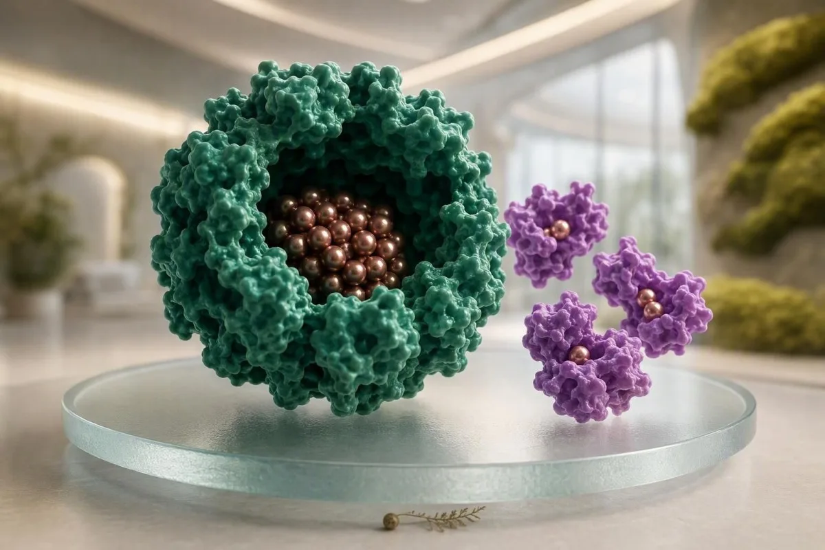
Ferritin is usually the better indicator of stored iron, while serum iron is a fast-moving snapshot of iron circulating today. Reading them together prevents many false conclusions.


- Ferritin below 15 ng/mL confirms iron deficiency in most otherwise healthy adults; it reflects stored iron better than serum iron.
- Serum iron can shift by 30% or more across a day and may rise sharply after an iron tablet or iron-rich meal.
- Inflammation can raise ferritin despite low usable iron; a ferritin below 100 ng/mL with transferrin saturation below 20% can still support deficiency in chronic disease.
- Transferrin saturation below 20% suggests too little iron is available for red-cell production, especially when paired with low ferritin.
- Test timing matters: a morning, fasting sample before the daily iron dose is usually the most comparable repeat measurement.
- Ferritin above 300 ng/mL in men or 200 ng/mL in women deserves clinical context, but does not alone diagnose iron overload.
- CRP and soluble transferrin receptor can clarify an apparently normal ferritin when inflammation is present.
Which result actually shows iron stores?
Ferritin is the test that best estimates iron stores, whereas serum iron measures iron temporarily circulating on the day of the draw. A low ferritin is far more diagnostically stable than a single low serum iron result.
In practical terms, ferritin is the warehouse inventory and serum iron is the number of delivery vans currently on the road. In an adult without active inflammation, ferritin below 15 ng/mL is highly specific for absent or near-absent stores; many clinicians investigate symptoms and bleeding sources once it falls below 30 ng/mL. Serum iron may be 45 µg/dL one morning and 110 µg/dL on another without any meaningful change in body iron.
The more useful companion to serum iron is transferrin saturation (TSAT), calculated as serum iron divided by total iron-binding capacity. TSAT below 20% indicates restricted iron availability, while values above 45% on repeat fasting studies raise the question of iron loading. Our iron studies guide explains why these values belong on the same page.
Kantesti is an AI blood test analyzer that reads ferritin, iron, TIBC, TSAT, haemoglobin, MCV and inflammatory markers as a pattern rather than declaring a result from one number. As of August 20, 2026, that distinction remains central to safe interpretation: ferritin stores change over weeks to months; serum iron fluctuations can occur within hours.
What ferritin and serum iron measure biologically
Ferritin measures stored iron inside cells, while serum iron measures transferrin-bound iron in circulation. They answer different biological questions and should not be treated as interchangeable.
Ferritin is a protein shell capable of holding thousands of iron atoms, mainly in liver cells, macrophages and bone marrow. A small amount leaks or is secreted into serum, giving us a useful indirect readout of total-body reserves. One ng/mL of ferritin roughly corresponds to 8-10 mg of stored iron in healthy adults, although this rough conversion fails during inflammation or liver-cell injury.
Serum iron is almost entirely iron attached to transferrin, the transport protein that delivers iron to marrow for haemoglobin production. It is not a measure of all iron in the body. A person with ferritin of 8 ng/mL can have a temporarily normal serum iron after breakfast, and a person with ferritin of 400 ng/mL can have low serum iron during pneumonia.
When I review a panel, I also look at the low transferrin saturation pattern and red-cell indices. MCV often drops only after stores have been low for some time, so normal haemoglobin and normal MCV do not rule out iron depletion.
Why the body separates storage from transport
The body keeps free iron extremely low because unbound iron can drive oxidative chemical reactions. Ferritin locks iron away; transferrin carries it in a controlled form. That separation is why ferritin vs serum iron disagreement is often physiology, not a laboratory error.
Why ferritin and serum iron commonly disagree
Ferritin and serum iron disagree because serum iron changes rapidly, whereas ferritin is altered by storage, inflammation and liver-cell release. The apparent contradiction often becomes clear once timing and CRP are known.
A classic example is serum iron of 35 µg/dL with ferritin of 75 ng/mL during a chest infection. Hepcidin, a liver-made hormone, blocks iron release from macrophages and gut cells, so circulating iron falls even though ferritin is normal or high. That is functional iron deficiency, meaning iron exists but is not reaching marrow efficiently.
The opposite pattern—serum iron 180 µg/dL with ferritin 12 ng/mL—often follows a 65 mg elemental-iron tablet, an iron-containing multivitamin or a meat-rich meal shortly before testing. This is why a high serum iron result does not reliably prove healthy stores. For preparation details, see iron and fasting advice.
Dr. Thomas Klein has seen this mismatch repeatedly in people tested after an endurance event: transient muscle stress, dehydration and a morning supplement can make the panel look far more reassuring than the underlying ferritin. A repeat, standardized sample is usually more informative than arguing over a single draw.
How inflammation can make ferritin look falsely reassuring
Inflammation raises ferritin because ferritin is an acute-phase reactant, so a normal ferritin cannot reliably exclude iron deficiency during illness. CRP, TSAT and sometimes soluble transferrin receptor help separate storage from inflammatory sequestration.
Interleukin-6 increases hepcidin, and hepcidin closes ferroportin—the cellular export channel for iron. The result is low serum iron, low or normal TIBC, low TSAT, and ferritin that may be 50, 100 or 300 ng/mL despite limited iron for erythropoiesis. A CRP above 5 mg/L makes ferritin less specific for stores.
The World Health Organization recommends using a ferritin threshold of less than 70 µg/L in adults with infection or inflammation to identify iron deficiency (WHO, 2020). In chronic kidney disease, heart failure and inflammatory bowel disease, several specialty guidelines use ferritin below 100 ng/mL plus TSAT below 20% as a practical deficiency pattern, though the exact threshold remains debated.
Kantesti is an AI biomarker interpretation platform that flags ferritin alongside CRP, ESR, kidney function and liver enzymes, because an isolated ferritin can mislead in either direction. Readers with a high CRP may find our guide to ferritin and CRP useful before assuming stores are adequate.
How meals, supplements and time of day shift serum iron
Recent meals, oral iron and morning-versus-afternoon sampling can alter serum iron substantially without changing iron stores. Ferritin is less sensitive to these short-term exposures, although it still should be trended under similar conditions.
Serum iron is commonly highest in the morning and tends to decline later in the day, although individual variation is wide enough that timing alone should not diagnose deficiency. In one classic clinical study, fasting did not eliminate all variation, but samples collected after 12 hours of fasting could show higher iron values than shorter fasts. Consistency matters more than chasing a perfect clock time.
For a planned iron panel, I generally advise a morning sample, usual water intake, and no oral iron for at least 24 hours unless the prescribing clinician says otherwise. This is a testing convention, not a reason to stop prescribed treatment without guidance. A multivitamin with 18 mg iron can be enough to distort serum iron for that visit.
A 29-year-old vegetarian patient once had serum iron of 156 µg/dL after a supplement, yet ferritin of 9 ng/mL and TSAT of 32%. Her repeat pre-dose TSAT was 12%, which fit the symptoms far better. Foods with heme and non-heme iron can support intake, but dietary change alone may not correct an ongoing loss.
Patterns that point to true iron deficiency
True iron deficiency usually produces low ferritin and low TSAT, often before haemoglobin falls. The most convincing pattern is ferritin below 30 ng/mL with TSAT below 20%, after considering inflammation.
Early deficiency can show ferritin 18 ng/mL, haemoglobin 13.2 g/dL and MCV 87 fL—technically no anaemia, but no comfortable reserve either. Symptoms such as exertional fatigue, restless legs, hair shedding or reduced exercise tolerance are real but non-specific; they deserve a search for cause rather than reflex supplementation. See low ferritin without heavy periods for the overlooked gastrointestinal and dietary clues.
Once iron-restricted erythropoiesis develops, RDW often rises before MCV falls, reflecting mixed older normal-sized and newer smaller red cells. A reticulocyte haemoglobin measurement below about 28 pg, where available, can show insufficient recent iron delivery to marrow. Laboratory cutoffs vary by assay and population.
Camaschella's review in the New England Journal of Medicine describes ferritin as the most accurate single marker of iron stores in uncomplicated deficiency, while stressing that inflammatory states complicate its interpretation (Camaschella, 2015). That nuance is the difference between treating a number and understanding a pattern.
Patterns that suggest iron restriction from chronic disease
Inflammation-related iron restriction usually causes low serum iron and TSAT with ferritin that is normal or elevated. TIBC is often low or normal because transferrin falls during the acute-phase response.
In uncomplicated iron deficiency, transferrin production usually rises, making TIBC high—often above 400 µg/dL—while ferritin falls. In inflammatory iron restriction, transferrin is downregulated, so TIBC may be below 300 µg/dL despite low serum iron. That opposite TIBC direction is clinically useful and often missed.
Soluble transferrin receptor (sTfR) is less affected by inflammation than ferritin and can help when ferritin is 50-150 ng/mL with a raised CRP. A high sTfR suggests cellular iron need or increased red-cell production; it is not completely immune to confounding, particularly with haemolysis or thalassaemia. Our soluble transferrin receptor guide covers when it is worth requesting.
In my experience, clinicians should not label this pattern “anaemia of chronic disease” and stop there. The chronic condition may coexist with absolute deficiency from menstrual loss, gastrointestinal blood loss, reduced absorption or frequent donation—two processes can be true at once.
When a high ferritin does not mean iron overload
High ferritin is more often caused by inflammation, fatty liver, alcohol exposure, metabolic disease or cell injury than by iron overload. Transferrin saturation is the essential second test when ferritin is elevated.
Ferritin above 200 ng/mL in women or 300 ng/mL in men is outside many laboratory reference intervals, but reference intervals are not diagnoses. TSAT persistently above 45% is more suggestive of increased iron absorption or overload than ferritin alone. A serum iron drawn after supplements, however, can make TSAT falsely impressive.
Ferritin may exceed 500 ng/mL in metabolic dysfunction-associated steatotic liver disease, acute infection, heavy alcohol use or poorly controlled diabetes. Liver enzymes, CRP, fasting glucose and alcohol history change the interpretation. Review liver-support evidence rather than assuming a “detox” will explain or fix a high number.
Ferritin above 1,000 ng/mL warrants timely medical assessment, especially with raised ALT or AST, unexplained fatigue, skin pigmentation, joint symptoms or a family history of iron overload. It is rarely an emergency by itself, but it should not sit unreviewed for six months.
Why pregnancy, endurance sport and blood donation need different context
Pregnancy, endurance training and blood donation can lower ferritin before standard CBC values become abnormal. These groups need trend-based interpretation because changing plasma volume and iron demand can mask depletion.
During pregnancy, plasma volume expansion lowers haemoglobin by dilution, while fetal and placental demands increase iron needs. Many obstetric services treat ferritin below 30 ng/mL as iron deficiency in pregnancy, but local protocols differ. For other pregnancy-specific reference shifts, consult our pregnancy lab guide.
Endurance athletes can lose iron through gastrointestinal micro-loss, sweat, dietary restriction and foot-strike haemolysis; low ferritin may impair training quality even with haemoglobin in range. Testing immediately after a marathon is poor practice because inflammation and fluid shifts cloud ferritin and iron. A quieter training week gives a cleaner baseline.
Whole-blood donation removes roughly 200-250 mg of iron per donation. Ferritin can remain low for months even after haemoglobin passes donor screening, so frequent donors should consider a scheduled ferritin check and discuss replacement with their clinician. Our post-donation ferritin timing guide gives a practical framework.
Medicines and gut conditions that change the iron picture
Acid-suppressing medicines, coeliac disease, bariatric surgery and ongoing gastrointestinal loss can cause low ferritin despite an apparently iron-rich diet. Serum iron may still look normal on a supplement day.
Non-heme iron needs an acidic, reducing environment and dedicated intestinal transporters, so long-term proton-pump inhibitor use can contribute to poor absorption in susceptible people. Coeliac disease may present first as iron deficiency, sometimes without obvious bowel symptoms. Unexplained iron deficiency in adult men and postmenopausal women needs a cause-focused medical work-up, not just tablets.
Calcium taken with iron can reduce absorption during that dose, while vitamin C-containing food may improve non-heme iron uptake modestly. The effect is smaller than many marketing claims suggest; the practical obstacle is often nausea, constipation or an unrecognized source of loss. Iron bisglycinate versus sulfate reviews tolerability differences.
Dr. Thomas Klein advises patients not to self-treat a ferritin of 6 ng/mL indefinitely without asking why it fell. If periods are not heavy, the history should include donation, diet, NSAID use, bowel symptoms, prior surgery and family history; the answer is occasionally simple, but not always.
How to prepare for the most reliable iron panel
For the most comparable iron panel, use the same laboratory, test in the morning when possible, avoid the pre-test iron dose, and record recent illness. Ferritin changes slowly, but serum iron needs controlled conditions to be meaningful.
Ask for a CBC, ferritin, serum iron, TIBC or transferrin, and calculated TSAT; add CRP when infection, inflammatory disease or obesity-related inflammation is plausible. A fasting period of 8-12 hours is commonly used for iron studies, although laboratories differ, so follow the laboratory's specific instructions. Water is fine unless told otherwise.
Do not schedule a routine iron panel during fever, within several days of a hard endurance event, or immediately after an iron infusion unless the treating team specifically needs it. Intravenous iron can elevate ferritin for weeks, making early rechecks hard to interpret. Our blood-test timing guide details other short-term influences.
Kantesti is an AI-powered blood test analysis tool that can compare prior ferritin, TSAT and CBC values after you upload a report, highlighting whether a change exceeds ordinary noise. The useful record is not only the number: save test time, fasting status, supplement dose, menstrual timing, recent illness and donation date.
What should happen after iron treatment starts
Effective iron treatment usually raises reticulocytes within 7-10 days and haemoglobin by about 1-2 g/dL over 2-4 weeks if the diagnosis and absorption are correct. Ferritin replenishes later than haemoglobin.
A good response is not judged from serum iron the morning after a tablet. Serum iron may spike immediately, while the clinically meaningful response is a reticulocyte rise followed by a sustained haemoglobin increase. Failure of haemoglobin to rise by about 1 g/dL after 2-4 weeks should trigger review of adherence, ongoing loss, absorption and the original diagnosis.
Ferritin often takes several months to rebuild, particularly if the cause continues. Many clinicians continue oral iron for around 3 months after haemoglobin normalizes, then recheck ferritin, but treatment length should be individualized for tolerance, pregnancy, kidney disease and the source of deficiency. See iron treatment retest timing for a safer monitoring plan.
High RDW during early treatment is not necessarily bad news; it can reflect new iron-replete red cells entering circulation alongside older microcytic cells. A rising RDW with a rising reticulocyte count is different from worsening anaemia without a marrow response.
When ferritin or serum iron results need prompt review
Seek prompt medical review for severe symptoms, falling haemoglobin, black stools, chest pain, breathlessness at rest, or ferritin above 1,000 ng/mL. Iron studies guide evaluation but do not replace an assessment of bleeding, infection or organ disease.
Anaemia can become urgent when it causes fainting, chest pressure, new breathlessness at rest, a racing heartbeat or marked weakness. Haemoglobin below 8 g/dL (80 g/L) is often clinically significant, but urgency depends on symptoms, speed of decline, pregnancy, cardiac disease and active loss. Black tarry stools or vomiting material that resembles coffee grounds require urgent care.
A low ferritin in an adult man or a person after menopause should prompt discussion of gastrointestinal evaluation, because chronic blood loss must be excluded. In contrast, a single low serum iron with normal ferritin, normal TSAT and recent illness commonly needs repeat testing rather than panic. Pale-skin and anaemia clues can help readers recognize the broader symptom picture.
Kantesti AI does not diagnose or prescribe; it organizes the questions a clinician needs to answer. Our medical review standards are described through the Medical Advisory Board, where physician oversight remains the safeguard when results and symptoms do not line up.
The best next tests when ferritin and iron conflict
When ferritin and serum iron conflict, repeat a standardized full iron panel and add CRP; soluble transferrin receptor, reticulocyte haemoglobin or haemoglobin electrophoresis may be appropriate next tests. The right follow-up depends on the pattern, not the isolated flag.
Low serum iron plus ferritin 80 ng/mL should usually lead to TSAT, TIBC and CRP before an iron diagnosis is made. Low ferritin plus normal serum iron usually needs a cause assessment, not reassurance. Ferritin below 30 ng/mL is generally sufficient evidence of low stores in an otherwise well adult, even if the serum iron happened to be normal.
If MCV is low but ferritin and TSAT are not, consider thalassaemia trait, chronic inflammation, lead exposure in relevant settings or less common causes. A high red-cell count with disproportionately low MCV is one clue that haemoglobin electrophoresis may be more useful than escalating iron. The MCV and MCH comparison explains this distinction.
Kantesti AI interprets discordant ferritin vs serum iron results through linked biomarkers and longitudinal changes, while our clinical validation framework describes the limits of automated interpretation. The final decision belongs with the clinician who can examine you, assess bleeding risk and order targeted follow-up.
A practical bottom line for reading an iron panel
Use ferritin to estimate stored iron, use serum iron and TSAT to assess current availability, and use CRP to judge whether inflammation is distorting the picture. No single iron marker is definitive in every clinical setting.
The most reliable everyday sequence is simple: confirm whether ferritin is low, then inspect TSAT, TIBC, CBC and CRP for the reason it is low—or for the reason it only appears adequate. A ferritin of 10 ng/mL deserves attention even with normal haemoglobin; a serum iron of 35 µg/dL after an infection often deserves context first. That is the honest answer to ferritin vs serum iron.
Kantesti, an AI lab test interpretation service, helps users preserve the contextual details that make repeat tests clinically interpretable across time. For people with multiple reports, our lab trend analysis guide shows why a 6-month ferritin slope is often more meaningful than one serum iron result.
Research publications: Klein, T. (2026). Clinical Validation Framework v2.0 (Medical Validation Page). Zenodo. https://doi.org/10.5281/zenodo.17993721. ResearchGate: https://www.researchgate.net/; Academia.edu: https://www.academia.edu/. Klein, T. (2026). AI Blood Test Analyzer: 2.5M Tests Analyzed | Global Health Report 2026. Zenodo. https://doi.org/10.5281/zenodo.18175532. ResearchGate: https://www.researchgate.net/; Academia.edu: https://www.academia.edu/.
Frequently Asked Questions
Is ferritin more accurate than serum iron for iron deficiency?
Ferritin is more accurate than serum iron for estimating iron stores in people without active inflammation. A ferritin below 15 ng/mL strongly supports iron deficiency, and many clinicians regard values below 30 ng/mL as depleted or borderline-low stores when symptoms or risk factors are present. Serum iron can change after food, supplements and time of day, so a normal result does not exclude low stores. During inflammation, ferritin can rise artificially, making TSAT and CRP necessary companions.
Can serum iron be low while ferritin is normal?
Yes, low serum iron with normal or high ferritin commonly occurs during infection, autoimmune disease, chronic kidney disease and other inflammatory states. Hepcidin reduces iron release into plasma, so serum iron and TSAT may fall below 20% even while ferritin is 50-300 ng/mL. This pattern is called iron restriction or functional iron deficiency. A CRP above 5 mg/L and a low or normal TIBC make inflammation more likely.
Should I fast for ferritin and serum iron testing?
Fasting is not usually crucial for ferritin, but an 8-12 hour fast can make serum iron and TSAT easier to compare with prior results. Many clinicians prefer a morning sample because serum iron often declines later in the day. Do not take the morning oral iron dose before a planned iron panel unless your clinician has directed otherwise; holding it for about 24 hours is commonly advised. Always follow the instructions supplied by the laboratory that performs the test.
Can an iron supplement raise serum iron but not ferritin?
Yes, an oral iron dose can temporarily raise serum iron within hours while ferritin remains low because stores take weeks to months to replenish. For example, a person with ferritin of 12 ng/mL may show a normal or high serum iron after taking 65 mg elemental iron that morning. That does not mean the deficiency has resolved. Treatment response is better judged by symptoms, reticulocytes, haemoglobin and ferritin on a properly timed recheck.
What ferritin level is too high?
Ferritin above 200 ng/mL in women or 300 ng/mL in men is above many reference ranges, but it does not automatically mean iron overload. Inflammation, liver disease, alcohol exposure and metabolic disease frequently raise ferritin. Persistent TSAT above 45% makes iron overload more plausible, while ferritin above 1,000 ng/mL warrants timely clinician-led evaluation. The cause is determined from the full panel, symptoms, liver tests and sometimes genetic or imaging assessment.
Can I have iron deficiency with normal haemoglobin?
Yes, iron deficiency often develops before haemoglobin becomes low. Ferritin can fall below 30 ng/mL while haemoglobin remains above 12 g/dL in women or 13 g/dL in men and MCV remains normal. Some people report fatigue, reduced exercise capacity, restless legs or hair shedding at this stage, although those symptoms have many causes. The priority is confirming the pattern and identifying why iron stores are falling.
Get AI-Powered Blood Test Analysis Today
Join over 2 million users worldwide who trust Kantesti for instant, accurate lab test analysis. Upload your blood test results and receive comprehensive interpretation of 15,000+ biomarkers in seconds.
📚 Referenced Research Publications
Klein, T., Mitchell, S., & Weber, H. (2026). Clinical Validation Framework v2.0 (Medical Validation Page). Kantesti AI Medical Research.
Klein, T., Mitchell, S., & Weber, H. (2026). AI Blood Test Analyzer: 2.5M Tests Analyzed | Global Health Report 2026. Kantesti AI Medical Research.
📖 External Medical References
World Health Organization (2020). WHO guideline on use of ferritin concentrations to assess iron status in individuals and populations. World Health Organization.
⚕️ Medical Disclaimer
This article is for educational purposes only and does not constitute medical advice. Always consult a qualified healthcare provider for diagnosis and treatment decisions.
E-E-A-T Trust Signals
Experience
Physician-led clinical review of lab interpretation workflows.
Expertise
Laboratory medicine focus on how biomarkers behave in clinical context.
Authoritativeness
Written by Dr. Thomas Klein with review by Dr. Sarah Mitchell and Prof. Dr. Hans Weber.
Trustworthiness
Evidence-based interpretation with clear follow-up pathways to reduce alarm.