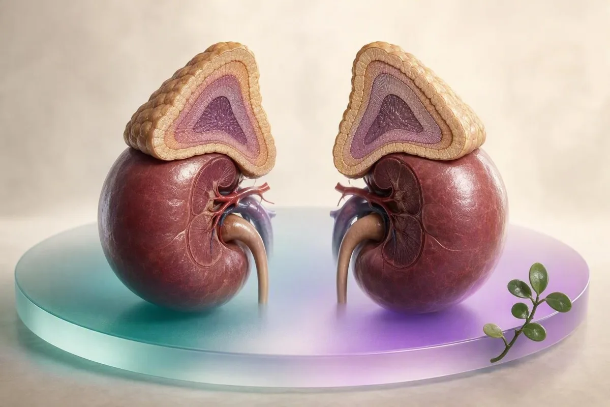
Cortisol has no single universal normal range. The collection clock time, specimen, assay and reporting unit can change what a result means.


- Morning serum cortisol is commonly about 5-25 µg/dL (138-690 nmol/L) at 6-8 am, but the printed laboratory interval is the only valid comparator.
- Evening cortisol levels should be lower than morning values; a 4 pm result cannot be judged against an 8 am reference interval.
- Unit conversion is reliable for the same analyte: 1 µg/dL cortisol equals 27.59 nmol/L and 10 ng/mL.
- Serum cortisol usually measures total, protein-bound plus free cortisol; salivary and urine tests answer different clinical questions.
- A morning cortisol below 3 µg/dL (83 nmol/L) can strongly support adrenal insufficiency in the right setting, while values above 15 µg/dL (414 nmol/L) often make it less likely.
- Late-night salivary cortisol and 24-hour urinary free cortisol are screening tools for cortisol excess, not substitutes for a morning serum cortisol.
- Modern immunoassays and LC-MS/MS can produce different cortisol values, particularly at low concentrations and after synthetic steroid exposure.
- Medication history matters: hydrocortisone, prednisolone, inhaled steroids, estrogen, anticonvulsants and biotin can each alter interpretation.
- Trend comparisons are most useful when collection time, specimen and laboratory method are matched.
- Urgent symptoms such as fainting, repeated vomiting, severe weakness, confusion or low blood pressure need same-day clinical assessment, regardless of a home interpretation.
Why there is no single normal range for cortisol
The normal range for cortisol depends first on collection time, then on specimen and assay. As of August 19, 2026, many laboratories quote an adult 6-8 am total serum interval near 5-25 µg/dL (138-690 nmol/L), yet an evening value in that range could be unexpectedly high rather than reassuring.
Cortisol normally peaks shortly after waking and declines across the day, so a result is inseparable from its timestamp. A serum cortisol of 8 µg/dL (221 nmol/L) may be unremarkable at 4 pm but deserves context if collected at 8 am during acute illness. In my 15 years of clinical practice, the missing collection time has caused more avoidable alarm than modest numerical abnormalities.
Laboratories build reference intervals from their own population, instrument and calibration materials; they are not universal treatment targets. Kantesti is an AI blood test analyzer that reads cortisol alongside collection time, units, specimen and adjacent adrenal markers rather than colouring one number red. This is particularly useful when people compare reports from two countries or two laboratories.
A single cortisol result does not diagnose Cushing syndrome or adrenal insufficiency. The Endocrine Society advises against using random serum cortisol to screen for Cushing syndrome because the normal circadian swing is too wide (Nieman et al., 2008). For related hormone context, see our guide to cortisol and ACTH patterns.
The clock time belongs beside the result
A cortisol report without a collection time is clinically incomplete for most outpatient questions. Record whether you woke at 5 am or worked overnight, because the biological morning peak follows the sleep-wake cycle more closely than the wall clock in shift workers.
Morning cortisol levels: useful intervals and diagnostic cutoffs
Morning cortisol levels are highest near 6-8 am, and many adult serum laboratories use roughly 5-25 µg/dL or 138-690 nmol/L. These values describe total cortisol in a reference population; they are not a stand-alone pass-or-fail test of adrenal health.
For suspected adrenal insufficiency, an 8-9 am cortisol below 3 µg/dL (83 nmol/L) is strongly concerning when symptoms and ACTH fit; a value above 15 µg/dL (414 nmol/L) often indicates adequate reserve in stable outpatients. Values between 3 and 15 µg/dL are an indeterminate zone, not a diagnosis, and usually prompt an ACTH stimulation test or specialist review.
The old stimulated cortisol target of 18 µg/dL (500 nmol/L) cannot simply be carried into every modern assay. Javorsky and colleagues found that assay-specific stimulated cutoffs near 14-15 µg/dL may prevent false diagnoses of adrenal insufficiency with newer assays (Javorsky et al., 2021). That technical detail changes real lives; I have seen patients unnecessarily started on glucocorticoids after an outdated threshold was applied.
If your report says 7.2 µg/dL at 07:45, do not convert it into a verdict before checking illness, sleep, steroid exposure and albumin. The practical next comparison is usually a properly timed repeat with ACTH sample handling planned in advance, because ACTH is far less stable than cortisol.
Evening cortisol levels should be lower than morning values
Evening cortisol levels normally fall well below morning values because the hypothalamic-pituitary-adrenal axis follows a daily rhythm. A common late-afternoon serum interval is approximately 2-11 µg/dL (55-303 nmol/L), although laboratories differ substantially.
At 4-6 pm, a total serum cortisol of 12 µg/dL (331 nmol/L) can sit above a laboratory's expected interval even though that same number would be ordinary at 8 am. This is why comparing an evening draw to a morning screenshot online is unsafe. The direction of change across the day can matter as much as the absolute value.
Late-night testing is used mainly when clinicians suspect cortisol excess, not because every tired person needs a nighttime cortisol check. Late-night salivary cortisol is normally very low near bedtime; each laboratory sets its own upper limit, often around 0.09-0.15 µg/dL (2.5-4.1 nmol/L) depending on assay and collection device.
An overnight worker who sleeps from 09:00 to 16:00 may have a shifted cortisol rhythm, making conventional clock-time intervals less dependable. In these cases I ask for sleep timing, recent travel and light exposure before interpreting results; our article on jet lag and test timing explains why this history changes the plan. Kantesti is an AI blood test interpretation platform that preserves this timing context when comparing hormone reports.
Cortisol lab units: µg/dL, nmol/L and ng/mL
Cortisol lab units can be converted precisely only when the specimen and analyte are the same. For total cortisol, 1 µg/dL equals 27.59 nmol/L and equals 10 ng/mL; conversion does not make a serum result comparable with saliva or urine.
The conversion formula is simple: µg/dL multiplied by 27.59 equals nmol/L, while nmol/L divided by 27.59 equals µg/dL. Thus 10 µg/dL is 276 nmol/L, and 500 nmol/L is 18.1 µg/dL. A report using ng/mL shows 10 µg/dL as 100 ng/mL, which looks ten times larger but is the same concentration.
Do not apply that formula blindly to urinary free cortisol reporting in µg per 24 hours or nmol per 24 hours. Those units describe total hormone excreted over a timed collection, not a concentration at one moment. A collection of 60 µg/24 hours may be within one laboratory's limit and above another's, depending on methodology and local validation.
A surprisingly common error is confusing µg/dL with µg/L; they differ by a factor of 10. Dr. Thomas Klein recommends saving the original PDF rather than manually copying values into notes, especially when reviewing a hormone panel with other DHEA-S clues.
Serum, saliva and urine cortisol measure different things
Serum, saliva and urine cortisol tests are not interchangeable because they sample different fractions and time windows. Serum usually reports total cortisol at one time point, saliva estimates free cortisol at collection, and urine reflects free cortisol excretion over 24 hours.
Total serum cortisol includes cortisol bound to corticosteroid-binding globulin, albumin and the small free fraction. High estrogen states raise corticosteroid-binding globulin and can raise total cortisol without true cortisol excess. Low albumin or markedly low binding globulin can do the reverse, leaving a total serum value deceptively low.
Salivary cortisol is useful near bedtime because only free hormone diffuses into saliva, but contamination matters. Brushing teeth, oral bleeding, steroid creams transferred from fingers, tobacco and eating close to collection can distort a low-level sample. Our detailed comparison of saliva and urine cortisol tests covers when each specimen is sensible.
Twenty-four-hour urinary free cortisol averages secretion across a day and is generally interpreted against the collection's upper limit of normal, often around 50 µg/24 hours (138 nmol/24 hours) in a given laboratory. Kantesti AI interprets cortisol results by separating total serum, late-night salivary and urinary free cortisol rather than merging them into one score.
Why test methods produce different cortisol ranges
Cortisol reference intervals vary by test method because immunoassays and LC-MS/MS do not detect exactly the same chemical signal. Immunoassays can cross-react with related steroids, while mass spectrometry generally provides greater analytical specificity.
Automated immunoassays are fast and widely available, but prednisolone and some steroid metabolites may interfere depending on the manufacturer. LC-MS/MS separates molecules by mass and is especially helpful when results conflict with symptoms or when exogenous steroid exposure is plausible. Neither method is inherently “wrong”; the lab-specific interval must match the method used.
Assay bias becomes most visible at low cortisol concentrations, exactly where late-night salivary cortisol and adrenal testing decisions can be sensitive. A 2 µg/dL difference around a stimulated cutoff may change whether someone is labelled deficient. Bornstein and colleagues advise assay-aware interpretation in suspected primary adrenal insufficiency rather than treating one historical cutoff as universal (Bornstein et al., 2016).
Kantesti AI flags method changes when they are visible on a report and avoids declaring a trend when an immunoassay has been replaced by LC-MS/MS. Readers can review our clinical validation approach to see why source ranges and collection metadata matter as much as optical character recognition.
Sleep, stress and illness can change a cortisol result quickly
Poor sleep, acute illness, pain and intense exercise can raise cortisol within hours, whereas chronic illness may flatten its usual daily fall. These effects can move a result across a reference boundary without proving an adrenal disorder.
Cortisol begins to rise about 2-3 hours before usual waking, then often increases a further 38-75% during the first 30-45 minutes after awakening. That cortisol awakening response is normal physiology, which is why an 06:30 draw after a 04:30 awakening cannot be interpreted exactly like an 08:00 draw after an undisturbed night.
Fever, major dental procedures, a marathon, panic, severe calorie restriction and hospitalization can all increase serum cortisol. Conversely, severe critical illness can alter binding proteins so total cortisol and free cortisol diverge. I see this pattern often after poor sleep; our practical guide to labs after sleepless nights helps distinguish a repeat-worthy result from an urgent one.
For a planned baseline test, keep wake time as usual, avoid unusually hard training for 24 hours and sit quietly for 15-30 minutes before collection if the laboratory permits. Do not try to “lower cortisol” by skipping prescribed medicine or fasting longer than instructed; that can create a less useful, not more accurate, result.
How to compare cortisol results without creating a false trend
A cortisol change is most believable when collection time, specimen, laboratory method and medication status are matched. A 30% difference between two unmatched cortisol draws may be normal biological variation rather than a change in adrenal function.
Start with four fields: exact collection time, reported unit, specimen type and reference interval printed beside the result. Add wake time, recent steroid doses, pregnancy status, acute illness and whether the laboratory changed. This small audit often explains an apparent rise from 9 to 16 µg/dL without any new disease.
If repeats are being used to evaluate an endocrine problem, try to collect at the same clock time within 30-60 minutes and use the same lab. A morning serum result from one laboratory and a late-night saliva result from another should be treated as complementary tests, not a rising or falling series. Our side-by-side result comparison guide gives a patient-friendly record format.
Kantesti is an AI-powered blood test analysis tool that compares cortisol only after checking whether the results share a meaningful clinical context. Its trend view cannot replace dynamic endocrine testing, but it can surface the question a clinician needs answered: “Were these actually comparable samples?”
Pregnancy and estrogen can raise total cortisol without Cushing syndrome
Pregnancy and oral estrogen commonly raise total serum cortisol by increasing corticosteroid-binding globulin. A high total cortisol value in these settings may reflect more binding protein rather than excessive free cortisol action.
During pregnancy, total serum cortisol can reach roughly 2-3 times non-pregnant values by the third trimester, and the daily rhythm is also less sharply defined. Standard non-pregnant serum reference intervals are therefore poor tools for screening cortisol excess in pregnancy. Clinicians usually choose pregnancy-aware specialist testing when symptoms are compelling.
Combined oral contraceptives and oral estrogen can increase binding globulin over several weeks; transdermal estrogen usually has a smaller hepatic binding effect. In contrast, nephrotic protein loss, severe liver disease and critical illness may lower binding proteins and reduce total cortisol. The result is a biochemical illusion unless the context is visible.
A person taking an estrogen-containing pill with a morning cortisol of 28 µg/dL (773 nmol/L) does not automatically have Cushing syndrome. I would ask about weight change, bruising, new diabetes, blood pressure and physical findings before choosing a test. Hormone timing also matters in men, as discussed in our morning hormone testing guide.
Steroids and common medicines can distort cortisol testing
Hydrocortisone, prednisolone and many inhaled, topical or injected steroids can suppress the body’s cortisol production or interfere with measurement. Never stop a prescribed steroid abruptly just to prepare for a cortisol test.
Hydrocortisone is chemically identical to cortisol and will generally appear as cortisol in serum testing. Prednisolone may cross-react in some immunoassays, while dexamethasone usually has little direct assay cross-reactivity but can suppress ACTH and endogenous cortisol. The exact washout plan depends on the indication, dose and clinical risk, so it belongs with the prescriber or endocrinologist.
Inhaled steroids, potent skin preparations, joint injections and “natural” supplements can cause clinically relevant suppression, particularly with repeated or high-dose exposure. Enzyme-inducing drugs such as carbamazepine, phenytoin and rifampicin can speed cortisol metabolism and complicate replacement therapy. Ritonavir can have the opposite practical problem by increasing exposure to some inhaled steroids.
The term “adrenal fatigue” is not an accepted endocrine diagnosis, and supplements marketed for it can contain undisclosed steroids or stimulants. Before purchasing products to alter a laboratory number, read our evidence-based review of adrenal supplements.
When a low cortisol result needs urgent medical review
Low cortisol becomes urgent when it occurs with low blood pressure, fainting, repeated vomiting, severe weakness, confusion, low sodium or high potassium. These features can signal adrenal crisis, which requires immediate medical treatment rather than outpatient retesting.
A morning cortisol below 3 µg/dL (83 nmol/L) with compatible symptoms should prompt urgent clinician contact, especially after recent steroid withdrawal, pituitary disease, adrenal surgery or severe infection. In a suspected adrenal crisis, clinicians draw cortisol and ACTH if this does not delay care, then administer stress-dose glucocorticoid treatment. Waiting for a repeat home sample is unsafe.
The pattern matters: low cortisol plus high ACTH, low sodium and high potassium supports primary adrenal insufficiency, while low or inappropriately normal ACTH suggests a pituitary or hypothalamic cause. Potassium may remain normal in secondary adrenal insufficiency because aldosterone is often preserved. That distinction guides both imaging and replacement treatment.
If symptoms are milder but persistent, arrange timely review rather than assuming fatigue alone proves cortisol deficiency. Our article on Addison disease clues explains the symptom-and-lab pattern that merits active follow-up.
A high cortisol result rarely confirms Cushing syndrome by itself
A high morning serum cortisol does not diagnose Cushing syndrome because stress, estrogen and binding-protein changes commonly elevate total cortisol. Clinicians usually confirm suspected cortisol excess with two abnormal screening assessments rather than one serum value.
The Endocrine Society recommends late-night salivary cortisol, 24-hour urinary free cortisol or a 1 mg overnight dexamethasone suppression test as initial options when clinical suspicion is genuine (Nieman et al., 2008). Typical concerning features include progressive central weight gain, wide purple striae, easy bruising, proximal muscle weakness, new diabetes and difficult hypertension. Fatigue, anxiety and a high single morning cortisol are not enough.
For urinary free cortisol, laboratories often consider values above the assay-specific upper limit on at least two complete collections more persuasive than one elevated collection. A result more than 3 times the upper limit of normal is harder to dismiss, but alcohol excess, uncontrolled diabetes, major depression and acute illness can still create a pseudo-Cushing pattern.
A 52-year-old runner with a cortisol of 27 µg/dL after an overnight emergency department visit needs a different conversation from someone with repeated abnormal late-night saliva samples and worsening diabetes. For the broader differential, see causes of high cortisol.
Dynamic cortisol tests answer questions a baseline value cannot
ACTH stimulation and dexamethasone suppression tests assess cortisol response, not merely a resting concentration. Their accuracy depends on exact medication timing, sample collection and an assay-specific interpretation threshold.
In a standard ACTH stimulation test, cortisol is sampled before and usually 30 and 60 minutes after synthetic ACTH administration. With newer assays, a peak around 14-15 µg/dL may be adequate, whereas some laboratories retain a cutoff near 18 µg/dL; use the test laboratory's protocol. Baseline ACTH should ideally be collected before treatment when safe.
A 1 mg overnight dexamethasone suppression test normally suppresses next-morning serum cortisol to below 1.8 µg/dL (50 nmol/L) in many protocols. Estrogen, poor dexamethasone absorption, enzyme-inducing drugs and missed doses can yield false-positive results. This is one area where a specialist's pre-test instructions are far more valuable than improvisation.
A 24-hour urine collection needs every void from the start time through the finish time, stored exactly as directed. Under-collection is common and can falsely reassure; review our 24-hour urine collection checklist before repeating a borderline result.
Seven mistakes people make when comparing cortisol reports
The most common cortisol comparison mistake is treating every flagged result as the same test of the same hormone state. A laboratory flag indicates departure from that laboratory’s reference group, not a confirmed diagnosis or a universal target.
First, do not compare a 17:00 serum cortisol with an 08:00 interval. Second, do not convert a late-night saliva value into an “equivalent” serum value. Third, do not assume a change from 250 to 300 nmol/L is clinically meaningful if sleep, stress or assay changed between draws.
Fourth, do not omit steroid exposure because a cream, inhaler or injection seems unrelated. Fifth, do not use a single cortisol test to explain nonspecific tiredness without checking anemia, thyroid disease, sleep, mood, glucose and medication effects. Sixth, do not self-treat a low value with glandular extracts or unregulated corticosteroids.
Seventh, do not confuse “out of range” with dangerous. A result can be statistically unusual yet medically minor, while a value inside range can still be concerning if it is inappropriately low during severe illness. Our guide to out-of-range flags is a useful reset when a portal label feels alarming.
Research sources, AI context and the next safe step
The next safe step after an unusual cortisol result is to verify collection details, review medicines and decide with a clinician whether repeat or dynamic testing is needed. No AI output, reference interval or web article can diagnose an adrenal emergency from a number alone.
Dr. Thomas Klein’s rule in clinic is simple: retain the original report, write down wake time and all steroid exposures, then bring the complete pattern to the appointment. Kantesti is an AI lab test interpretation service that organizes cortisol with ACTH, sodium, potassium, glucose and prior results, while leaving diagnosis and treatment decisions to licensed clinicians. This prevents a unit mismatch from becoming a frightening story.
Our medical team reviews clinical standards and safety boundaries through the Medical Advisory Board, and the organization’s remit is described on our About Us page. If a report is scanned from a photo, confirm that the extracted unit, decimal point and collection time match the original before relying on any interpretation.
Related Kantesti publications provide methodology context for laboratory interpretation: Kantesti LTD. (2026). BUN/Creatinine Ratio Explained: Kidney Function Test Guide. Zenodo. https://doi.org/10.5281/zenodo.18207872; ResearchGate record and Academia.edu record. Kantesti LTD. (2026). Urobilinogen in Urine Test: Complete Urinalysis Guide 2026. Zenodo. https://doi.org/10.5281/zenodo.18226379; ResearchGate record and Academia.edu record.
Frequently Asked Questions
What is the normal range for cortisol in the morning?
The normal range for cortisol in an adult morning serum sample is often about 5-25 µg/dL, which equals 138-690 nmol/L, when collected near 6-8 am. Each laboratory must provide its own reference interval because assay calibration and local validation differ. In suspected adrenal insufficiency, an 8-9 am value below 3 µg/dL (83 nmol/L) is concerning, while a value above 15 µg/dL (414 nmol/L) often makes clinically significant deficiency less likely in a stable outpatient. Results between those values commonly need ACTH and sometimes stimulation testing.
What should cortisol be in the evening?
Evening cortisol levels should be lower than morning values, and many laboratories use a late-afternoon serum interval around 2-11 µg/dL or 55-303 nmol/L. The exact limit depends on the collection time, laboratory method and the person’s sleep schedule. A cortisol of 10 µg/dL can be ordinary in the morning but relatively high at 5 pm. Late-night salivary cortisol has separate, much lower cutoffs and should never be compared directly with serum cortisol.
How do I convert cortisol from µg/dL to nmol/L?
To convert total cortisol from µg/dL to nmol/L, multiply the result by 27.59. For example, 10 µg/dL equals 276 nmol/L, and 18 µg/dL equals 497 nmol/L. To convert nmol/L to µg/dL, divide by 27.59. This calculation applies only when the specimen and analyte are identical; it does not make serum, saliva and 24-hour urine cortisol results interchangeable.
Is a morning cortisol of 6 µg/dL low?
A morning cortisol of 6 µg/dL, or about 166 nmol/L, is not automatically diagnostic but often falls into an indeterminate zone for adrenal reserve. The meaning depends on collection time, wake time, symptoms, ACTH, steroid exposure, albumin and the reporting laboratory’s interval. Many clinicians investigate values between 3 and 15 µg/dL with repeat early-morning sampling or an ACTH stimulation test when suspicion is present. Severe weakness, vomiting, fainting or low blood pressure changes the urgency and warrants same-day clinical assessment.
Why is my cortisol high when I feel stressed?
Stress, pain, poor sleep, fever, intense exercise and acute illness can raise cortisol, particularly in a morning serum sample. A one-off high total cortisol above a laboratory limit does not establish Cushing syndrome, because estrogen and pregnancy can also raise cortisol-binding protein and total cortisol. Guidelines generally use late-night salivary cortisol, 24-hour urinary free cortisol or dexamethasone suppression testing when cortisol excess is genuinely suspected. Repeated abnormal screening tests plus compatible clinical features are more informative than one stressed-day result.
Can birth control raise cortisol blood test results?
Combined oral contraceptives can raise total serum cortisol by increasing corticosteroid-binding globulin, often without a comparable increase in free cortisol activity. This effect develops over weeks and can make a morning total cortisol appear high relative to a non-pregnant reference interval. Oral estrogen has a stronger binding-protein effect than transdermal estrogen in many patients. Tell the ordering clinician about all hormonal medicines before a dexamethasone suppression or adrenal evaluation is planned.
Get AI-Powered Blood Test Analysis Today
Join over 2 million users worldwide who trust Kantesti for instant, accurate lab test analysis. Upload your blood test results and receive comprehensive interpretation of 15,000+ biomarkers in seconds.
📚 Referenced Research Publications
Klein, T., Mitchell, S., & Weber, H. (2026). Kantesti LTD. (2026). BUN/Creatinine Ratio Explained: Kidney Function Test Guide. Zenodo.. Kantesti AI Medical Research.
Klein, T., Mitchell, S., & Weber, H. (2026). Kantesti LTD. (2026). Urobilinogen in Urine Test: Complete Urinalysis Guide 2026. Zenodo.. Kantesti AI Medical Research.
📖 External Medical References
⚕️ Medical Disclaimer
This article is for educational purposes only and does not constitute medical advice. Always consult a qualified healthcare provider for diagnosis and treatment decisions.
E-E-A-T Trust Signals
Experience
Physician-led clinical review of lab interpretation workflows.
Expertise
Laboratory medicine focus on how biomarkers behave in clinical context.
Authoritativeness
Written by Dr. Thomas Klein with review by Dr. Sarah Mitchell and Prof. Dr. Hans Weber.
Trustworthiness
Evidence-based interpretation with clear follow-up pathways to reduce alarm.