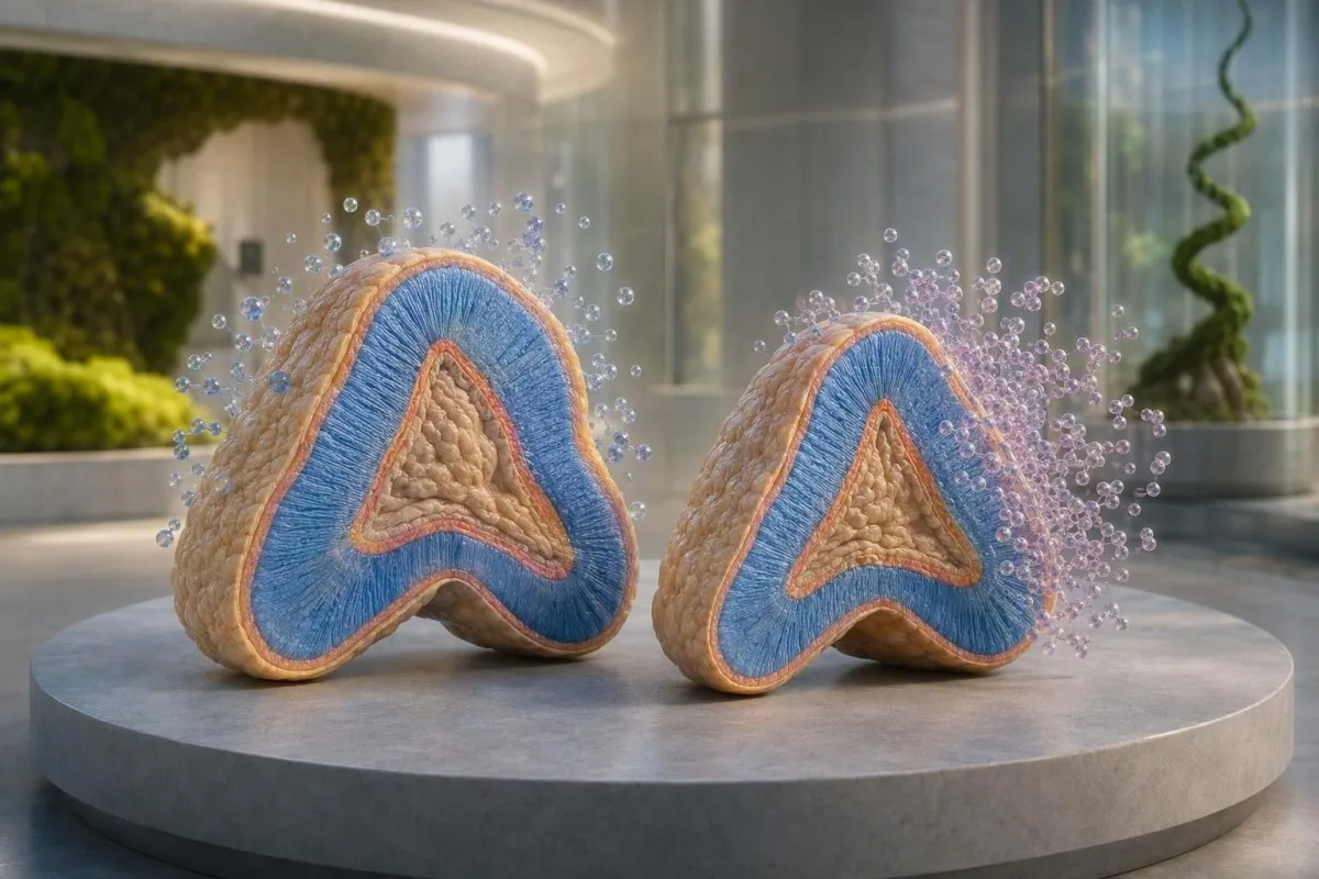
DHEA and DHEA-S are related adrenal hormones, but they answer different clinical questions. The more stable result is usually the one that gives clinicians the useful clue.


- Best first test: DHEA-S is usually preferred because its 7-10 hour half-life makes one result far less sensitive to a brief daily hormone pulse.
- DHEA half-life: Unconjugated DHEA may change substantially within 15-30 minutes, so a single DHEA blood test is harder to interpret.
- High DHEA-S: A value above about 700 µg/dL in an adult woman raises concern for an adrenal source and needs timely clinical review.
- PCOS clue: Mild high DHEA-S can occur in polycystic ovary syndrome, but it does not diagnose PCOS by itself.
- Follow-up test: Early-morning 17-hydroxyprogesterone is often the next test when high DHEA-S accompanies irregular periods or excess terminal hair growth.
- Supplements matter: DHEA products can raise DHEA-S, testosterone, and estradiol; document the dose and timing before interpreting a result.
- Reference ranges: DHEA-S normally falls with age, so a result must be compared with the laboratory's sex- and age-specific interval.
- Urgent symptoms: New deepening voice, rapidly progressive coarse hair growth, or marked muscle changes with high DHEA-S deserve prompt assessment.
Why DHEA-S Usually Gives the More Useful Adrenal Clue
For most people having a DHEA blood test for acne, irregular cycles, excess hair growth, or suspected adrenal hormone excess, DHEA-S is the better first test. DHEA-S is the sulfated storage form of DHEA and remains in circulation for roughly 7-10 hours, while unconjugated DHEA can fluctuate over 15-30 minutes. That stability makes DHEA-S a more dependable marker of average adrenal androgen production from a single sample.
DHEA-S vs DHEA is mostly a question of time scale. DHEA is the active parent steroid made largely in the adrenal zona reticularis, whereas DHEA-S is DHEA joined to a sulfate group that creates a much larger circulating reservoir. DHEA-S concentrations are commonly 100-1,000 times higher than DHEA concentrations in serum, which helps laboratories measure it consistently.
Kantesti is an AI blood test analyzer that reads a DHEA-S result alongside age, sex, medicines, testosterone, and cycle context rather than treating a single flag as a diagnosis. In my clinical work, a mildly high DHEA-S with normal total testosterone often leads to a very different discussion than the same DHEA-S value paired with rapidly changing physical features. For the basic terminology behind the test, see our guide to what DHEA means.
A normal DHEA-S result does not rule out every androgen-related condition, and a high result does not automatically mean a serious adrenal disorder. It is a source clue, not a stand-alone diagnosis. As Dr. Thomas Klein, I find that framing useful: the number tells us where to look next, not what conclusion to jump to.
How the Adrenal Glands Turn DHEA Into DHEA-S
DHEA and DHEA-S are produced mainly by the adrenal cortex, but DHEA-S is formed after DHEA undergoes sulfation through the enzyme SULT2A1. This conversion gives DHEA-S its longer half-life and makes it a practical marker of adrenal androgen output.
About 90% or more of circulating DHEA-S is adrenal in origin, whereas DHEA has contributions from the adrenals, gonads, and peripheral tissues. That distinction is why an elevated DHEA-S result points clinicians more specifically toward the adrenal glands than an elevated DHEA result does. The adrenal glands sit above the kidneys, but their hormone pattern is best understood with cortisol and ACTH rather than by location alone.
ACTH stimulates adrenal androgen production, although DHEA-S does not track minute-to-minute ACTH variation as closely as cortisol. A low DHEA-S can sometimes support chronic ACTH suppression, such as after prolonged glucocorticoid exposure, while a high level suggests increased adrenal androgen production. Our comparison of cortisol and DHEA patterns explains why these two results can move in opposite directions.
The sulfate group is not just biochemical decoration. It creates a negatively charged hormone that binds strongly to albumin and is cleared more slowly by the kidneys, providing a circulating reserve that can later be converted back to DHEA in peripheral tissues. This is one reason DHEA-S is useful for mapping adrenal output but is not a direct measure of hormone activity inside a particular tissue.
Which Hormone Is More Stable on a Blood Test?
DHEA-S is substantially more stable in the body than DHEA, with a half-life of roughly 7-10 hours versus approximately 15-30 minutes for DHEA. Clinicians therefore usually select DHEA-S when they need a single, interpretable estimate of adrenal androgen production.
DHEA has a pronounced diurnal rhythm and pulsatile secretion, particularly in younger adults, so the draw time can materially affect an isolated result. DHEA-S also varies somewhat across the day, but the change is smaller because its long half-life smooths those peaks and troughs. When following a trend, I still prefer the same laboratory and a similar morning appointment time.
Sample handling matters more for certain companion hormones than for DHEA-S. ACTH requires chilled, rapid processing and can become unreliable when pre-analytic handling is poor; DHEA-S is generally more forgiving. If ACTH was drawn with your panel, our practical guide to ACTH timing and handling is worth reading before comparing results.
Kantesti AI does not treat a 10% difference in DHEA-S from two different laboratories as proof that your adrenal function changed. Analytical method, calibration, and the reference interval can produce a modest apparent shift. A repeat that is more than 25-30% different, obtained under similar conditions, is usually more clinically persuasive than a tiny change around the upper limit.
DHEA-S Reference Ranges Depend Heavily on Age and Sex
DHEA-S peaks in late adolescence or early adulthood and then declines steadily, often by 70-80% from young adulthood to advanced age. There is no single universal normal range, so the laboratory's age- and sex-specific interval should always outrank an internet cutoff.
In many laboratories, a typical DHEA-S range for women aged 18-29 is approximately 65-380 µg/dL, while a common interval for women aged 50-59 is about 15-170 µg/dL. Common male intervals are higher, often roughly 280-640 µg/dL at ages 18-29 and 80-560 µg/dL at ages 50-59. These are examples only; local assays differ.
To convert DHEA-S from µg/dL to µmol/L, multiply by 0.0271. Thus, 300 µg/dL is approximately 8.1 µmol/L. The unit conversion is exact enough for comparison, but the decision limit remains laboratory- and patient-specific rather than mathematical.
Immunoassays and liquid chromatography-tandem mass spectrometry can yield non-identical steroid results, especially near a cutoff. This is one reason a report may be marked high in one country and fall within range elsewhere. Our review of why lab ranges differ by sex helps put these flagged values in perspective.
Why Clinicians Usually Order a DHEA-S Test First
Clinicians usually order a DHEA-S test first when they need to determine whether androgen excess may have an adrenal contribution. It is especially useful for new terminal hair growth, resistant acne, irregular periods, reduced scalp hair density, or unexpectedly high testosterone.
A mild DHEA-S elevation is common enough in polycystic ovary syndrome that it should not cause alarm by itself. The 2018 Endocrine Society hirsutism guideline recommends evaluating androgen excess in women with clinically meaningful hirsutism, rather than screening everyone with minor cosmetic hair growth (Martin et al., 2018). PCOS remains a clinical diagnosis based on a wider pattern, not one adrenal marker.
The reason we worry more about high DHEA-S combined with rapid symptom change is that the combination can suggest a high-output androgen source. A woman developing coarse facial or body hair, scalp hair loss, voice deepening, or increased muscle mass over 3-6 months needs a more urgent assessment than someone whose symptoms evolved slowly over several years. This is one of those areas where symptom velocity can matter more than a borderline number.
Total testosterone, free testosterone or calculated free testosterone, SHBG, and sometimes prolactin and TSH provide the surrounding context. If free testosterone is high but DHEA-S is normal, an ovarian or medication-related source may be more likely than an adrenal one. Our article on high free testosterone in women covers that distinction, and our women's hormone guide explains why cycle history still matters.
When a Direct DHEA Test Adds Information
A direct DHEA measurement is most useful when a specialist needs a closer look at immediate steroid production or is monitoring prescribed DHEA therapy. For routine evaluation of possible adrenal androgen excess, DHEA usually adds less than DHEA-S because it is more variable.
Direct DHEA may be considered in a broader steroid profile for suspected congenital adrenal hyperplasia, unusual adrenal enzyme patterns, or research-oriented endocrine workups. It can also be useful when DHEA-S and symptoms disagree sharply, although that is uncommon. A normal DHEA-S with a single high DHEA value often reflects timing, a supplement, or assay variation before it reflects disease.
DHEA testing has a narrower role in evaluating low libido, fatigue, or so-called adrenal fatigue. Adrenal fatigue is not a recognized medical diagnosis, and neither DHEA nor DHEA-S can validate it. In patients with genuine concern for adrenal insufficiency, clinicians prioritize an 8 a.m. cortisol and, where indicated, ACTH stimulation testing; see our guide to cortisol and ACTH patterns.
Do not self-start 25 mg or 50 mg DHEA because a single DHEA value seems low. Supplementation can alter androgen and estrogen pathways, worsen acne or hair loss, and complicate an endocrine evaluation. The evidence for symptom benefit outside specific medical settings is honestly mixed.
What Counts as High DHEA-S and When Follow-Up Is Needed
A DHEA-S value modestly above the laboratory range is usually rechecked and interpreted with symptoms and other androgens, while a value above about 700 µg/dL in an adult woman warrants prompt evaluation for a significant adrenal source. This threshold is a clinical warning sign, not a diagnosis of an adrenal growth.
A value less than 1.5 times the upper limit of normal, particularly without progressive symptoms, is often repeated before imaging is considered. A value more than twice the upper limit or a repeat level around 700-800 µg/dL in women deserves endocrine review, particularly if testosterone is also markedly high. Reference limits for men are higher, so the laboratory's upper limit remains central.
Dr. Thomas Klein's practical rule is simple: ask whether the result matches the body. A 29-year-old with gradual acne and irregular cycles plus DHEA-S of 420 µg/dL may need an outpatient PCOS-focused workup; a 48-year-old with a previously normal cycle pattern, voice changes, and DHEA-S of 850 µg/dL should be assessed much faster. Those are not interchangeable scenarios.
New virilizing features, severe new headaches with visual symptoms, or symptoms of cortisol excess such as easy bruising and proximal muscle weakness should not wait for a routine annual appointment. For less urgent cycle-related symptoms, our guide to blood tests for irregular periods outlines what clinicians commonly check next.
The Main Causes of High DHEA-S Beyond PCOS
High DHEA-S most often reflects PCOS, a DHEA-containing supplement, medication effects, or less commonly congenital adrenal hyperplasia or an adrenal androgen-secreting growth. The clinical picture and degree of elevation separate these possibilities more reliably than DHEA-S alone.
PCOS usually produces mild to moderate elevation, not extreme values, and often coexists with irregular ovulation, acne, or insulin resistance. DHEA-S can be normal in PCOS, so a normal result does not exclude it. The 2018 international PCOS guideline emphasizes excluding thyroid dysfunction, hyperprolactinemia, and non-classic congenital adrenal hyperplasia when symptoms suggest androgen excess (Teede et al., 2018).
Non-classic 21-hydroxylase deficiency is screened with an early-morning follicular-phase 17-hydroxyprogesterone measurement. A basal value above roughly 200 ng/dL or 6 nmol/L typically leads to ACTH stimulation testing, though the assay method changes the exact threshold. The Endocrine Society congenital adrenal hyperplasia guideline supports this targeted approach rather than indiscriminate genetic testing (Speiser et al., 2018).
Less common explanations include danazol, certain antiseizure medicines, laboratory interference, and adrenal growths. Weight change alone does not usually explain a very high DHEA-S result. When cycles and fertility are part of the story, review the separate role of low AMH results rather than assuming every reproductive hormone abnormality has the same cause.
Does Low DHEA-S Mean Anything Clinically Important?
Low DHEA-S is often a normal age-related finding and usually does not require treatment in isolation. It can become clinically relevant when it accompanies low morning cortisol, high ACTH, pituitary disease, long-term steroid use, or symptoms suggesting adrenal insufficiency.
DHEA-S falls progressively after its peak in the 20s, and a level near the lower range at age 65 is not comparable with the same number at age 25. In older adults, a low DHEA-S often reflects expected physiology rather than a deficiency that needs replacing. I do not recommend treating a laboratory low flag without a defined clinical indication.
Chronic prednisone, dexamethasone, and other systemic glucocorticoids can suppress ACTH and lower DHEA-S. In central adrenal insufficiency, DHEA-S may be low because the pituitary is not adequately stimulating the adrenal cortex. However, DHEA-S cannot diagnose adrenal insufficiency; cortisol-based testing does that work.
Fatigue, nausea, unintentional weight loss, salt craving, low blood pressure, or skin darkening need medical assessment, especially when paired with low sodium or abnormal cortisol results. Our guide to low cortisol symptoms explains when same-day evaluation is appropriate.
How to Prepare for a More Reliable DHEA-S Test
DHEA-S testing usually does not require fasting, but a morning draw, consistent timing, and a complete supplement and medication list make interpretation more reliable. The most useful preparation step is to tell the clinician about every hormone product, including creams and online supplements.
A morning sample between about 7 and 10 a.m. is often chosen when cortisol, testosterone, 17-hydroxyprogesterone, or ACTH are being collected simultaneously. DHEA-S itself is relatively stable, but matching the draw time helps compare future results. Fasting is unnecessary unless your clinician ordered glucose, lipids, or other fasting-dependent tests too.
DHEA supplements, compounded hormone creams, testosterone products, and some fertility treatments can change the result. Do not stop prescribed therapy on your own; instead, ask the prescriber whether a pause is medically safe and how long it should be. A 48-hour supplement hold is sometimes suggested by laboratories, but there is no universal rule because dose, formulation, and the clinical question differ.
Biotin can interfere with some immunoassays, although its effect on DHEA-S depends on the laboratory method. Bring the exact product or a photograph of the label to your appointment. Our detailed guide to supplements before blood tests can help you create a useful checklist.
The Hormone Pattern That Makes DHEA-S More Meaningful
DHEA-S becomes clinically informative when it is interpreted with testosterone, SHBG, 17-hydroxyprogesterone, prolactin, TSH, cortisol, and menstrual or medication history. No single hormone result can reliably distinguish PCOS, adrenal disease, and medication effects.
High DHEA-S plus high testosterone suggests broader androgen excess and should prompt a careful symptom review and focused source assessment. High DHEA-S with normal testosterone more often supports an adrenal-predominant pattern, though it still does not identify the cause alone. Very high testosterone may require a different urgency pathway than a modest isolated DHEA-S elevation.
Kantesti is an AI biomarker interpretation platform that compares DHEA-S with linked hormone results, medication entries, and prior values so that a single out-of-range result is not overcalled. In our review workflow, an absent cycle date or unrecorded DHEA supplement can change the interpretation more than a 15 µg/dL numerical difference. The way our clinical logic joins related results is described in our AI technology guide.
An 8 a.m. 17-hydroxyprogesterone above the laboratory screening threshold points toward follow-up for non-classic congenital adrenal hyperplasia, while a high prolactin or abnormal TSH can explain irregular periods through a separate pathway. See our clinician-style hormone panel guide for the common combinations.
What Follow-Up Usually Looks Like After an Abnormal Result
The usual next step after an unexpected DHEA-S result is confirmation and targeted companion testing, not immediate imaging. Repeat testing is most useful when the result is mildly abnormal, symptoms are stable, and supplement or timing effects are plausible.
For a mildly high result, clinicians commonly repeat DHEA-S and add total testosterone, SHBG, early-morning 17-hydroxyprogesterone, and pregnancy testing when appropriate. If menstrual changes are present, prolactin and TSH may be included. Imaging is generally reserved for striking biochemical elevations, virilization, or a pattern that remains unexplained after first-line testing.
If DHEA-S is markedly elevated on repeat testing, an endocrinologist may consider adrenal imaging after biochemical confirmation. A scan should answer a focused question, not serve as a fishing expedition for every slightly high result. Incidental adrenal findings are common enough that premature imaging can create more uncertainty than clarity.
Bring the original laboratory report, not just a screen capture, to the appointment. Units, assay method, reference interval, draw time, and the exact DHEA-S analyte all matter. If you need help organizing the conversation, use our blood test second-opinion checklist.
Why DHEA-S Trends Beat One Isolated Result
A DHEA-S trend collected under similar conditions is more useful than one borderline result because it separates meaningful endocrine change from ordinary biological and laboratory variation. A progressive rise over months deserves more attention than a stable value just above range.
DHEA-S normally declines with age, so a sustained upward trajectory in adulthood is more notable than the same stable result measured once. Conversely, an apparent fall after starting estrogen-containing contraception may reflect treatment effect rather than adrenal failure. A useful trend record includes date, laboratory, menstrual context, medication changes, and supplement dose.
Kantesti AI can compare prior results and flag the context around a new DHEA-S change, but an algorithm cannot replace examination for rapid virilization or suspected adrenal insufficiency. Our approach is deliberately conservative: a result is a prompt for clinical reasoning, not permission to self-diagnose. This is particularly helpful when reports arrive months apart from different laboratories.
Avoid testing DHEA-S every few weeks for nonspecific fatigue. For a stable mild abnormality, a clinician may repeat it after correcting a known confounder or at a planned endocrine review; the interval varies with symptoms and the degree of elevation. Our guide to longitudinal blood-test analysis explains how to preserve the details that make trends trustworthy.
Five DHEA-S Misconceptions That Can Lead You Astray
The biggest DHEA-S misconception is that a high result proves an adrenal growth or that a low result proves adrenal exhaustion. DHEA-S is a useful adrenal androgen marker, but it is neither a cancer test nor a measure of daily stress resilience.
First, high DHEA-S is not the same as high cortisol. These hormones are both adrenal products but can move independently because they arise from related yet distinct pathways and are regulated differently. A normal cortisol result does not erase a high DHEA-S result, and a high cortisol result does not predict high DHEA-S.
Second, DHEA-S is not a menopause test. Ovarian aging is better assessed through symptoms, cycle history, and, in selected situations, FSH or estradiol; DHEA-S declines with age in all adults. Third, low DHEA-S does not mean that buying an over-the-counter hormone will restore energy safely.
Finally, a reference-range flag is not a severity scale. A value just above range may be clinically trivial, whereas a within-range result can still need attention if it is very different from your own baseline and symptoms are concerning. Kantesti is an AI lab test interpretation service designed to explain that difference in plain language while directing urgent patterns to clinician review; our Medical Advisory Board oversees the clinical standards behind this work.
Research Notes, Clinical Limits, and a Safe Next Step
As of August 10, 2026, the safest interpretation of DHEA-S remains pattern-based: confirm the assay and reference range, assess symptoms and medicines, then order focused follow-up rather than treating the number alone. This article supports informed discussion with a qualified clinician and does not replace endocrine assessment.
Kantesti is an AI-powered blood test analysis tool used to organize laboratory reports in clinical context across multilingual health records. Our system can identify a DHEA-S flag and related results in about 60 seconds, but it cannot determine whether you have PCOS, congenital adrenal hyperplasia, or an adrenal growth without clinical evaluation. The safety boundary matters.
Klein, T., et al. (2026). Women's Health Guide: Ovulation, Menopause & Hormonal Symptoms. Figshare. https://doi.org/10.6084/m9.figshare.31830721. ResearchGate record search. Academia.edu record search.
Klein, T., et al. (2026). Multilingual AI Assisted Clinical Decision Support for Early Hantavirus Triage: Design, Engineering Validation, and Real-World Deployment Across 50,000 Interpreted Blood Test Reports. Figshare. https://doi.org/10.6084/m9.figshare.32230290. ResearchGate record search. Academia.edu record search.
Frequently Asked Questions
Is DHEA-S or DHEA better to test?
DHEA-S is usually the better first test for suspected adrenal androgen excess because its half-life is approximately 7-10 hours, compared with roughly 15-30 minutes for DHEA. The longer half-life makes DHEA-S less vulnerable to short hormone pulses and draw-time variation. Clinicians commonly choose DHEA-S for acne, excess terminal hair growth, irregular periods, or a possible adrenal source of high androgens. Direct DHEA testing is more often reserved for specialist steroid profiling or monitoring DHEA treatment.
What DHEA-S level is dangerously high?
A DHEA-S level above approximately 700 µg/dL in an adult woman is high enough to warrant prompt clinical evaluation for a significant adrenal androgen source, especially if symptoms are progressing quickly. A value more than twice the laboratory upper limit also deserves timely review. These cutoffs do not diagnose an adrenal growth, because reference ranges differ by sex, age, and assay method. New voice deepening, rapid coarse hair growth, or marked muscle changes make the assessment more urgent.
Can PCOS cause high DHEA-S?
PCOS can cause a mild to moderate DHEA-S elevation because some people with PCOS have increased adrenal androgen production. DHEA-S may also be normal in PCOS, so it cannot confirm or exclude the diagnosis alone. PCOS assessment combines menstrual history, clinical or biochemical androgen excess, ovarian features where relevant, and exclusion of alternatives such as thyroid disease, high prolactin, and non-classic congenital adrenal hyperplasia. A basal early-morning 17-hydroxyprogesterone above about 200 ng/dL often leads to further testing for congenital adrenal hyperplasia.
Do I need to fast for a DHEA-S test?
Fasting is not usually required for a DHEA-S test. A morning collection between approximately 7 and 10 a.m. is often preferred when DHEA-S is being drawn with cortisol, testosterone, ACTH, or 17-hydroxyprogesterone. The practical preparation priority is documenting DHEA supplements, testosterone products, hormone creams, and prescription steroids. Do not stop prescribed hormones without asking the clinician who manages them.
What can falsely raise DHEA-S?
DHEA supplements, compounded hormone products, some medications, and laboratory-method differences can produce or exaggerate a high DHEA-S result. A result may also be difficult to interpret if the sample was taken during a changing hormone regimen or compared with a previous result from a different laboratory. Biotin can affect selected immunoassays, although the effect depends on the test method and dose. Repeating a mildly abnormal result with the same laboratory and documented supplement status is often sensible.
Should low DHEA-S be treated with supplements?
Low DHEA-S should not automatically be treated with over-the-counter DHEA, particularly when it is an age-appropriate finding. DHEA-S commonly declines by 70-80% from young adulthood to older age, and a low result alone does not diagnose adrenal insufficiency. DHEA products can worsen acne, alter testosterone and estradiol levels, and complicate assessment of hormone-related conditions. Low DHEA-S with low morning cortisol, abnormal ACTH, weight loss, low blood pressure, or long-term steroid use deserves medical review instead of self-treatment.
Get AI-Powered Blood Test Analysis Today
Join over 2 million users worldwide who trust Kantesti for instant, accurate lab test analysis. Upload your blood test results and receive comprehensive interpretation of 15,000+ biomarkers in seconds.
📚 Referenced Research Publications
Klein, T., Mitchell, S., & Weber, H. (2026). Women's Health Guide: Ovulation, Menopause & Hormonal Symptoms. Kantesti AI Medical Research.
Klein, T., Mitchell, S., & Weber, H. (2026). Multilingual AI Assisted Clinical Decision Support for Early Hantavirus Triage: Design, Engineering Validation, and Real-World Deployment Across 50,000 Interpreted Blood Test Reports. Kantesti AI Medical Research.
📖 External Medical References
⚕️ Medical Disclaimer
This article is for educational purposes only and does not constitute medical advice. Always consult a qualified healthcare provider for diagnosis and treatment decisions.
E-E-A-T Trust Signals
Experience
Physician-led clinical review of lab interpretation workflows.
Expertise
Laboratory medicine focus on how biomarkers behave in clinical context.
Authoritativeness
Written by Dr. Thomas Klein with review by Dr. Sarah Mitchell and Prof. Dr. Hans Weber.
Trustworthiness
Evidence-based interpretation with clear follow-up pathways to reduce alarm.