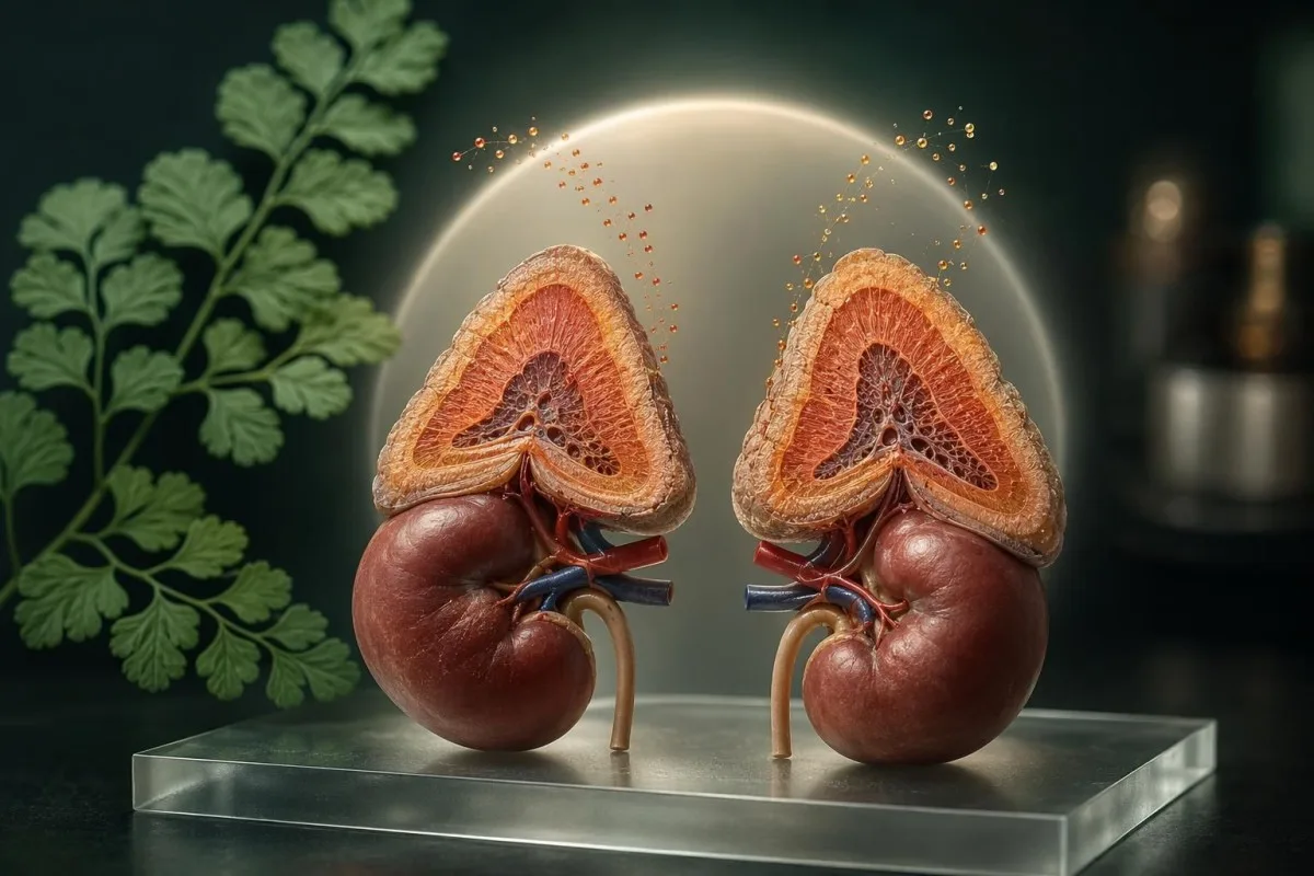
Cortisol and DHEA-S are made by the adrenal glands, but a single result rarely diagnoses a stress problem. The useful question is whether their timing, test method, medications, symptoms, and companion results form a coherent endocrine pattern.


- Cortisol timing matters: serum cortisol is normally highest around 8 am and falls toward midnight, so a random daytime value is often non-diagnostic.
- Morning cortisol below 3 mcg/dL (83 nmol/L) strongly suggests adrenal insufficiency in the right clinical setting, while values above 15 mcg/dL (414 nmol/L) often make it less likely; assay-specific cutoffs apply.
- DHEA-S is preferred over DHEA for most adrenal-androgen interpretation because DHEA-S has a longer half-life and less hour-to-hour variation.
- Cortisol DHEA ratio has no universally validated diagnostic cutoff for burnout, adrenal fatigue, Cushing syndrome, or adrenal insufficiency.
- High cortisol low DHEA-S can occur with aging, illness, glucocorticoid exposure, or chronic ACTH suppression, but it is not a diagnosis by itself.
- Low cortisol high DHEA-S should prompt a check of sampling time, supplements, assay method, ACTH, and possible androgen excess rather than an assumption of adrenal recovery.
- DHEA-S above 700 mcg/dL (19.0 micromol/L) in women generally warrants prompt evaluation for an adrenal source, especially with new virilising symptoms.
- Steroid medicines including tablets, injections, inhalers, skin creams, eye drops, and nasal sprays can suppress the hypothalamic-pituitary-adrenal axis.
- Repeat testing is most informative when done at the same time of day, through the same laboratory, with sleep, exercise, illness, and medication changes recorded.
Why Cortisol and DHEA Need a Time-Sensitive Reading
Cortisol vs DHEA is best interpreted as a time-stamped adrenal pattern, not as two isolated scores. Cortisol can change substantially over hours, whereas DHEA-S changes more slowly; a 4 pm cortisol and a DHEA-S collected without wake-time context cannot reliably describe chronic stress or adrenal reserve.
Cortisol is the adrenal cortex's principal glucocorticoid, while DHEA and DHEA-S are adrenal androgens that decline with age. Serum cortisol normally peaks near waking and approaches its daily low point around midnight; DHEA-S has a half-life of roughly 7 to 10 hours, making it steadier but still age-, sex-, and assay-dependent.
A person waking at 5 am for hospital work and tested at 8 am has a very different physiological context from someone tested at 8 am after sleeping until 7:30 am. In my clinical experience, the lab requisition's collection time is often recorded, but the patient's wake time, night shift, acute illness, and recent exercise are not—and those omissions can change the interpretation.
Kantesti is an AI blood test analyzer that places cortisol, DHEA-S, ACTH, electrolytes, glucose, and medication context into one chronological review rather than treating a flagged hormone as a stand-alone verdict. Patients can also use our hormone biomarker guide to check the specimen type and reference interval printed by their own laboratory.
Cortisol, DHEA, and DHEA-S Are Different Measurements
DHEA-S is usually the more useful companion test to cortisol because it is more stable in circulation than DHEA. A DHEA result can vary with collection time and laboratory technique, while DHEA-S offers a more dependable estimate of adrenal androgen production over days rather than minutes.
DHEA is converted to DHEA-S mainly in the adrenal zona reticularis, then can be converted back in peripheral tissues. Adult DHEA-S reference intervals are broad: many laboratories list about 40 to 325 mcg/dL for younger adult women and 110 to 510 mcg/dL for younger adult men, with markedly lower expected values after age 60.
A result can look "low" against a general adult range yet be unremarkable for a 72-year-old. Conversely, a DHEA-S of 420 mcg/dL may be ordinary in a young man but clearly above many female reference intervals; this is why DHEA test meaning starts with age, sex, and the reporting laboratory.
Cortisol is commonly reported in mcg/dL or nmol/L, DHEA-S in mcg/dL or micromol/L, and DHEA in ng/dL or nmol/L. Dividing values with unlike units or unlike specimen matrices—such as salivary cortisol and serum DHEA-S—creates a ratio that may look precise but has no coherent biological interpretation.
Morning Cortisol Ranges: Useful Thresholds and Their Limits
An 8 am serum cortisol below 3 mcg/dL usually needs urgent endocrine assessment, while a value above 15 mcg/dL often argues against clinically significant adrenal insufficiency. Results between those values commonly require ACTH and a dynamic stimulation test rather than a diagnosis from one draw.
Many laboratories report an early-morning serum cortisol reference interval of roughly 5 to 25 mcg/dL (138 to 690 nmol/L), but the interval is not a pass-fail test for adrenal reserve. Modern cortisol assays read lower than older immunoassays in some settings, so an endocrinologist may use a stimulation-test peak around 14 to 18 mcg/dL depending on the assay.
The Endocrine Society guideline led by Bornstein et al. recommends a standard 250 mcg cosyntropin stimulation test when clinical suspicion persists and the morning cortisol is indeterminate (Bornstein et al., 2016). An ACTH sample should ideally be collected before steroid treatment when safely possible, because hydrocortisone can make subsequent interpretation difficult.
A low result alongside dizziness, vomiting, weight loss, salt craving, low sodium, high potassium, or darkening of the skin deserves prompt attention; it is not a wellness finding. Our guide to low cortisol warning signs explains when symptoms shift the issue from outpatient retesting to same-day medical care.
What the Cortisol DHEA Ratio Can—and Cannot—Tell You
The cortisol DHEA ratio has no guideline-approved cutoff for diagnosing adrenal fatigue, burnout, Cushing syndrome, or adrenal insufficiency. It can be a descriptive observation only when both values come from the same collection, validated assay context, and correctly matched units.
Ratios amplify laboratory noise when the denominator is small. For example, a cortisol of 18 mcg/dL divided by a DHEA-S of 45 mcg/dL produces a very different number from 18 divided by 90, even though both DHEA-S results may be normal for an older adult; this is a mathematical effect, not proof of adrenal dysfunction.
A cortisol DHEA ratio also ignores cortisol-binding globulin, which rises with oral oestrogen and pregnancy and can raise total serum cortisol without increasing free cortisol. The Endocrine Society Cushing guideline does not endorse a cortisol-to-DHEA or cortisol-to-DHEA-S ratio; it recommends late-night salivary cortisol, 24-hour urinary free cortisol, or the 1 mg overnight dexamethasone suppression test for appropriate patients (Nieman et al., 2008).
Dr. Thomas Klein's practical rule is simple: use ratios to generate a question, never to settle one. A cortisol and ACTH pattern review is much more informative because ACTH indicates whether the pituitary is stimulating the adrenal glands.
High Cortisol With Low DHEA-S: Common Explanations
High cortisol with low DHEA-S most often reflects timing, age, illness, medication exposure, or altered ACTH signaling—not a proven state of adrenal exhaustion. The pattern becomes more meaningful when it persists on properly timed testing and fits symptoms, ACTH, and other adrenal findings.
Acute pain, fever, major sleep loss, surgery, panic, and strenuous endurance exercise can raise cortisol temporarily. I have seen a marathon runner with a morning cortisol of 27 mcg/dL after a red-eye flight and poor sleep; a repeat after 10 days of routine sleep was 15 mcg/dL, illustrating why one stressed sample should not be overread.
DHEA-S normally falls across adulthood, by roughly 2% to 4% per year after early adulthood in many longitudinal observations, so the same cortisol DHEA ratio can rise with age even if cortisol is unchanged. Chronic inflammatory disease, poorly controlled diabetes, and critical illness may also lower DHEA-S through pathways that are not fully resolved.
A persistent high cortisol result needs targeted testing only when the clinical picture fits—easy bruising, proximal muscle weakness, new diabetes, difficult hypertension, osteoporosis, or characteristic weight redistribution. Read more about high cortisol causes before assuming that everyday stress has caused an endocrine disease.
Low Cortisol With High DHEA-S: What to Check First
Low cortisol with high DHEA-S is not a standard recovery pattern and should first trigger a check of sampling time, supplements, assay interference, and androgen symptoms. If both values were collected correctly, ACTH and an endocrine review can clarify whether the values reflect physiology or two unrelated signals.
A 4 pm cortisol of 4 mcg/dL may be physiologically normal because cortisol should decline later in the day; it is not comparable with an 8 am diagnostic threshold. DHEA-S remains relatively stable, so collecting it beside an afternoon cortisol can falsely suggest a low-cortisol high-DHEA-S pattern.
Over-the-counter DHEA can raise DHEA-S substantially, and doses of 25 to 50 mg daily are common enough that I ask directly rather than assuming a laboratory anomaly. New acne, scalp hair thinning, excess facial hair, deepening voice, or rapidly changing muscle mass alongside high DHEA-S merits clinician review rather than self-adjusting supplements.
In women, DHEA-S above 700 mcg/dL (19.0 micromol/L) raises concern for an adrenal androgen-secreting source and generally warrants prompt evaluation. For broader androgen context, see high free testosterone in women; ovarian and adrenal patterns are not interchangeable.
Medicines and Supplements That Distort Adrenal Tests
Glucocorticoid medicines are the most consequential cause of misleading cortisol results because they can suppress ACTH and the adrenal glands. Tablets are not the only concern: repeated injections, potent skin preparations, inhalers, nasal sprays, eye drops, and some complementary products can all matter.
Prednisone, prednisolone, hydrocortisone, methylprednisolone, and dexamethasone differ in assay cross-reactivity and duration of adrenal suppression. Hydrocortisone is chemically cortisol and can directly affect a serum cortisol measurement; dexamethasone often does not cross-react in cortisol immunoassays but can still suppress endogenous ACTH and cortisol.
Oral oestrogen increases cortisol-binding globulin and can increase total serum cortisol by 30% to 100% without the same rise in free cortisol. Biotin supplements can interfere with some immunoassays, while anticonvulsants such as carbamazepine may alter steroid metabolism; the exact direction depends on the test method.
Kantesti AI is an AI lab test interpretation service that flags medication-result conflicts for clinician discussion, but it cannot tell anyone to stop steroid treatment. Never pause, taper, or skip prescribed glucocorticoids for a test unless the prescribing clinician or endocrinology team has supplied a specific plan; abrupt withdrawal can be dangerous. Our adrenal supplement safety guide covers common product pitfalls.
Sleep, Exercise, Illness, and Shift Work Change the Pattern
Poor sleep, acute illness, and intense exercise can shift cortisol enough to make a single result unrepresentative. DHEA-S usually moves less over 24 hours, so a disrupted cortisol rhythm can artificially exaggerate a cortisol DHEA ratio.
The cortisol awakening response commonly rises during the first 30 to 45 minutes after waking, although its size varies between people and methods. For a night-shift worker, an "8 am cortisol" after working overnight may be poorly aligned with the biological morning; many endocrinologists instead anchor testing to habitual wake time and the clinical question.
Heavy resistance exercise, a long run, or high-intensity intervals in the prior 12 to 24 hours can increase cortisol and alter glucose, creatine kinase, and white cell counts. A useful laboratory note records sleep duration, wake time, fever or infection, alcohol, training load, and whether the sample was collected before usual medications.
There is no universal requirement to fast for serum cortisol or DHEA-S, but a consistent morning routine improves comparison between visits. If insomnia is part of the story, our sleep-related lab guide explains why iron, thyroid tests, glucose, and sleep-disordered breathing clues may be more actionable than an isolated cortisol result.
When DHEA-S Suggests Adrenal Androgen Excess
Markedly high DHEA-S suggests increased adrenal androgen production, especially when accompanied by rapidly progressive androgenic symptoms. Mild elevation is more often seen with common conditions such as polycystic ovary syndrome, medications, or laboratory variation than with an adrenal mass.
DHEA-S is produced almost exclusively by the adrenal glands, unlike testosterone, which has substantial gonadal production. In a premenopausal woman with irregular cycles and gradual excess hair growth, modest DHEA-S elevation can occur in polycystic ovary syndrome; a level over 700 mcg/dL or rapid symptom progression warrants a faster search for an adrenal cause.
The 2018 Endocrine Society hirsutism guideline advises androgen testing when hirsutism is moderate to severe, rapidly progressive, or accompanied by menstrual disturbance or virilisation (Azziz et al., 2018). Total testosterone, free testosterone using a reliable method, 17-hydroxyprogesterone, and sometimes imaging may be selected based on the history—not ordered indiscriminately.
Kantesti AI interprets DHEA-S beside testosterone, SHBG, LH, FSH, prolactin, and metabolic markers because one androgen cannot identify the source. Readers dealing with cycle changes can compare this with our irregular period testing guide.
When a Low DHEA-S Result Matters Clinically
Low DHEA-S is common with aging and is not, by itself, evidence of adrenal insufficiency. It becomes clinically relevant when it accompanies low morning cortisol, low or inappropriately normal ACTH, unexplained fatigue, low blood pressure, or other pituitary-adrenal abnormalities.
In primary adrenal insufficiency, destruction or impaired function of the adrenal cortex can reduce cortisol, aldosterone, and DHEA-S, while ACTH rises. Typical accompanying clues include sodium below 135 mmol/L, potassium above 5.0 mmol/L, low blood pressure, and increased skin pigmentation, although no single clue is present in every person.
In central adrenal insufficiency, ACTH is low or inappropriately normal, and DHEA-S may fall early because the zona reticularis depends on ACTH stimulation. A normal DHEA-S can make substantial chronic ACTH deficiency less likely in some adults, but a low DHEA-S cannot confirm it because age, illness, and medicines are major confounders.
The practical next step is not DHEA replacement based on a low number; it is confirmation of adrenal function through an endocrine pathway. See our detailed Addison disease clues if low cortisol occurs with salt loss, persistent vomiting, or faintness.
Serum, Saliva, and Urine Tests Answer Different Questions
Serum cortisol, late-night salivary cortisol, and 24-hour urinary free cortisol should not be interpreted as interchangeable tests. Each measures a different fraction of cortisol and is chosen for a distinct clinical question, so a ratio built across test types is invalid.
Serum cortisol usually measures total cortisol, including protein-bound hormone, while salivary cortisol reflects free cortisol that diffuses into saliva. Late-night salivary cortisol is useful because the normal circadian nadir should be very low; smoking, shift work, oral contamination, and irregular sleep can reduce its reliability.
A 24-hour urinary free cortisol collection estimates unbound cortisol excreted over a whole day, but under-collection and kidney impairment can distort results. Urine creatinine helps laboratories judge collection completeness, and an eGFR below about 60 mL/min/1.73 m² may reduce the test's usefulness for Cushing screening.
A single random serum cortisol should not screen for Cushing syndrome, and a normal late-night saliva result does not exclude adrenal insufficiency. If your report contains several specimen types, our 24-hour urine collection guide can help you spot collection errors before drawing conclusions.
Why “Adrenal Fatigue” Is Not a Medical Diagnosis
“Adrenal fatigue” is not an accepted endocrine diagnosis because no validated cortisol or DHEA pattern proves that everyday stress has worn out the adrenal glands. The risk is not semantic: the label can delay detection of true adrenal insufficiency, depression, sleep apnea, anaemia, thyroid disease, medication effects, or diabetes.
Fatigue is real, but it is diagnostically broad. In a patient with fatigue, unintentional weight loss, dizziness, and a morning cortisol of 2.8 mcg/dL, I would prioritise adrenal insufficiency testing; in someone with normal morning cortisol, loud snoring, and haemoglobin A1c of 6.2%, sleep and glucose pathways may be more fruitful.
Dr. Thomas Klein, MD, advises patients not to accept a saliva-panel colour score as a diagnosis when symptoms are significant. Proper evaluation starts with a careful history, orthostatic blood pressure when indicated, medication reconciliation, CBC, electrolytes, glucose or HbA1c, thyroid testing, ferritin when appropriate, and focused endocrine tests.
Kantesti is an AI-powered blood test analysis tool that can organise those cross-panel clues and show whether the change is new or persistent, but final diagnosis belongs with the treating clinician. Our burnout lab review explains which common tests can help without medicalising ordinary stress.
How to Repeat Cortisol and DHEA-S Testing More Reliably
A repeat adrenal hormone test is most useful when collected at the same laboratory, near the same wake-relative time, with medication and illness details documented. Repeating a random result under different circumstances often adds noise rather than clinical clarity.
For suspected adrenal insufficiency, clinicians often request serum cortisol and ACTH between 7 and 9 am before taking the morning hydrocortisone dose, but the timing plan must be individualised. Do not copy another patient's medication-hold instructions; people taking replacement steroids, prednisone, or dexamethasone need clinician-specific advice.
Record the exact collection date and time, usual wake time, hours slept, fever or infection, vigorous exercise within 24 hours, steroid exposure within the previous 3 months, DHEA use, oral oestrogen, and pregnancy status. These details are often more explanatory than a 10% to 20% movement within a laboratory reference interval.
Trend interpretation needs apples-to-apples data, especially when reference ranges differ between laboratories. A longitudinal lab trend review can preserve each laboratory's interval and collection context instead of plotting incompatible raw values as if they were identical.
How Kantesti Reviews an Adrenal Hormone Pattern
Kantesti reviews cortisol and DHEA-S alongside collection time, laboratory range, age, sex, ACTH, electrolytes, glucose, and documented medicines rather than calculating an unsupported wellness score. That approach can identify questions for a clinician, but it does not diagnose Cushing syndrome or adrenal insufficiency from an uploaded report.
Kantesti is an AI biomarker interpretation platform used by more than 2 million people across 127+ countries to organise laboratory data in clinical context. When a report shows cortisol at 4.5 mcg/dL, our interpretation must distinguish an afternoon sample from an 8 am sample before it can meaningfully flag a possible follow-up.
A pattern that includes low cortisol, sodium of 131 mmol/L, potassium of 5.5 mmol/L, and a high ACTH deserves a higher-priority clinical prompt than low DHEA-S alone. Conversely, cortisol of 23 mcg/dL during acute illness with normal electrolytes and no Cushing features usually calls for recovery and context, not panic.
Kantesti's AI technology guide describes how our system retains reference intervals and trend data, while medical validation standards explains the limits of automated interpretation. Good AI output should make the next medical conversation sharper, not replace it.
When Cortisol or DHEA Results Need Prompt Medical Care
Urgent care is appropriate for suspected adrenal crisis symptoms, not merely for an out-of-range DHEA-S result. Severe weakness, fainting, persistent vomiting, confusion, severe abdominal pain, dehydration, low blood pressure, or an inability to keep prescribed steroids down needs same-day emergency assessment.
Adrenal crisis can present with low blood pressure, low sodium, high potassium, low glucose, abdominal symptoms, and profound fatigue, but laboratory confirmation should never delay emergency treatment when the clinical picture is convincing. People prescribed emergency hydrocortisone should follow their individual sick-day rules and seek urgent help if oral medication cannot be retained.
Specialist endocrinology review is reasonable for repeated abnormal morning cortisol, abnormal ACTH, DHEA-S over 700 mcg/dL in women, rapid virilising symptoms, suspected pituitary disease, or possible Cushing features. A laboratory flag alone is not an emergency, but the combination of a number, symptoms, and trajectory can be.
As of July 20, 2026, the safest message is still pattern before ratio, and diagnosis before supplements. Our Medical Advisory Board oversees clinical content standards, and patients with concerning symptoms should use their own clinician or emergency service rather than relying on digital interpretation alone.
Frequently Asked Questions
What is a normal cortisol DHEA ratio?
There is no universally accepted normal cortisol DHEA ratio for adults because cortisol and DHEA-S use different units, have different daily rhythms, and vary substantially by age and laboratory method. A ratio cannot diagnose adrenal fatigue, Cushing syndrome, or adrenal insufficiency at any validated cutoff. An 8 am cortisol below 3 mcg/dL (83 nmol/L) is clinically more actionable than a ratio, particularly when symptoms or abnormal ACTH are present. DHEA-S should be compared with the laboratory's age- and sex-specific interval before any ratio is considered.
Does high cortisol and low DHEA mean adrenal fatigue?
High cortisol with low DHEA-S does not prove adrenal fatigue, which is not a recognised endocrine diagnosis. This combination may occur with age-related DHEA-S decline, acute illness, sleep loss, glucocorticoid medication exposure, altered ACTH signalling, or a sample collected under stressful conditions. A cortisol result above about 25 mcg/dL (690 nmol/L) requires clinical context, because oral oestrogen can also increase total serum cortisol by 30% to 100%. Persistent abnormalities should be reviewed with ACTH, collection time, symptoms, and medication history.
Can low cortisol and high DHEA-S happen together?
Low cortisol and high DHEA-S can occur, but it is often explained by an afternoon cortisol collection, DHEA supplementation, assay differences, or unrelated androgen excess rather than a single adrenal state. A 4 pm cortisol of 4 mcg/dL may be normal, whereas that same result at 8 am can be concerning. DHEA-S above 700 mcg/dL (19.0 micromol/L) in women generally requires assessment for an adrenal androgen source, especially with rapid acne, hair changes, or voice changes. ACTH, total testosterone, 17-hydroxyprogesterone, and repeat timed testing may be needed.
Should cortisol and DHEA-S be tested in the morning?
Morning testing is usually preferable when evaluating possible adrenal insufficiency because cortisol is expected to be near its daily peak between roughly 7 and 9 am. DHEA-S is more stable throughout the day, but collecting it at the same time as cortisol makes the pair easier to interpret. Shift workers need a personalised plan because a clock-time 8 am sample after an overnight shift may not reflect their biological morning. The laboratory report should be interpreted alongside usual wake time, sleep duration, illness, exercise, and steroid medicines.
Which medications can lower cortisol or DHEA-S?
Glucocorticoid medications can lower endogenous cortisol and DHEA-S by suppressing ACTH, even when delivered through inhalers, injections, nasal sprays, potent creams, or eye drops. Hydrocortisone can directly affect a cortisol test because it is cortisol, while dexamethasone may suppress the axis without appearing as cortisol in many assays. Opioids and some medications used for epilepsy can also affect hypothalamic-pituitary-adrenal function or steroid metabolism. Patients should never stop prescribed steroids before testing without instructions from the prescribing clinician.
Is a low DHEA-S level dangerous?
A low DHEA-S level is usually not dangerous by itself, particularly after age 50, because DHEA-S naturally declines throughout adulthood. It matters more when paired with a low 8 am cortisol, abnormal ACTH, low sodium below 135 mmol/L, high potassium above 5.0 mmol/L, low blood pressure, or symptoms suggestive of adrenal insufficiency. Low DHEA-S cannot independently diagnose pituitary or adrenal disease. The appropriate next step is usually a clinician-guided adrenal evaluation rather than self-prescribed DHEA.
Can DHEA supplements affect my adrenal hormone test results?
Yes. Over-the-counter DHEA can raise measured DHEA-S and other androgen results, making it harder to assess your natural adrenal production. Bring the bottle or dose information to your appointment and ask whether it should be paused before testing.
Get AI-Powered Blood Test Analysis Today
Join over 2 million users worldwide who trust Kantesti for instant, accurate lab test analysis. Upload your blood test results and receive comprehensive interpretation of 15,000+ biomarkers in seconds.
📚 Referenced Research Publications
Klein, T., Mitchell, S., & Weber, H. (2026). Serum Proteins Guide: Globulins, Albumin & A/G Ratio Blood Test. Kantesti AI Medical Research.
Klein, T., Mitchell, S., & Weber, H. (2026). C3 C4 Complement Blood Test & ANA Titer Guide. Kantesti AI Medical Research.
📖 External Medical References
⚕️ Medical Disclaimer
This article is for educational purposes only and does not constitute medical advice. Always consult a qualified healthcare provider for diagnosis and treatment decisions.
E-E-A-T Trust Signals
Experience
Physician-led clinical review of lab interpretation workflows.
Expertise
Laboratory medicine focus on how biomarkers behave in clinical context.
Authoritativeness
Written by Dr. Thomas Klein with review by Dr. Sarah Mitchell and Prof. Dr. Hans Weber.
Trustworthiness
Evidence-based interpretation with clear follow-up pathways to reduce alarm.