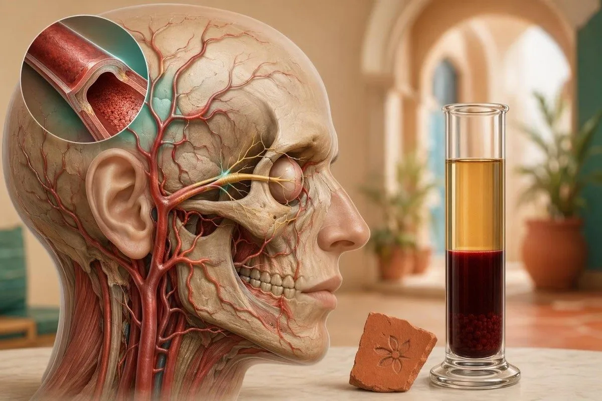
A high ESR can be the laboratory clue that turns a headache into an urgent medical evaluation. The number matters most when it appears beside jaw claudication, scalp tenderness, or vision changes.


- Sed rate above 50 mm/h supports urgent evaluation for giant cell arteritis when new headache, jaw pain, scalp tenderness, or visual symptoms are present.
- ESR blood test results cannot diagnose giant cell arteritis alone because infection, cancer, anemia, kidney disease, and many autoimmune conditions can also raise ESR.
- Vision symptoms such as temporary vision loss, double vision, or a curtain-like shadow require same-day emergency care, even before lab confirmation.
- Normal ESR does not rule out giant cell arteritis; about 4% of biopsy-proven cases may have both ESR and CRP in the normal range.
- CRP plus ESR is more useful than either test alone; CRP sensitivity for giant cell arteritis was 86.9% and ESR sensitivity was 84.1% in Kermani et al., 2012.
- Treatment timing matters because suspected giant cell arteritis is usually treated immediately with high-dose glucocorticoids, often prednisone 40-60 mg/day, while confirmation is arranged.
- Jaw claudication means chewing-triggered jaw fatigue or pain, and it is one of the most specific symptoms for giant cell arteritis in adults over 50.
- High ESR trend after steroids should fall over days to weeks, but symptoms and vision risk guide decisions more than the number alone.
Why a High Sed Rate Changes the Urgency
A sed rate above 50 mm/h with a new headache, jaw pain while chewing, scalp tenderness, or vision symptoms should trigger urgent assessment for giant cell arteritis. ESR supports the suspicion; it does not prove the diagnosis. In clinic, I treat this pattern as time-sensitive because untreated giant cell arteritis can threaten vision within hours to days.
Giant cell arteritis is a large-vessel vasculitis that almost always occurs after age 50, with the highest incidence in people aged 70-80 years. A high ESR is common because inflammatory proteins make red cellular elements settle faster, but the symptom pattern decides the urgency, not the lab result by itself.
I am Thomas Klein, MD, and the situation that worries me is the 72-year-old who says, “My temple hurts and my jaw gets tired halfway through breakfast.” If that patient has an ESR of 82 mm/h, I do not file it under routine abnormal labs; I ask about vision immediately and review whether the result meets criteria for critical lab follow-up.
Kantesti is an AI blood test analyzer that reads ESR in the context of age, sex, CRP, hemoglobin, platelets, and the symptoms entered by the user. Our AI can flag a dangerous pattern, but suspected giant cell arteritis still needs urgent clinician review because treatment decisions depend on examination, imaging, and sometimes temporal artery tissue examination.
What the ESR Blood Test Actually Measures
The ESR blood test measures how many millimetres red cellular elements fall in a vertical tube over 1 hour. A faster sedimentation rate usually reflects increased inflammatory proteins, especially fibrinogen, but anemia and age can also push the number upward.
A typical adult ESR reference range is roughly 0-15 mm/h for younger men and 0-20 mm/h for younger women, but many laboratories allow higher cutoffs after age 50. A practical age-adjusted upper limit is age divided by 2 for men and age plus 10 divided by 2 for women, though not every lab reports it that way.
The sedimentation rate rises when fibrinogen, immunoglobulins, and other acute-phase proteins reduce the usual repulsion between red cellular elements. That is why ESR can be high in giant cell arteritis, polymyalgia rheumatica, pneumonia, endocarditis, kidney disease, and some malignancies; the tube cannot tell those apart.
If your report lists ESR in mm/hr, mm/h, or Westergren units, it is usually referring to the same measurement method. For a deeper reference-range discussion by age and sex, see our ESR range guide.
Symptoms That Make ESR More Concerning
A high ESR becomes more concerning for giant cell arteritis when it appears with a new localized headache, jaw claudication, scalp tenderness, fever, shoulder or hip stiffness, or visual disturbance. The same ESR value is less specific when it follows a chest infection or recent surgery.
The headache of giant cell arteritis is usually new after age 50, often temporal, and may feel different from migraines the patient had for 30 years. I ask about pain when combing hair, discomfort from resting glasses on the scalp, and jaw fatigue after 2-5 minutes of chewing because patients rarely volunteer those details unless asked.
Jaw claudication is not ordinary jaw soreness. It is exertional pain or fatigue of the jaw muscles during chewing, and in my experience it is one of the most useful bedside clues when ESR is above 50 mm/h.
Headache workups can easily drift toward sinus disease, migraine, or tension pain, especially if the first exam is brief. Our headache lab guide explains how anemia, thyroid disease, and inflammatory markers can overlap, but giant cell arteritis remains a same-day question when cranial symptoms and ESR elevation line up.
Vision Symptoms Are the Emergency Line
Vision symptoms with suspected giant cell arteritis are an emergency, even if the sed rate has not returned yet. Temporary vision loss, double vision, new blurred vision, or a curtain-like shadow can precede permanent ischemic optic nerve injury.
The eye risk in giant cell arteritis comes from reduced flow through branches supplying the optic nerve and retina. Once permanent visual loss occurs in one eye, the second eye can be affected quickly, which is why clinicians often start treatment before every confirmatory test is complete.
A normal eye chart reading does not fully reassure me if the patient reports 10-minute episodes of gray vision or new double vision. Those transient symptoms may be ischemic warning shots, particularly when ESR is 70-100 mm/h and the patient is over 60.
Blurred vision has many causes, including glucose swings, B12 deficiency, thyroid disease, and medication effects. Still, if blurred vision arrives with headache, jaw claudication, or scalp tenderness, use our broader blurred vision labs only after emergency causes have been addressed.
Why ESR Alone Cannot Diagnose GCA
ESR alone cannot diagnose giant cell arteritis because it is sensitive to inflammation but not specific to the inflamed artery. A sed rate of 90 mm/h may fit GCA, but it may also fit pneumonia, lymphoma, rheumatoid arthritis, kidney disease, or severe anemia.
In Kermani et al., 2012, ESR sensitivity for biopsy-proven giant cell arteritis was 84.1%, while CRP sensitivity was 86.9%; the combination performed better than either marker alone. The uncomfortable truth is that a test can be fairly sensitive and still be poor at naming the disease.
ESR rises slowly and can stay elevated for weeks because fibrinogen and immunoglobulins have longer biological effects. CRP often changes faster, sometimes within 6-8 hours of an inflammatory trigger, which is why I almost always want both ESR and CRP when GCA is on the table.
A CRP result may be standard CRP or high-sensitivity CRP, and they are not interchangeable for acute vasculitis questions. Our CRP versus hs-CRP explainer shows why a cardiovascular hs-CRP report can mislead patients who are trying to interpret a possible vasculitis flare.
When ESR Is High but CRP Is Normal
A high ESR with normal CRP weakens but does not eliminate the possibility of giant cell arteritis. ESR can stay elevated from age, anemia, kidney disease, monoclonal proteins, or previous inflammation after CRP has already settled.
I have seen ESR values of 60-80 mm/h in older adults with anemia and no vasculitis at all. The reason is mechanical: fewer red cellular elements and altered plasma proteins can accelerate sedimentation even when CRP is modest.
Giant cell arteritis with normal CRP is less common, but it happens. Kermani et al., 2012 reported that about 4% of patients with biopsy-proven giant cell arteritis had both ESR and CRP in the normal range, which is exactly why symptoms cannot be ignored.
If your report shows this mixed pattern, compare it with hemoglobin, MCV, creatinine, albumin, and total protein before guessing. We cover the practical differential in our guide to high ESR, normal CRP.
CBC Patterns That Strengthen the Suspicion
CBC patterns can strengthen suspicion for giant cell arteritis when ESR is elevated. Mild normocytic anemia, high platelets, and a raised CRP together suggest an inflammatory process more convincingly than ESR alone.
Anemia of inflammation often produces hemoglobin around 9-12 g/dL with a normal or near-normal MCV. Platelet counts may rise above 400 x 10^9/L because interleukin-6 stimulates liver and marrow responses during active vasculitis.
The pattern matters more than any single flag. ESR 78 mm/h, hemoglobin 10.8 g/dL, platelets 510 x 10^9/L, and CRP 58 mg/L tells a different story than ESR 78 mm/h with normal CBC, normal CRP, and recent iron infusion.
Patients sometimes focus only on the red ESR flag and miss the supporting CBC clues. Our article on high ESR and low hemoglobin walks through the inflammatory anemia pattern that often accompanies vasculitis, chronic infection, or malignancy.
What Doctors Do When GCA Is Suspected
When giant cell arteritis is suspected, clinicians usually act before every test is final. The typical workup includes ESR, CRP, CBC, liver enzymes, kidney function, urgent eye assessment if symptoms are present, and rapid referral for imaging or temporal artery tissue examination.
According to the 2018 EULAR recommendations, suspected active giant cell arteritis should receive high-dose glucocorticoid therapy immediately to reduce ischemic complications (Dejaco et al., 2018). In practice, that often means prednisone 40-60 mg/day for cranial symptoms, while visual symptoms may require intravenous methylprednisolone 500-1000 mg/day for 3 days depending on local protocol.
Kantesti is an AI biomarker interpretation platform that can identify ESR-CRP-CBC clusters suggesting urgent review, but it does not tell a patient to self-start steroids. Steroids can alter glucose, blood pressure, infection risk, mood, and bone health within days, so the prescribing decision belongs with a clinician.
A practical inflammation panel for suspected GCA includes ESR, CRP, CBC with platelets, CMP, alkaline phosphatase, and sometimes fibrinogen. For a broader comparison of inflammatory tests and their blind spots, see our guide to inflammation blood tests.
How GCA Is Confirmed After ESR Raises Suspicion
Giant cell arteritis is confirmed with clinical assessment plus vascular ultrasound, temporal artery tissue examination, MRI, CT angiography, or PET-CT when appropriate. ESR raises suspicion; confirmation looks for arterial wall thickening, halo sign, luminal change, or characteristic tissue findings.
The British Society for Rheumatology guideline emphasizes that treatment should not be delayed while awaiting temporal artery tissue examination when the clinical picture is strong (Dasgupta et al., 2010). That point matters: steroids may begin first, and confirmation can still be pursued in the following days.
Ultrasound can show a non-compressible halo around the temporal artery, but accuracy depends heavily on operator skill and timing after steroid initiation. Some European fast-track clinics use same-day ultrasound because it can shorten diagnostic delay from weeks to hours.
GCA also overlaps with polymyalgia rheumatica, which causes shoulder and hip girdle stiffness lasting more than 45 minutes in the morning. If symptoms are more systemic or multi-joint, our autoimmune panel guide explains why ANA and rheumatoid factor may not answer the vasculitis question directly.
False Positives, False Negatives, and Lab Timing
False positives and false negatives occur with ESR because the test is affected by plasma proteins, red cellular element shape, anemia, age, pregnancy, and technical handling. A single sed rate should be interpreted as a clue, not a verdict.
ESR may be falsely high when the tube is tilted, the sample is delayed too long, or room temperature is unusually high. It may be falsely low in polycythemia, marked leukocytosis, abnormal red cellular element shapes, or very high protein states that alter settling dynamics unpredictably.
Timing also matters after treatment starts. CRP often falls within days of effective steroid treatment, while ESR may lag for 1-3 weeks, so a still-high ESR after 5 days does not automatically mean treatment failure.
When a result does not fit the patient, repeating the test is reasonable, but not when vision symptoms are active. For non-emergency discrepancies, our repeat abnormal labs guide explains when same-week retesting helps and when it only creates noise.
Reading Your ESR Report Without Missing Red Flags
Read an ESR report by checking the number, units, age-adjusted expectation, symptoms, CRP, CBC, and recent clinical events. A high ESR is most useful when paired with a timeline: what changed, when it changed, and whether vision or jaw symptoms appeared.
Do not compare ESR from different labs without checking method and units. Most modern reports use Westergren-based mm/h, but small method differences can make a 10-15 mm/h shift less meaningful than patients expect.
Kantesti AI flags possible lab-format issues, unit mismatches, and odd result clusters during review, which is useful when a scanned report has multiple pages. Our guide to AI lab error checks explains why OCR accuracy and clinical context both matter.
If you upload a photo, make sure the reference range, collection date, and units are visible. The same practical advice appears in our blood test photo scan guide because cropped images are a surprisingly common reason patients misread ESR or CRP.
What to Do Today if Your Symptoms Fit
If you are over 50 and have new headache, jaw claudication, scalp tenderness, or vision symptoms with a high ESR, seek same-day medical care. If vision is changing now, use emergency services rather than waiting for an outpatient appointment.
Bring the exact ESR value, CRP value, collection date, and a symptom timeline. A useful note is simple: headache began 6 days ago, jaw fatigue began 3 days ago, scalp tenderness began yesterday, and vision blurred for 8 minutes this morning.
Do not take leftover steroids, antibiotics, or anti-inflammatory tablets to see what happens before being assessed. Steroids can partially mask fever and alter inflammatory markers, while non-steroidal anti-inflammatory drugs may complicate kidney function or gastrointestinal bleeding risk in older adults.
If your results are scattered across portals, organize them before the visit so the clinician can see trend and context. Our online results guide explains how to verify dates, units, and reference ranges before sharing lab data.
How Kantesti Interprets ESR in Context
Kantesti interprets ESR by combining the sedimentation rate with CRP, CBC indices, platelets, liver enzymes, kidney function, age, sex, and symptom entries. The safest AI interpretation is pattern-based, because ESR alone is too nonspecific for a diagnosis.
Kantesti is an AI blood test interpretation platform used by patients in 127+ countries, and our clinical rule layer treats suspected GCA symptoms differently from a routine high ESR in an asymptomatic person. A 76-year-old with ESR 92 mm/h and jaw claudication deserves a different alert than a 32-year-old recovering from pneumonia.
Our medical validation work includes specialty-specific rubric testing, trap cases, and clinician review rather than simple reference-range matching. Readers who want the methodology can review our clinical validation standards and the pre-registered Kantesti AI Engine benchmark on Figshare.
Kantesti’s neural network also maps ESR to related biomarkers rather than treating it as a standalone inflammation score. The broader marker library is described in our biomarker guide, which covers more than 15,000 lab markers and common unit variations.
Bottom Line for Patients and Families
The bottom line is simple: a high sed rate supports urgent evaluation for giant cell arteritis only when the symptoms fit, and it cannot diagnose the condition alone. New headache, jaw claudication, scalp tenderness, or vision symptoms in someone over 50 should not be watched passively.
Ask three direct questions: could this be giant cell arteritis, do my vision symptoms require emergency eye assessment, and should treatment begin before imaging or tissue examination? Those questions are more useful than asking whether the ESR is “high enough,” because some true cases sit below 50 mm/h.
Thomas Klein, MD, reviews Kantesti medical content with clinician colleagues because inflammatory marker interpretation is a common source of both overreaction and dangerous delay. You can see the physician oversight behind our work on the medical advisory board page.
Kantesti is an AI-powered blood test analysis tool that can help you organize ESR, CRP, CBC, and trend data before a consultation. If you want to test how your report is structured, use the free blood test review option, but do not delay urgent care for vision symptoms while waiting for any digital interpretation.
Frequently Asked Questions
What sed rate level suggests giant cell arteritis?
A sed rate above 50 mm/h supports suspicion for giant cell arteritis when a person over 50 has new headache, jaw claudication, scalp tenderness, or vision symptoms. Many patients with active GCA have ESR values between 50 and 100 mm/h, but the level is not diagnostic by itself. ESR below 50 mm/h does not rule out GCA, especially if CRP is high or visual symptoms are present.
Can a normal ESR rule out giant cell arteritis?
A normal ESR cannot fully rule out giant cell arteritis. In Kermani et al., 2012, about 4% of biopsy-proven GCA cases had both ESR and CRP in the normal range. Doctors therefore treat the symptom pattern as critical, particularly new visual symptoms, jaw claudication, or temporal headache in adults over 50.
Is CRP better than ESR for giant cell arteritis?
CRP is often more responsive than ESR because it can rise and fall faster during acute inflammation. In Kermani et al., 2012, CRP sensitivity for giant cell arteritis was 86.9% and ESR sensitivity was 84.1%, so neither test is perfect. Clinicians usually order both ESR and CRP because the combination is more informative than either marker alone.
What symptoms with high ESR need urgent care?
High ESR needs urgent care when it occurs with new headache after age 50, jaw pain or fatigue while chewing, scalp tenderness, double vision, temporary vision loss, or a curtain-like shadow in vision. Vision symptoms should be treated as an emergency because giant cell arteritis can cause permanent visual loss. A same-day assessment is safer than waiting for repeat labs.
Can infection cause a high sed rate like giant cell arteritis?
Yes, infection can raise sed rate to the same range seen in giant cell arteritis, including values above 50 or even 100 mm/h. Pneumonia, endocarditis, tuberculosis, urinary infection, and inflammatory complications after infection can all elevate ESR. That is why ESR supports inflammation but cannot identify whether the cause is vasculitis, infection, cancer, or another inflammatory disease.
How quickly should ESR fall after treatment for giant cell arteritis?
ESR often falls over 1 to 3 weeks after effective treatment for giant cell arteritis, while CRP may improve within days. Symptoms such as headache, jaw claudication, fever, and polymyalgia stiffness often guide early response more reliably than ESR alone. A persistently high ESR after several days of steroids does not automatically mean treatment failure, but worsening vision or new neurologic symptoms needs immediate reassessment.
What side effects should I watch for when starting prednisone for suspected giant cell arteritis?
Prednisone can quickly raise blood sugar and blood pressure and may cause insomnia, mood changes, indigestion, or fluid retention. Contact the prescribing team promptly for severe mood symptoms, black stools, fever, or troublesome glucose readings, and do not stop it abruptly unless they advise.
Why might my clinician order an ultrasound or scan of my chest and abdomen after GCA is diagnosed?
GCA can affect the aorta and other large arteries even when head symptoms are the main concern. Ultrasound, CT, MRI, or PET imaging may identify large-vessel inflammation and provide a baseline for monitoring aortic narrowing or enlargement.
Can giant cell arteritis cause symptoms outside the head and eyes?
Yes. Some people also have shoulder or hip aching from polymyalgia rheumatica, fever, weight loss, or arm pain with use if larger arteries are involved. Tell your clinician about these symptoms, especially if you are over 50 with a new headache or raised ESR.
Get AI-Powered Blood Test Analysis Today
Join over 2 million users worldwide who trust Kantesti for instant, accurate lab test analysis. Upload your blood test results and receive comprehensive interpretation of 15,000+ biomarkers in seconds.
📚 Referenced Research Publications
Klein, T., Mitchell, S., & Weber, H. (2026). AI Blood Test Analyzer: 2.5M Tests Analyzed | Global Health Report 2026. Kantesti AI Medical Research.
Klein, T., Mitchell, S., & Weber, H. (2026). RDW Blood Test: Complete Guide to RDW-CV, MCV & MCHC. Kantesti AI Medical Research.
📖 External Medical References
⚕️ Medical Disclaimer
This article is for educational purposes only and does not constitute medical advice. Always consult a qualified healthcare provider for diagnosis and treatment decisions.
E-E-A-T Trust Signals
Experience
Physician-led clinical review of lab interpretation workflows.
Expertise
Laboratory medicine focus on how biomarkers behave in clinical context.
Authoritativeness
Written by Dr. Thomas Klein with review by Dr. Sarah Mitchell and Prof. Dr. Hans Weber.
Trustworthiness
Evidence-based interpretation with clear follow-up pathways to reduce alarm.