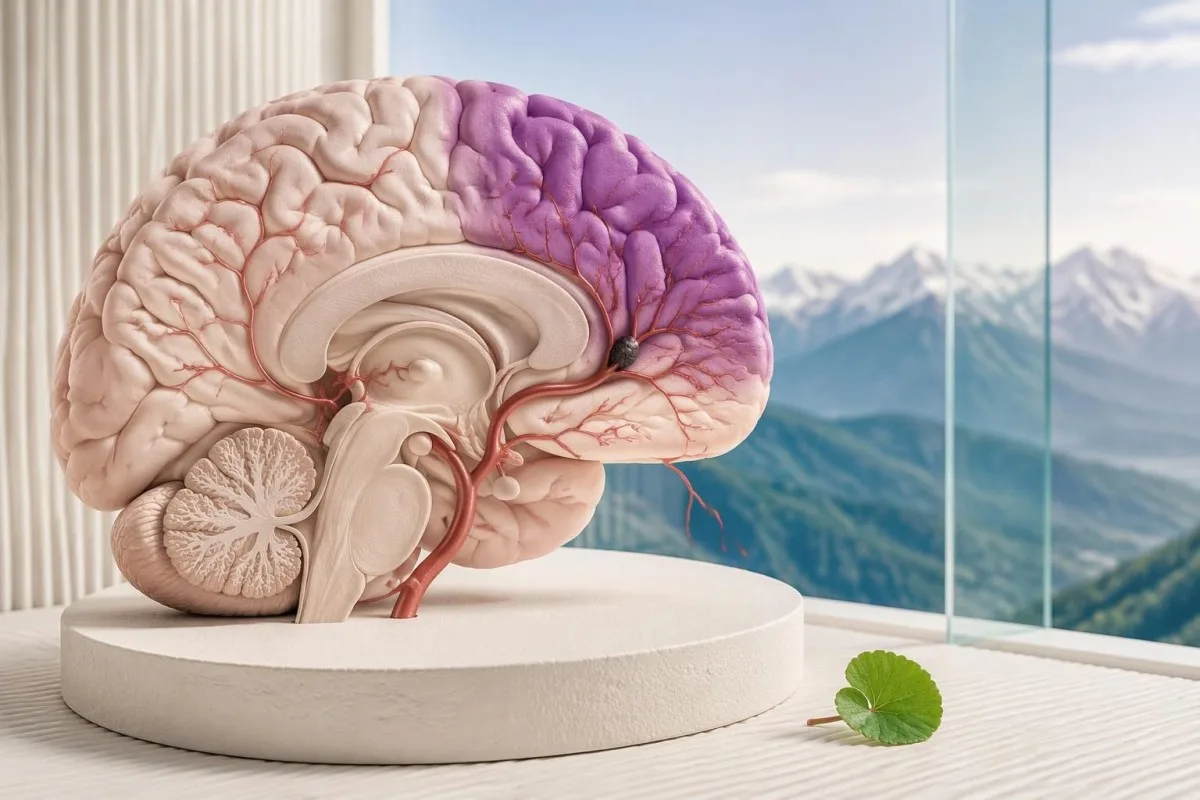
Nagła utrata równowagi, zaburzenia widzenia, opadanie kącika ust, osłabienie ręki lub trudności z mówieniem mogą świadczyć o udarze. Natychmiast zadzwoń pod lokalny numer alarmowy — nawet jeśli objawy ustąpią lub rutynowe badania krwi będą prawidłowe; ani poprawa, ani prawidłowe wyniki laboratoryjne nie oznaczają, że można bezpiecznie czekać.

Najważniejsze wnioski
- BE-FAST oznacza równowagę (Balance), oczy (Eyes), twarz (Face), ręce (Arms), mowę (Speech) i czas (Time); jeden nagły objaw ostrzegawczy może uzasadniać wezwanie pogotowia.
- Numery alarmowe to 999 lub 112 w Wielkiej Brytanii oraz 911 w USA; w innych krajach należy skorzystać z lokalnego numeru alarmowego.
- Ustępujące objawy mogą wskazywać na przemijający atak niedokrwienny (TIA) i nadal wymagają pilnej oceny.
- Prawidłowe wyniki badań krwi nie mogą wykluczyć ani udaru niedokrwiennego, ani udaru spowodowanego krwawieniem.
- Ostatni moment, gdy stan pacjenta był prawidłowy oznacza ostatnią chwilę, w której na pewno funkcjonował na swoim zwykłym poziomie neurologicznym — a nie moment, w którym ktoś zauważył objawy.
- Okna terapeutyczne zazwyczaj obejmują dożylne leczenie trombolityczne w ciągu 4,5 godziny u kwalifikujących się pacjentów; wybrani pacjenci mogą zostać poddani trombektomii do 24 godzin.
- Objawy ostrzegawcze u kobiet obejmują te same objawy BE-FAST co u mężczyzn; ciąża lub okres poporodowy zmieniają kontekst kliniczny, ale nie zmniejszają potrzeby natychmiastowego działania.
- Nie czekaj na wynik badania laboratoryjnego, interpretację AI, kolejny epizod ani poprawę przed wezwaniem pomocy.
Które objawy udaru wymagają wezwania pogotowia?
Nagłe objawy udaru wymagają natychmiastowego wezwania pomocy: nowe zaburzenia równowagi, utrata wzroku lub podwójne widzenie, opadanie kącika ust, osłabienie ręki lub trudności z mową. Zastosuj BE-FAST, ale nie czekaj na wystąpienie wszystkich 6 objawów; ustąpienie objawów i prawidłowe rutynowe badania krwi nie wykluczają udaru.
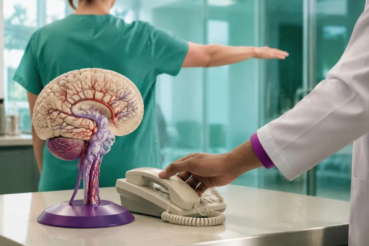
Udar występuje, gdy tętnica mózgowa zostaje zablokowana lub dochodzi do pęknięcia naczynia mózgowego; te 2 mechanizmy wymagają odmiennego leczenia. Nie można wiarygodnie odróżnić ich na podstawie objawów w domu, dlatego przyjmowanie aspiryny lub samodzielne obniżanie ciśnienia przed profesjonalną oceną może być niebezpieczne.
Praktyczną wskazówką jest nagła zmiana w stosunku do zwykłego funkcjonowania danej osoby, a nie to, czy wygląda ona na poważnie chorą. Ktoś może pozostawać przytomny, wejść do pokoju lub opisywać tylko 1 osłabioną rękę, a mimo to mieć udar powodujący niepełnosprawność; ból ani utrata przytomności nie są konieczne.
Wyobraź sobie na przykład osobę, która o 10:12 mówi normalnie, a o 10:14, podczas jedzenia śniadania, zaczyna mówić niewyraźnie. Natychmiast zadzwoń i podaj obie godziny; nie poświęcaj kolejnych 10 minut na sprawdzanie symetrii twarzy, szukanie objawów w internecie ani organizowanie rutynowego badania krwi.
Kantesti to analizator wyników badań krwi oparty na AI, służący do interpretacji raportów laboratoryjnych, a nie usługa diagnostyczna w nagłych przypadkach udaru. Naszą organizacja i zakres kliniczny należy rozumieć z uwzględnieniem tego ograniczenia: nawet interpretacja dostarczona w około 60 sekund nie może nigdy opóźniać wezwania pomocy.
Co oznacza sześć objawów udaru ujętych w BE-FAST?
Objawy udaru BE-FAST obejmują równowagę (Balance), oczy (Eyes), twarz (Face), ręce (Arms), mowę (Speech) i czas wezwania pomocy (Time). Ten 6-elementowy mnemonik uzupełnia FAST o nagłe zaburzenia równowagi i zmiany widzenia, pomagając rozpoznać udary, które nie zaczynają się od wyraźnego opadania kącika ust ani osłabienia ręki.
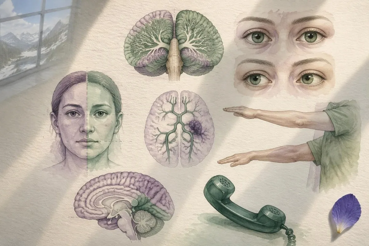
Równowaga oznacza nagłą niestabilność, utratę koordynacji lub trudności ze staniem bądź chodzeniem; oczy oznaczają nagłą utratę wzroku, podwójne widzenie lub ubytek części pola widzenia. Każdy z tych objawów może wystąpić bez pozostałych 3 fizycznych objawów FAST, więc prawidłowy uśmiech nie oznacza, że można bezpiecznie je zlekceważyć.
Twarz oznacza nowy asymetryczny uśmiech lub opadanie części twarzy; ręce oznaczają nowe osłabienie, opadanie lub drętwienie, zwłaszcza po 1 stronie ciała. Zaburzenia mowy obejmują niewyraźną mowę, używanie niewłaściwych słów, niemożność zrozumienia prostego zdania lub niemożność wypowiedzenia słów — nie tylko nieznany akcent czy ciche mówienie.
Czas oznacza: dzwoń teraz, nie kończ najpierw listy kontrolnej. W retrospektywnym badaniu obejmującym 736 udarów niedokrwiennych FAST nie wykrył 14.1% przypadków; dodanie objawów zaburzeń równowagi i widzenia zmniejszyło odsetek niewykrytych przypadków do 4.4% (Aroor et al., 2017), ale wyniki z jednego ośrodka nie określają uniwersalnej dokładności domowych testów przesiewowych.
U osoby, u której objawy dotyczą tylko ręki, nadal trzeba zwrócić uwagę na czas ich wystąpienia i sprawność. Nasz przewodnik dotyczący wzorców drętwienia poszczególnych palców opisuje częstą przyczynę obwodową, ale nagłej utraty sprawności 1 ręki nie wolno bez oceny uznawać za zespół cieśni nadgarstka.
Kiedy nagłe zawroty głowy lub zaburzenia równowagi mogą świadczyć o udarze?
Nagłe, silne zaburzenia równowagi mogą sygnalizować udar krążenia tylnego, zwłaszcza jeśli towarzyszą im podwójne widzenie, zmiany mowy, osłabienie lub niemożność chodzenia. Udary te obejmują struktury takie jak móżdżek i pień mózgu, dlatego wszystkie 3 fizyczne testy FAST mogą dawać prawidłowy wynik.
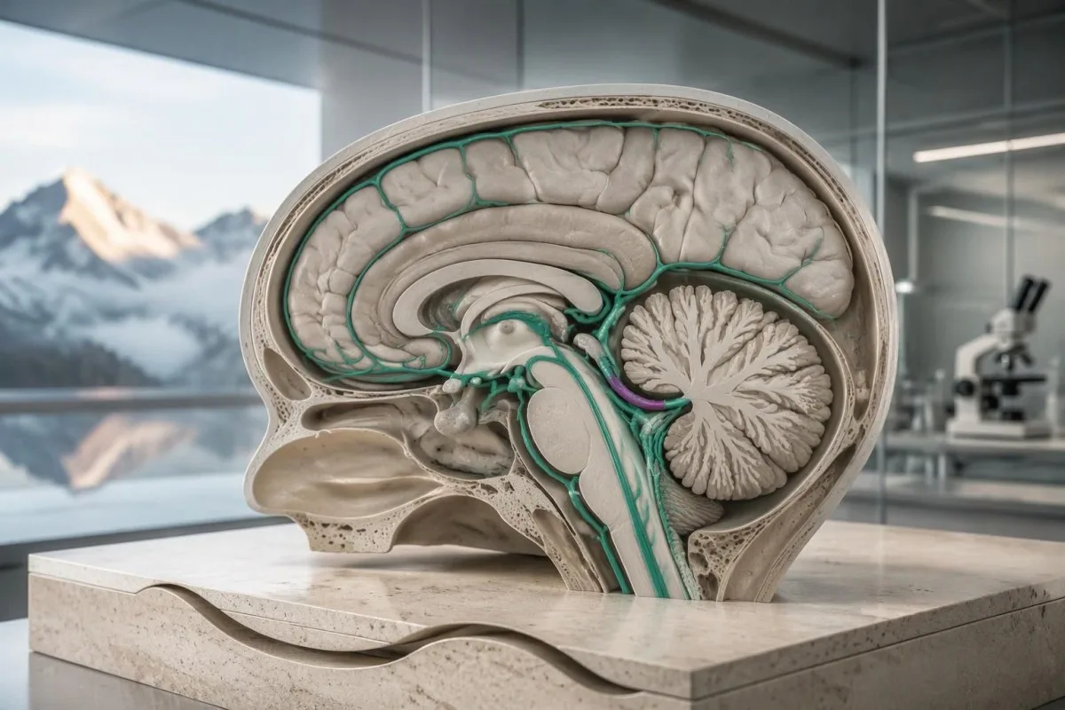
Not every dizzy spell is a stroke; inner-ear disorders and faintness are common alternatives. The concerning combination is a new, persistent balance disturbance plus loss of a previously easy function—for example, being unable to take 2 unsupported steps or suddenly needing furniture to remain upright.
A positional trigger does not automatically prove an inner-ear cause, and nausea does not rule out stroke. If a person develops abrupt vertigo with 1 new neurological feature, such as double vision or slurred speech, call emergency services rather than testing repeated head movements at home.
The HINTS examination is a clinician-performed assessment used in a specific acute vestibular syndrome, not a 3-step self-test for every dizzy patient. Incorrect technique or use in the wrong setting can falsely reassure; an online demonstration cannot substitute for trained examination and appropriate imaging.
Orthostatic symptoms usually have a different pattern, often linked to standing and recurring over time. Our standing-related dizziness guide explains that distinction, but even someone with established POTS needs emergency assessment for 1 sudden, unfamiliar neurological deficit.
Które nagłe zaburzenia widzenia mogą ostrzegać przed udarem?
Sudden vision loss, new double vision, or loss of one side of the visual field can be stroke symptoms. A problem affecting just 1 eye can also represent interrupted retinal circulation, which requires emergency assessment even when facial movement, arm strength, and speech seem normal.
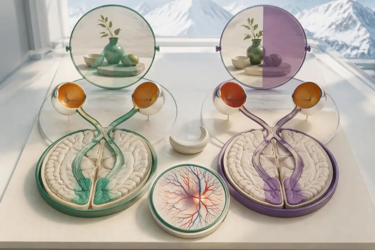
Visual-field loss is different from blurred vision: a person may miss everything to the left or right despite both eyes being open. For example, they might see only 1 half of a plate or repeatedly bump into objects on the same side, without realizing that vision—not attention—is the problem.
New double vision can reflect impaired eye alignment from brainstem dysfunction, especially when paired with imbalance or speech changes. Tell the dispatcher whether the symptom involves 1 eye, both eyes, or a missing side of the scene, but do not delay the call to perform a detailed eye examination.
A brief, painless blackout in 1 eye can be a retinal TIA rather than an ordinary eye complaint. The fact that sight returns after several minutes is not evidence that circulation is safe; the episode may require brain, vessel, and cardiac assessment in addition to an eye examination.
Routine eye screening answers a different question from sudden visual loss. Our explanation of pressure and visual-field testing describes longer-term assessment, but an abrupt change over seconds or minutes should not wait for an optician appointment scheduled 1 week later.
Czy wczesne objawy udaru pojawiają się przed głównym incydentem?
Early signs of stroke are often the stroke itself, not a predictable warning phase lasting days. Some people have a preceding TIA, but there is no reliable 24-hour symptom sequence; sudden loss of speech, strength, vision, or coordination deserves action at its first appearance.
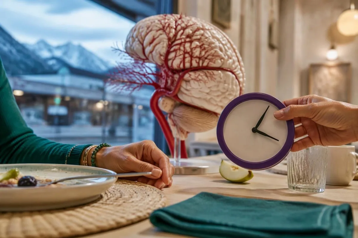
Last known well is the last time someone was definitely at their usual neurological baseline. If a person went to sleep normally at 22:30 and woke with weakness at 06:30, both times matter; reporting 06:30 as the definite onset would incorrectly shorten the possible duration.
Wake-up symptoms are still an emergency, even when several hours may have passed. Some patients with unknown onset can receive treatment selected by imaging, so assuming that the usual 4.5-hour intravenous treatment window has already closed may wrongly prevent access to useful care.
A sudden severe headache reaching maximum intensity within about 1 minute is a thunderclap headache and warrants emergency assessment, particularly with vomiting, neck stiffness, or neurological changes. Many ischemic strokes cause no headache, so its absence must not become a reason to wait.
The speed of change helps distinguish some neurological patterns, but it cannot establish a diagnosis alone. Our objawy ostrzegawcze postępującego osłabienia discuss a different emergency pattern; weakness evolving over 2 days and weakness appearing in seconds both need appropriate clinical evaluation.
Czy objawy udaru u kobiet są inne?
Stroke symptoms in women usually include the same BE-FAST warning signs as in men. Some observational reports describe more accompanying confusion, headache, nausea, or generalized symptoms, but there is no separate reliable 6-sign checklist for women, and these nonspecific features cannot diagnose stroke alone.

A useful distinction is between a focal deficit and an accompanying symptom: inability to use 1 arm is more localizing than fatigue alone. If a woman has sudden nausea plus double vision and inability to walk, the neurological combination—not whether the symptoms sound stereotypically female—should drive the emergency call.
Pregnancy and the postpartum period change the possible causes of headache and neurological symptoms, including hypertensive complications and cerebral venous thrombosis. Tell emergency clinicians the pregnancy stage or delivery date; a new severe headache with 1 focal deficit should not be attributed to exhaustion or migraine without assessment.
Normal or previously reassuring pregnancy bloodwork cannot exclude a neurological emergency. Our explanation of przewodnik po wynikach badań laboratoryjnych w stanie nagłym HELLP addresses one obstetric complication, but neither a normal platelet count nor normal liver enzymes excludes all causes of postpartum headache or stroke.
Consider a hypothetical woman 10 days after delivery who develops abrupt word-finding difficulty and a headache unlike her usual migraines. The safe action is an emergency call with the delivery date and last known well—not waiting for a repeat CBC, checking whether the headache improves, or asking an app to classify symptoms.
Dlaczego ustępujące objawy nadal mogą wskazywać na TIA?
Stroke-like symptoms that disappear may represent a TIA or a stroke with improving deficits, and both need urgent assessment. A modern TIA diagnosis concerns transient ischemia without acute infarction, not simply symptoms lasting under 24 hours; only professional evaluation can make that distinction.
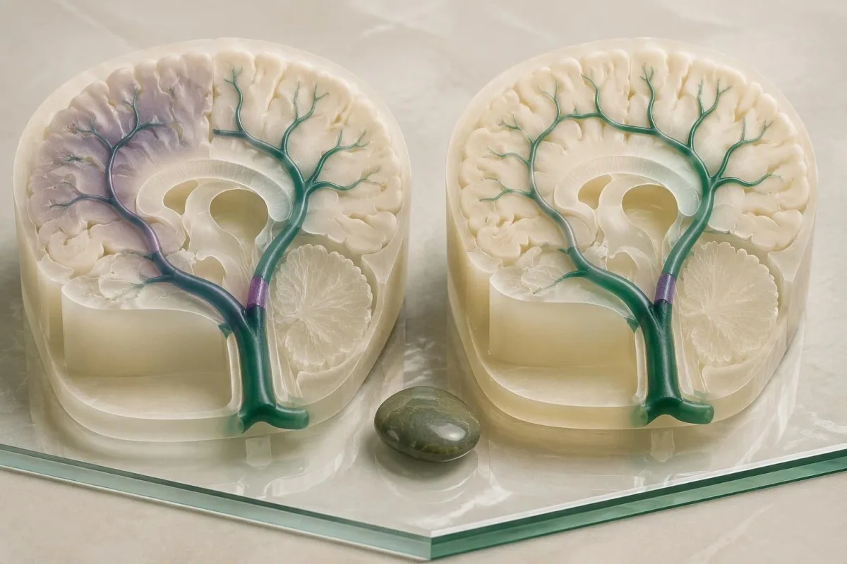
Amin et al. (2023), in an American Heart Association scientific statement, reported 90-day stroke-risk estimates after TIA as high as 17.8%, with almost half of those strokes occurring within 2 days. Those figures describe particular study populations, not an individual prediction; prompt assessment and prevention can change the risk.
If weakness resolves while an ambulance is coming, do not cancel because the person now feels normal. Record when symptoms started, when they stopped, which side was affected, and whether any episode recurred; even 1 brief focal event may identify an unstable arterial or cardiac source.
Risk scores such as ABCD2 cannot safely replace vessel imaging, cardiac assessment, and clinical judgment. A low score may overlook important causes, while a normal examination after 10 minutes of resolved symptoms documents recovery rather than proving that the original event was harmless.
At Kantesti, we separate long-term laboratory risk review from assessment of a new neurological event. Our family-history stroke testing guide is relevant after urgent care, but a preventive panel next week is not a substitute for assessment of 1 suspected TIA today.
Czy prawidłowe rutynowe badania krwi mogą wykluczyć udar?
Normal routine bloodwork cannot rule out stroke or TIA. A CBC, metabolic panel, lipid panel, or coagulation panel measures selected cellular, chemical, or clotting properties; none shows whether 1 brain artery is blocked, whether brain tissue is injured, or whether bleeding has occurred inside the skull.
An INR of 1.0 and a normal aPTT do not prove that no clot exists. These tests assess aspects of coagulation under laboratory conditions, not the presence of an arterial obstruction; our guide to limitów prawidłowych testów krzepnięcia explains why reassuring values can coexist with clinically important clotting.
A normal cholesterol result answers a risk-factor question, not an emergency diagnostic question. For example, an LDL cholesterol of 85 mg/dL does not exclude an embolus from atrial fibrillation or an artery problem; our metabolic panel versus cholesterol guide also clarifies which tests a routine panel actually contains.
Kantesti is an AI blood test interpretation platform that explains reported laboratory findings without using normal values to exclude stroke. A hypothetical platelet count of 240 × 10⁹/L, glucose of 98 mg/dL, and creatinine of 0.9 mg/dL can all coexist with a new cerebral artery blockage.
A panel from 2 days ago has an additional limitation: it describes the time of collection, not current neurological function. Emergency teams may repeat selected tests for treatment safety, but a person should call for help based on sudden symptoms rather than wait for a new sample or compare old reference ranges.
Co należy zrobić, czekając na pomoc medyczną?
Call emergency services, report suspected stroke, and note the last known well time. Use 999 or 112 in the UK, 911 in the US, or your local emergency number; do not drive yourself, and do not wait for laboratory results before requesting help.
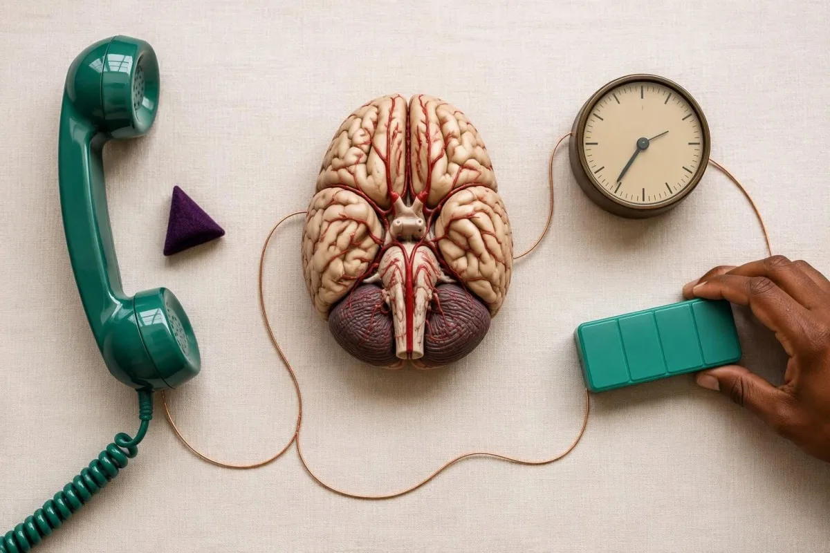
Keep the person safely supported and follow the dispatcher’s positioning instructions rather than making them walk to prove they are better. Do not offer food, drink, or tablets because stroke can impair swallowing; if consciousness or breathing changes, tell the dispatcher immediately and follow their resuscitation guidance.
Do not start aspirin, take an extra anticoagulant dose, or use extra blood-pressure medication on your own. The 2 major stroke mechanisms need different approaches, and sudden blood-pressure reduction can be harmful in some situations; clinicians may give aspirin for suspected TIA under a local pathway, but that is not a universal home-treatment instruction.
Gather medication names and the time of the last anticoagulant dose, especially for warfarin, apixaban, rivaroxaban, dabigatran, or edoxaban. Our wyjaśnienie wyniku INR helps clarify warfarin monitoring, but a normal INR does not reliably establish the absence of a direct oral anticoagulant’s effect.
For example, if symptoms began at 14:05 and a prescribed anticoagulant was taken at 08:00, communicate both exact times. Unlocking the door and having the medication list ready can help responders; finding an old report, charging a phone for an upload, or arranging private transport must not postpone the call.
Które badania w trybie nagłym służą do oceny podejrzenia udaru?
Suspected stroke is assessed with neurological examination, urgent brain imaging, and selected supporting tests—not routine bloodwork alone. A rapid glucose check identifies a treatable mimic; CT commonly checks for intracranial bleeding, while CT angiography or MRI may answer additional questions about arteries and brain tissue.
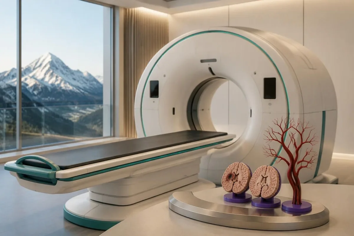
An early noncontrast CT may look normal during an ischemic stroke, so a reassuring initial scan is not always the end of assessment. MRI with diffusion-weighted imaging can detect many early infarcts, but early false-negative studies occur, particularly in posterior circulation events; persistent symptoms still need clinical reconciliation.
Low glucose can cause confusion, weakness, or speech difficulty, sometimes resembling a stroke. A glucose below 70 mg/dL, or 3.9 mmol/L, is hypoglycemia; below 54 mg/dL, or 3.0 mmol/L, is clinically significant hypoglycemia, and our low-glucose causes guide explains common medication and illness-related causes.
A severe metabolic disturbance can also complicate examination, but finding one does not automatically exclude simultaneous stroke. Our glucose-adjusted sodium explanation describes why marked hyperglycemia changes sodium interpretation; neither calculation should delay emergency assessment of 1 new focal deficit.
Kantesti AI can explain laboratory terminology after collection, but it cannot perform a neurological examination or interpret an evolving emergency as a safe exclusion of stroke. Powers et al. (2019) state that only glucose must precede intravenous thrombolysis in all patients; additional laboratory results may be needed when specific medication, bleeding, or coagulation concerns exist.
Dlaczego nie wolno czekać na rutynowe wyniki badań przed rozpoczęciem leczenia?
Stroke treatment is time-sensitive because threatened brain tissue may become permanently injured while testing is delayed. Established treatment pathways include intravenous thrombolysis within 4.5 hours for eligible patients and thrombectomy up to 24 hours in selected patients; those limits are eligibility windows, not safe periods for waiting.
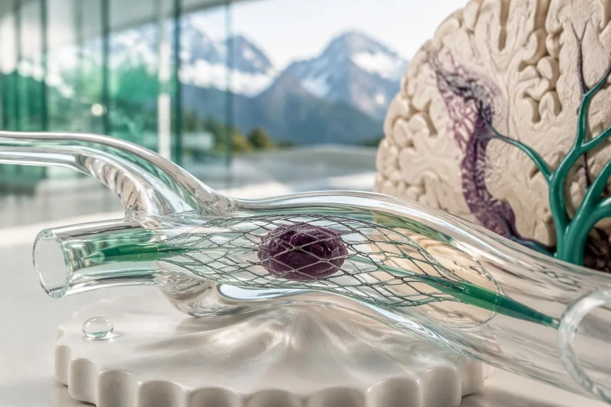
Thrombolysis uses medication to dissolve an eligible clot; thrombectomy uses a catheter-based device to remove selected artery blockages. The 2019 AHA/ASA guideline describes imaging-selected thrombectomy between 6 and 24 hours for certain patients (Powers et al., 2019), but anatomy, imaging findings, baseline function, and specialist judgment determine eligibility.
Blood pressure and medication exposure affect treatment safety without becoming reasons to stay home. An established pre-thrombolysis blood-pressure threshold is below 185/110 mmHg, and hospital teams can manage elevated readings; patients should not attempt to meet that threshold using extra tablets before transport.
Coagulation results matter when a specific safety concern is present, but they do not diagnose the blockage itself. A platelet count below 100 × 10⁹/L or warfarin-associated INR above 1.7 can affect standard intravenous thrombolysis eligibility; our clotting assay interpretation guide explains why different assays answer different questions.
Imagine someone first assessed 3 hours after definite symptom onset who waits another 90 minutes for a nonessential routine panel before reaching a stroke team. That delay may consume a conventional treatment window, although eligibility is never decided by time alone; the safer sequence is emergency assessment first, with necessary tests performed in parallel.
Czy migrena, napady padaczkowe lub biomarkery mózgowe mogą pomóc odróżnić udar?
Migraine, seizures, low glucose, and some vestibular disorders can mimic stroke, but they cannot be reliably distinguished at home. No routine report containing 1 neurological biomarker establishes that a sudden deficit is harmless; examination, symptom timing, and appropriate imaging remain central to assessment.
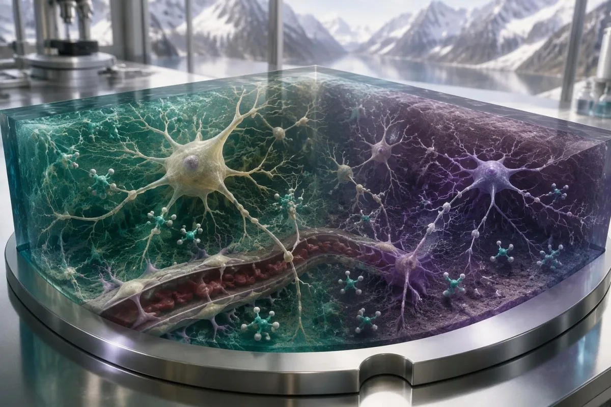
Migraine aura often spreads gradually across several minutes, whereas stroke often causes an abrupt loss of function, but the patterns overlap. A first aura, an unfamiliar aura, or a sudden negative symptom such as losing 1 side of vision should not be self-diagnosed as migraine just because headache is present.
A seizure can be followed by temporary weakness, sometimes called Todd paralysis, and a stroke can itself trigger a seizure. Reporting witnessed shaking or a 1-minute episode of unresponsiveness helps clinicians, but it does not make subsequent weakness safe to observe without urgent assessment.
Neurofilament light chain reflects neuronal injury in multiple conditions rather than functioning as a rapid stroke exclusion test. Our NfL result interpretation guide explains this nonspecificity; a single result, whether high or low, cannot replace assessment of 1 newly impaired neurological function.
Some GFAP-based testing pathways address selected traumatic brain injuries, not universal stroke screening. Our GFAP and head-injury guide discusses that separate use; even where research examines stroke biomarkers, assay timing, thresholds, and performance vary, so 1 normal value must not reassure a symptomatic person.
Jak badania krwi mogą pomóc po ocenie w trybie nagłym?
Blood tests help identify and monitor some stroke risk factors after urgent diagnosis, including diabetes and atherogenic cholesterol patterns. They do not replace blood-pressure measurement, cardiac rhythm assessment, or vascular imaging, and 1 reassuring panel does not guarantee that a person will not have another event.

A hypothetical follow-up panel with LDL cholesterol of 142 mg/dL and HbA1c of 7.2% identifies modifiable concerns, not the exact cause of the event. Our ApoB treatment-decision guide explains particle-related risk assessment; treatment targets depend on the diagnosed stroke mechanism, comorbidities, and the clinician’s prevention plan.
Hypertension needs direct measurement because a chemistry panel does not measure arterial pressure. Our secondary hypertension testing guide explains selected laboratory investigations, but 2 normal electrolyte results do not exclude common primary hypertension or replace repeated blood-pressure readings.
Atrial fibrillation may require ECG monitoring because 1 short recording can miss intermittent episodes. Similarly, broad inherited thrombophilia testing is not automatically useful after every arterial stroke; the history, age, imaging pattern, and suspected mechanism determine which additional investigations could change treatment.
Kantesti can support clinician-directed comparison of follow-up laboratory reports, but medication changes belong within the prescribed care plan. Whole-food eating patterns, smoking cessation, activity adapted to recovery, and consistent medication use address longer-term risk; none can reverse a new sudden deficit or justify waiting 1 more day for emergency assessment.
Publikacje naukowe, dowody i ograniczenia interpretacji wyników laboratoryjnych
The stroke evidence cited here supports emergency recognition and assessment; our 2 listed repository publications address general laboratory interpretation, not stroke diagnosis. Readers can review our validation methods and limits without treating technical evaluation as proof that an AI tool can exclude an acute neurological emergency.
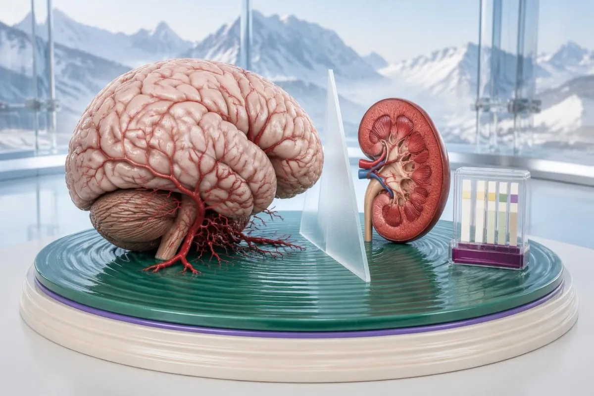
Our listed kidney-function interpretation guide explains BUN and creatinine in their own clinical context. Its Zenodo DOI identifies a repository publication; a creatinine of 0.9 mg/dL, for example, describes a laboratory finding and cannot establish that cerebral circulation is intact.
Towarzyszący materiał urinalysis interpretation publication addresses urine findings rather than acute brain assessment. These 2 repository references are included for transparency about related educational material, not as external stroke guidelines, peer-reviewed stroke trials, or evidence that urine testing can distinguish TIA from stroke.
Kantesti is an AI-powered blood test analysis tool whose educational role must remain separate from emergency triage and definitive diagnosis. Our standardy treści medycznych explain the editorial framework; the 3 external medical references below address BE-FAST, suspected TIA, and established acute ischemic stroke management.
Clinical follow-up may involve sharing reports with a treating team, but that should happen after emergency needs are addressed. Our polityka prywatności i zasady przetwarzania danych describe report handling; permission to review 1 family member’s laboratory report does not authorize access to another’s, and no upload should postpone an emergency call.
Często zadawane pytania
Jakie są pierwsze objawy ostrzegawcze udaru mózgu?
Pierwszymi objawami ostrzegawczymi udaru są często nagła utrata równowagi, zaburzenia widzenia, opadanie kącika ust, osłabienie ręki lub trudności z mową. BE-FAST porządkuje te objawy w 6 wskazówek, w tym: czas wezwać służby ratunkowe. Nie trzeba mieć wszystkich objawów, bólu głowy ani utraty przytomności, aby doznać udaru. W przypadku nowego, nagłego ubytku neurologicznego należy natychmiast wezwać pomoc i podać godzinę, o której dana osoba była ostatnio widziana w stanie bez objawów.
Czy objawy udaru mogą ustąpić po kilku minutach?
Objawy udaru mogą ustąpić po kilku minutach w przypadku TIA lub poprawy stanu po udarze, ale ich ustąpienie nie oznacza, że zagrożenie minęło. W oświadczeniu American Heart Association z 2023 roku dotyczącym TIA podano, że szacowane ryzyko udaru w ciągu 90 dni może wynosić nawet 17.8% w niektórych populacjach, przy czym prawie połowa udarów występuje w ciągu 2 dni. Szacunki te nie stanowią indywidualnej prognozy, a szybkie leczenie może zmniejszyć ryzyko. Należy pilnie zgłosić się na ocenę medyczną, nawet jeśli mowa, siła mięśniowa lub wzrok wrócą do normy.
Czy prawidłowe wyniki badań krwi mogą wykluczyć udar mózgu?
Rutynowe badania krwi nie mogą wykluczyć udaru, ponieważ nie pokazują bezpośrednio tętnic mózgowych, krwawienia wewnątrzczaszkowego ani uszkodzonej tkanki mózgowej. Liczba płytek krwi wynosząca 240 × 10⁹/L lub INR równy 1.0 mogą współistnieć z udarem niedokrwiennym. Ocena w nagłym przypadku opiera się na badaniu neurologicznym, obrazowaniu mózgu i wybranych badaniach pomocniczych. Nie czekaj na wynik laboratoryjny ani interpretację AI przed wezwaniem pomocy.
Czy nagłe zawroty głowy są oznaką udaru mózgu?
Nagłe zawroty głowy mogą być objawem udaru, zwłaszcza gdy towarzyszą im znaczne zaburzenia równowagi, podwójne widzenie, zmiany w mowie lub nowe osłabienie. Udary krążenia tylnego mogą nie powodować widocznych nieprawidłowości w 3 fizycznych testach FAST — ocenie twarzy (Face), ramion (Arms) i mowy (Speech). Wiele epizodów zawrotów głowy ma przyczyny inne niż udar, ale niemożność normalnego chodzenia lub nowy ubytek ogniskowy wymagają pilnej oceny. HINTS to badanie wykonywane przez przeszkolonego klinicystę, a nie wiarygodny test przesiewowy do samodzielnego wykonania w domu.
Czy objawy udaru mózgu u kobiet są inne?
Kobiety na ogół mają takie same objawy ostrzegawcze udaru BE-FAST jak mężczyźni, choć mogą im towarzyszyć ból głowy, nudności lub splątanie. Nie istnieje odrębna, wiarygodna lista sześciu objawów udaru u kobiet. Ciąża lub okres poporodowy wiążą się z dodatkowymi możliwymi przyczynami, o których należy poinformować personel medyczny pogotowia ratunkowego, zwłaszcza w przypadku nowego, silnego bólu głowy lub ogniskowego ubytku neurologicznego. Nagłych zmian neurologicznych nie należy przypisywać stresowi, migrenie ani wyczerpaniu bez przeprowadzenia oceny.
0 Czy powinienem przyjąć aspirynę, jeśli podejrzewam, że mam udar?
Nie rozpoczynaj samodzielnie przyjmowania aspiryny przy podejrzeniu udaru, chyba że pracownik służb ratunkowych wyraźnie zaleci jej przyjęcie. Dwa główne mechanizmy udaru — niedrożność tętnicy i krwawienie śródczaszkowe — wymagają odmiennego postępowania, a aspiryna może zaszkodzić w przypadku krwawienia. Lekarze mogą zalecić natychmiastowe podanie aspiryny przy podejrzeniu TIA w ramach lokalnej ścieżki klinicznej, ale nie jest to uniwersalne zalecenie do stosowania w domu. Najpierw wezwij służby ratunkowe i unikaj jedzenia, picia oraz przyjmowania tabletek, dopóki nie zostanie potwierdzone, że połykanie jest bezpieczne, i nie zostaną wyjaśnione zalecenia dotyczące leczenia.
Jak długo masz na uzyskanie pomocy w przypadku udaru?
Kwalifikujący się pacjenci zwykle otrzymują dożylną trombolizę w ciągu 4,5 godziny od znanego początku objawów, natomiast wybranym pacjentom można wykonać trombektomię do 24 godzin, w zależności od wyników badań obrazowych i spełnienia innych kryteriów. Niektórzy pacjenci z nieznanym czasem początku objawów lub udarem stwierdzonym po przebudzeniu również mogą kwalifikować się do leczenia wybranego na podstawie badań obrazowych. Te przedziały czasowe nie oznaczają bezpiecznego czasu oczekiwania, a wcześniejsza ocena na ogół pozwala zachować więcej możliwości leczenia. Zadzwoń natychmiast, nawet jeśli czas początku objawów jest niepewny lub minęło już kilka godzin.
Dowiedz się, jak czytać wyniki badań krwi
Prześlij wynik badania laboratoryjnego, a sztuczna inteligencja Kantesti w kilka sekund objaśni prostym językiem każdy biomarker. Z narzędzia korzysta ponad 2 miliony osób na całym świecie.
Odniesienia
- Klein, T., Mitchell, S. i Weber, H. (2026). Wyjaśnienie wskaźnika BUN/Creatynina: Przewodnik testu funkcji nerek. Badania medyczne z wykorzystaniem sztucznej inteligencji Kantesti. DOI
- Klein, T., Mitchell, S. i Weber, H. (2026). Test urobilinogenu w moczu: Kompletny przewodnik analizy moczu 2026. Badania medyczne z wykorzystaniem sztucznej inteligencji Kantesti. DOI
- Aroor S et al. (2017). BE-FAST (Balance, Eyes, Face, Arm, Speech, Time): Reducing the Proportion of Strokes Missed Using the FAST Mnemonic. Stroke. DOI
- Amin HP et al. (2023). Diagnosis, Workup, Risk Reduction of Transient Ischemic Attack in the Emergency Department Setting: A Scientific Statement From the American Heart Association. Stroke. DOI
- Powers WJ et al. (2019). Guidelines for the Early Management of Patients With Acute Ischemic Stroke: 2019 Update to the 2018 Guidelines for the Early Management of Acute Ischemic Stroke: A Guideline for Healthcare Professionals From the American Heart Association/American Stroke Association. Stroke. DOI
- 2 mln+Analizowane badania
- 127+Kraje
- 100+Języki
O zespole medycznym


Sygnały zaufania E-E-A-T
Doświadczenie
Kliniczna weryfikacja procesów interpretacji przez lekarza.
Ekspertyza
Medycyna laboratoryjna skupiona na tym, jak zachowują się biomarkery w kontekście klinicznym.
Autorytatywność
Recenzja medyczna: dr Thomas Klein, przy współpracy dr Sarah Mitchell i prof. dr. Hansa Webera.
Solidność
Interpretacja oparta na dowodach, z jasnymi ścieżkami dalszego postępowania, aby ograniczyć alarm.




Zastrzeżenie medyczne
Ten artykuł ma wyłącznie charakter edukacyjny i nie stanowi porady medycznej. Zawsze konsultuj decyzje dotyczące diagnozy i leczenia z wykwalifikowanym pracownikiem ochrony zdrowia.
Opublikowano · Skontaktuj się z zespołem redakcyjnym
Opublikowano przez Kantesti Ltd, zarejestrowaną w Anglii i Walii, numer firmy. 17090423, Londyn, Wielka Brytania.
