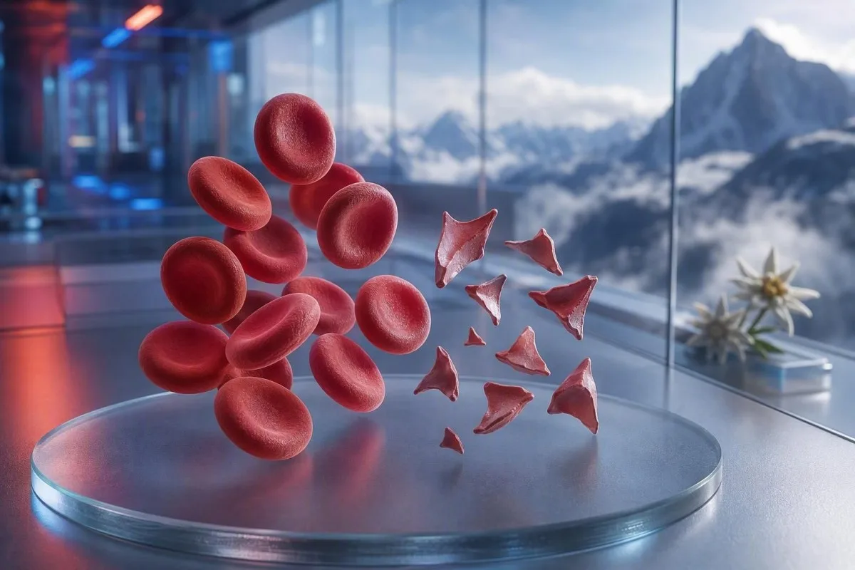
Шистоцитүүд нь аюултай жижиг судасны бүлэн үүсэлт байгааг илтгэж болох хуваагдсан улаан эсүүд боловч ганц түрхэцийн тайлбар нь онош биш. Яаралтай байдал нь хуваагдлын хувь, тромбоцитын тоо, гемоглобины өөрчлөлтийн чиглэл, бөөрний үйл ажиллагаа, гемолизийн маркерууд болон шинж тэмдгүүдээс хамаарна.


- Шистоцитын босго: Сайн бэлтгэгдсэн насанд хүрэгчдийн захын түрхэц дээрх 1%-ээс олон шистоцит нь бусад улаан эсийн хэлбэрийн өөрчлөлтгүй үед тромботик микроангиопати (TMA)-д сэжигтэй гэж үзнэ.
- Яаралтай бөөгнөрөл: Шистоцитүүд + тромбоцит 150 × 10^9/L-оос доош, гемоглобин буурч байгаа, LDH ихссэн, гаптоглобин бага байвал тухайн өдрийн дотор эмчийн үнэлгээ шаардлагатай.
- TTP-ийн сэжүүр: ADAMTS13-ийн идэвхжил 10%-ээс доош байх нь дархлааны тромботик тромбоцитопенийн пурпура (iTTP)-г дэмждэг; эмнэлзүйн сэжиг өндөр үед баталгаажуулах шинжилгээний хариу ирэхээс өмнө эмчилдэг.
- Бүх хуваагдал аюултай биш: Механик зүрхний хавхлагууд, хүнд хэлбэрийн гипертензи, диализийн хэлхээ, түлэгдэлт, дээжийн гэмтэл нь хуваагдсан улаан эсүүд үүсгэж болно.
- Түрхэц бол гар аргаар хийсэн нотолгоо: Автоматжуулсан анализатор нь шистоцитийг найдвартай оношлох боломжгүй гэж дохио өгдөг; туршлагатай морфологич слайдыг нягтлан шалгах ёстой.
- Жирэмслэлт эрсдэлийг нэмэгдүүлдэг: 20 долоо хоногоос хойш цусны даралт ихсэх, шээсэнд уураг илрэх, эсвэл тромбоцит багадахтай хавсарсан шистоцитүүдэд яаралтай эх барихын үнэлгээ шаардлагатай.
- Давтан шинжилгээ тусалдаг: Шинээр авсан EDTA дээж болон давтан түрхэц нь цуглуулгын алдаанаас үүдэлтэй үзэгдлийг үргэлжилж буй улаан эсийн задрал (fragmentation)-аас ялгаж салгаж чадна.
- Өөрөө эмчилж болохгүй: Төмөр, фолат, эсвэл нэмэлтүүд нь микроангиопатик гемолизийг эмчлэхгүй бөгөөд яаралтай үнэлгээнээс анхаарлыг сарниулж болно.
Хуваагдсан улаан эсүүд яаралтай эмчийн үзлэг шаардвал
Шистоцитүүд нь тромбоцитын тоо бага, цус багадалт, биохимийн гемолиз, бөөрний гэмтэл, мэдрэлийн шинж тэмдэг, халууралт, эсвэл хүнд хэлбэрийн гипертензитэй хамт илэрвэл тухайн өдөртөө яаралтай дахин үзүүлэх шаардлагатай. Зөвхөн түрхэцний тайлбар нь хоргүй эсвэл хиймэл (артефакт) байж болно; аюултай олдвор нь хэв шинж (pattern) юм. 2026 оны 8-р сарын 7-ны байдлаар би шинэ шистоцитүүд плюс тромбоцит 100 × 10^9/L-ээс доогуур байвал ердийн хяналтын шинжилгээ биш, “эмч рүү одоо залгах” дохио гэж үзнэ.
Тромбоцит бууралт нь ихэвчлэн хамгийн хурдан яаралтай байдлын дохио байдаг. Гемоглобин 92 г/Л, тромбоцит 68 × 10^9/L, мөн шинээр мэдээлэгдсэн шистоцитүүд нь гемоглобин 145 г/Л, тромбоцит тогтвортой 260 × 10^9/L-тэй 1% хэлтэрхийтэй харьцуулахад хамаагүй илүү санаа зовоох шинжтэй. Kantesti нь энэхүү кластерийг цусны ерөнхий шинжилгээ (complete blood count)-ний хүрээнд уншдаг AI цусны тест анализатор бөгөөд зөвхөн морфологийн тайлбарыг тусад нь нэмэгдүүлж тайлбарладаггүй.
Миний 15 жилийн клиникийн практикт хурдан гаргадаг дүгнэлтүүд маань хэдхэн хоногийн дотор өөрчлөгдсөн өвчтөнүүдийнх байдаг: гемоглобин буурах, креатинин өсөх, мөн лактат дегидрогеназа (LDH) өсөх. Доктор Томас Кляйны практик дүрэм энгийн: төөрөгдөл, шинэ сулрал, цээжний өвдөлт, амьсгаадах, маш бага шээс, эсвэл энэ хэв шинжтэй хавсарсан хүнд хэлбэрийн толгой өвдөх нь өнөөдөр яаралтай үнэлгээ шаардлагатай гэсэн үг.
Шистоцитүүд нь тухайн хүн өвдсөн бол давтан шинжилгээ хүлээх шалтгаан биш. Калийн хэвийн хэмжээ эсвэл цагаан эсийн тоо хэвийн байх нь тромботик микроангиопати (thrombotic microangiopathy)-ийн эрсдэлийг хүчингүй болгохгүй. Тоо бага байгааг ойлгох гэж буй өвчтөнүүд мөн манай гарын авлагыг үзэж болно. улаан цусны эсийн шинж тэмдэг багадах, гэхдээ хурц шинж тэмдгүүд нь онлайн тайлбараас үргэлж илүү чухал.
Дараах тохиолдолд яаралтай тусламжийн үйлчилгээнд одоо залгаарай:
Нэг талын шинэ сулрал, ухаан алдах, таталт, хүнд хэлбэрийн амьсгаадалт, цээжийг дарж өвдөх таагүй мэдрэмж, хар баас, эсвэл төөрөгдөл хурдан даамжрах нь клиникт мессеж бичихээс илүү яаралтай тусламжийн үйлчилгээг шаарддаг. Эдгээр шинж тэмдгүүд нь эрхтэн оролцож байгааг илэрхийлж болох бөгөөд шистоцитийн хувь хэмжээ нь үүнийг аюулгүйгээр үгүйсгэж чадахгүй.
Захын түрхэц дээрх шистоцитүүд яг юу гэсэн үг вэ
Шистоцитүүд нь эргэлдэж буй эсүүдийг физик байдлаар “хяргаж” гэмтээх үед үүсдэг жигд бус улаан эсийн хэлтэрхий бөгөөд ихэнхдээ жижиг судас дахь фибриний утаснууд эсвэл хэвийн бус өндөр-хяргах (high-shear) гадаргууудын нөлөөгөөр үүсдэг. Тэд ихэвчлэн хурц өнцөгтэй, дуулга шиг хэлбэртэй, эсвэл жижиг гурвалжин хэлбэртэй байдаг бөгөөд бүтэн улаан эсийн төвийн цайрал (central pallor)-ыг агуулдаггүй.
Жинхэнэ шистоцитүүд нь морфологийн олдвор бөгөөд стандарт CBC анализаторын гаргаж өгдөг “тоо” биш. Цус судлалын стандартчиллын олон улсын зөвлөл (International Council for Standardization in Haematology) тоон үнэлгээ чухал үед хамгийн оновчтой түрхэцийн бүсэд дор хаяж 1,000 улаан эсийг тоолохыг зөвлөдөг (Zini et al., 2021). Энэ нарийн зүйл нь нэг лаборатори “ховор хэлтэрхий” гэж мэдээлж байхад нөгөө нь хувь гэж мэдээлж болох шалтгааныг тайлбарладаг.
Сонгодог гол санаа зовоох зүйл нь микроангиопатик гемолиз бүхий цус багадалт (microangiopathic haemolytic anaemia), цусны эргэлтэд улаан эсүүд ялтас-ихтэй микротромбо болон фибринтэй нийлж таарах үед. Гэвч хүнд хэлбэрийн аортын нарийсал, механик хавхлага, экстракорпораль хэлхээ, мөн томоохон дулааны гэмтэл нь өөр механизмын улмаас жинхэнэ хэлтэрхий үүсгэж чадна. Тиймээс асуулт нь “Хэлтэрхий бодит уу?” биш, харин “Хэлтэрхийг юу үүсгэж байна вэ?” юм.”
Kantesti нь AI цусны шинжилгээний тайлал платформ бөгөөд байршуулах бүрэн цусны тоолол (CBC)-ыг CBC-д юу ордог тухай гарын авлага хажууд нь байрлуулж, билирубин, ретикулоцит, эсвэл гаптоглобин зэрэг дутуу хамтрагч үзүүлэлтүүдийг тодруулж өгнө. Энэ нь слайдыг өөрөө шалгаж чадахгүй; энэ ажил нь мэргэшсэн лабораторийн мэргэжилтний үүрэг хэвээр байна.
Насанд хүрэгчдийн түрхэц дээр шистоцитоз хэр их байвал санаа зовоох вэ
Насанд хүрэгчдийн захын түрхэц дээрх шистоцитын тоо 1%-оос дээш байвал, хэлтэрхий нь улаан эсийн зонхилох гаж хэлбэржилт бол тромботик микроангиопати сэжигтэй. 1%-оос доош тоо нь өвчнийг үгүйсгэхгүй, ялангуяа шингэн сэлбэлт, цус сэлбэлт, эсвэл хөгжиж буй өвчний дараа түрхэц бэлтгэсэн бол.
1%-ийн таслах утга нь оношилгооны сануулга бөгөөд хүндрэлийн шатлал биш. ICSH-ийн удирдамжид эрүүл насанд хүрэгчид болон хугацаандаа төрсөн нярайд 1%-оос доорх лавлах интервал хэрэглэдэг; дутуу төрсөн нярайд мөн адил утгаараа бишгээр 5% хүртэл хэлтэрхий байж болно (Zini et al., 2021). Хүнд тромбоцитопени бүхий 2%-ийн үр дүн нь механик цусны эргэлтийн дэмжих төхөөрөмж байрлуулсны дараах 4%-ээс илүү түгшүүртэй.
Лабораториуд хувь хэмжээгээр бус “аянгын,” “цөөн,” эсвэл “өндөр чадлын талбарт 1–2” гэж мэдээлж болно. Эдгээр хэллэгүүд нь харилцан адилгүй, учир нь талбайн хэмжээ, түрхэцийн зузаан, мөн тайлагналын бодлого өөр өөр байдаг. Лаборатори ICSH маягийн хувийн тоолол хийсэн эсэхийг асуух нь, та өөрөө тайлбарласан хэллэгийг хөрвүүлэх гэж оролдохоос ихэвчлэн илүү хэрэгтэй байдаг.
Өндөр RDW нь шистоцитийг хэмждэггүй. RDW нь эсийн хэмжээний хэлбэлзлийг илэрхийлдэг бөгөөд ретикулоцитоз, төмрийн дутагдал, цус сэлбэлт, эсвэл хэлтэрхий үүсэлттэй үед нэмэгдэж болно. Манай дэлгэрэнгүй RDW ба улаан эсийн индексийн гарын авлага нь өндөр RDW дангаараа TTP ч биш, мөн ямар нэг артефакт ч биш гэдгийг яагаад дэмждэггүйг тайлбарладаг.
Жинхэнэ гемолизийг дэмждэг цусны шинжилгээний хэв шинж
Жинхэнэ улаан эсийн хэлтэрхийжилт нь ихэвчлэн өндөр LDH, бага гаптоглобин, шууд бус билирубин нэмэгдэх, мөн цус багадалтын зэрэгцээ ретикулоцитын хариу урвал үүсгэдэг. Нэг ч маркер дангаараа тодорхой биш, учир нь элэгний өвчин, үрэвсэл, саяхан хийсэн цус сэлбэлт, мөн чөмөгний эмгэгүүд дүр зургийг гажуудуулж болно.
LDH нь мэдрэг боловч өвөрмөц биш. LDH нь 600 IU/L-ээс дээш байх нь зөв нөхцөлд идэвхтэй гемолизийг ихэвчлэн дэмждэг; гэхдээ булчингийн гэмтэл, элэгний гэмтэл, халдвар, мөн хавдар ч үүнийг нэмэгдүүлж болно. Лабораторийн доод хязгаараас доогуур, түгээмэл 0.3 g/L-ээс бага гаптоглобин нь чухал жин нэмдэг, учир нь энэ нь плазмд ялгарсан чөлөөт гемоглобиныг холбодог.
Ретикулоцитын тоо нь ихэвчлэн захын задралын үед нэмэгдэх ёстой, гэхдээ фолийн хүчил дутагдал, төмрийн дутагдал, бөөрний өвчин, эсвэл цочмог чөмөгний дарангуйлал үед бага хэвээр байж болно. Энэ илэрхий зөрчил амархан анзаарагдахгүй: өвчтөнд жинхэнэ гемолиз байж болох ч ретикулоцитын зохисгүй хариу бага байж болно. Тэгээд гаптоглобины үр дүнгийн удирдамж нь нийтлэг тайлбарын урхинуудыг хамардаг.
Kantesti AI нь ижил цагийн цэгээс авсан гемоглобин, MCV, тромбоцит, LDH, билирубин, креатинин, ретикулоцитуудыг харьцуулж хэлтэрхийлэгдсэн улаан эсийн хэв шинжийг тайлбарлана. 48–72 цагийн дотор 20 г/Л-ээс дээш буурсан гемоглобин нь өмнөх харьцуулалтгүйгээр бага зэрэг хэвийн бус утгаас клиникийн хувьд илүү мэдээлэлтэй.
Билирубин яагаад хэвийн байж болох вэ
Гемолизийн эхэн үед, судсаар шингэнээр шингэлсний дараа, эсвэл элэг билирубиныг үр ашигтай цэвэрлэж байвал шууд бус билирубин хэвийн байж болно. Тиймээс хэвийн билирубин нь итгэлтэй хэлтэрхийжилт, гаптоглобин бага, мөн буурч буй гемоглобиныг хүчингүй болгох ёсгүй.
Яагаад TTP-ийг гематологийн яаралтай тусламж гэж үздэг вэ
Тромботик тромбоцитопеник пурпура буюу TTP нь тромбоцитопени дагалдсан шистоцитууд, мөн өөр тодорхой хувилбаргүй үед микангиопатик гемолизийн цус багадалттай хавсарч илэрвэл сэжиглэнэ. Эмчлээгүй иммун TTP хурдан даамжирч болдог тул эмч нар ADAMTS13-ийн хариу ирэхээс өмнө плазма солилцоо болон иммун эмчилгээг эхлүүлж болно.
ADAMTS13-ийн идэвхжил 10%-ээс доогуур байх нь TTP-ийг хүчтэй дэмждэг, гэхдээ үр дүн нь хэдэн цаг эсвэл хэдэн өдөр шаардаж болно. ISTH-ийн оношлогооны удирдамж нь плазма эмчилгээ хийхээс өмнө (Zheng et al., 2020) дээж авахын зэрэгцээ PLASMIC оноо зэрэг клиникийн хэрэгслүүдээр урьдчилсан магадлалыг тооцоолохыг зөвлөдөг. Оношлогооны оноо нь шийдвэр гаргах гүүр бөгөөд мэргэжилтний дүгнэлтийг орлохгүй.
Халууралт, мэдрэлийн шинж, бөөрний үйл ажиллагааны алдагдал, тромбоцитопени, мөн гемолизийн цус багадалтын “пентад” нь олон бодит өвчтөнд бүрэн бус байдаг. George болон Nester нар тромботик микангиопати нь таван заавал байх хайрцаг биш, харин давхцсан шалтгаантай хам шинж гэж тодорхойлсон (George & Nester, 2014). Практикт бүх таван шинжийг хүлээх нь аюултай хуучин дадал юм.
Гемолиз дагалдсан 30 × 10^9/L-ээс доош тромбоцит нь гематологитой яаралтай хэлэлцэх шаардлагатай. Сэжигтэй TTP үед амь насанд аюултай цус алдалт байхгүй, эсвэл инвазив процедур хүлээж болохгүй тохиолдлоос бусад үед тромбоцитын сэлбэлтийг ихэвчлэн зайлсхийдэг. Холбогдох шинжилгээнд манай цус бүлэгнэлтийн шинжилгээний гарын авлага нь TTP үед PT ба aPTT яагаад хэвийн байж болохыг тайлбарладаг.
Эмч нарын шистоцит үүсгэж болох бусад аюултай шалтгаанууд
Шистоцитууд нь тархмал судсан доторх бүлэгнэл (DIC), Шига-токсинтай холбоотой гемолизийн уремийн хам шинж, хүнд хэлбэрийн гипертензи, жирэмслэлтэй холбоотой микангиопати, зарим эмүүд, мөн шилжүүлэн суулгалттай холбоотой тромботик микангиопати зэрэгт мөн тохиолддог. Холбогдох тромбоцит, коагуляцийн, бөөрний, мөн клиникийн илрэлүүд нь ихэвчлэн эдгээр нөхцлийг ялгаж салгадаг.
DIC нь ихэвчлэн хэлтэрхийжилттэй хамт PT уртассан, aPTT уртассан, фибриноген бага эсвэл буурч буй, мөн D-димер мэдэгдэхүйц өндөр байхыг хавсаргадаг. TTP нь ихэвчлэн ердийн коагуляцийн хугацаанууд харьцангуй хэвийн байдаг. Хүнд өвчтөнд фибриноген 1.5 g/L-ээс доош байх нь хэрэглээний коагулопати руу санааг шилжүүлдэг; гэхдээ фибриноген эхэндээ хэвийн харагдаж болно, учир нь цочмог үрэвслийн үед нэмэгддэг.
Хүнд хэлбэрийн гипертензи нь даралт микросудас гэмтээх үед улаан эсийг “тасалж” болно; толгой өвдөх, хараа өөрчлөгдөх, цээжээр өвдөх, эсвэл бөөрний үйл ажиллагааны алдагдалтай хамт 180/120 мм.муб-аас дээш эсвэл түүнтэй тэнцүү уншилт нь яаралтай тусламжийн нөхцөл юм. Иймээс цусны даралтын уншилт нь түрхэцийн тайлангийн хажууд байх ёстой. Өвчтөнүүд хоёрдогч гипертензи лабораторийн сэжүүрүүд шууд аюулыг арилгасны дараа.
Суулгалт дагаж шээсний ялгаралт буурах, цус багадалт, тромбоцитопени үүсэх нь гемолизын уремийн хам шинжийн талаар санаа зовох шалтгаан болдог. Өтгөний шинжилгээ нь Шига токсиныг илрүүлэхэд тусалж болох ч эхлээд яаралтай тусламжийг чиглүүлэхийн тулд шингэний байдал, креатинин, кали, мэдрэлийн үнэлгээ чухал. Өндөр лактат эсвэл тогтворгүй амин үзүүлэлтүүд нь “хүлээгээд үзэх” биш харин сепсис ба DIC-ийн үнэлгээг шаарддаг; эдгээрийг үзнэ үү сепсисийн маркерын хэв маяг.
Хуваагдсан улаан эсийн механик шалтгаанууд
Механик задрал нь TTP-гүйгээр ч бодит шистоцитүүд үүсгэж болно, ялангуяа хиймэл зүрхний хавхлаг, хүнд аортын нарийсал, ховдолын туслах төхөөрөмж, диализийн хэлхээ, эсвэл экстракорпораль мембран оксигенацийн үед. Эдгээр шалтгаанууд нь мөн эмнэлгийн хяналт шаарддаг ч эмчилгээ нь дархлааны TTP-ээс илүү механик эх үүсвэрийг онилдог.
Механик хавхлагын хэв шинжид ихэвчлэн архаг, бага зэргийн LDH нэмэгдэлт, тромбоцит тогтвортой байхтай хавсарсан бага гаптоглобин ордог. Шинээр үүссэн цус багадалт, амьсгаадалт нэмэгдэх, бараан өнгийн шээс, эсвэл хавхлагын дууны өөрчлөлт нь парахавхлагын алдагдал эсвэл төхөөрөмжийн үйл ажиллагааны доголдлыг илтгэж болох бөгөөд кардиологийн дахин үнэлгээг яаралтай хийх хэрэгтэй. Хийц (smear) нь сэжүүр өгнө; эхокардиографи нь эх үүсвэрийг олдог.
Тамирчид заримдаа марафон нь хэлтэрхийг тайлбарлаж чадах уу гэж асуудаг. Хөлний цохилтоос үүдэлтэй гемолиз нь холын зайн гүйлтийн дараа түр зуурын чөлөөт гемоглобин, бага гаптоглобин үүсгэж болох ч үнэмшилтэй шистоцитозыг дасгалтай “энгийнээр” холбож тайлбарлахгүй—ялангуяа тромбоцит буурч байвал. Илүү өргөн хүрээний марафон гүйгчийн лабораторийн удирдамж.
Хүнд түлэгдэлт болон томоохон судасны мэс засал нь бие махбодийн болон эндотелийн гэмтлээр дамжин хэлтэрхийлсэн эсүүд үүсгэж болно. Эдгээр нөхцөлд 6–24 цаг тутам лабораторийн чиг хандлага нь нэг smear-ээс илүү мэдээлэлтэй байж болно. Өдөөгч үйл явдлын дараах CBC тогтвортой байх нь тайвшруулах шинж; гемоглобин тасралтгүй буурч байвал тийм биш.
Цуглуулалт ба түрхэцийн артефакт шистоцит шиг хэрхэн дуурайдаг вэ
Дээжийг муу цуглуулах, smear бэлтгэлийг хойшлуулах, дутуу дүүргэсэн антикоагулянттай хоолой, мөн smear-ийн ирмэг гэмтэх нь цусны эргэлтийн түвшний гемолизыг тусгахгүйгээр шистоциттэй төстэй хэлбэрүүдийг үүсгэж болно. Цэвэрхэн цуглуулж, монослойд (monolayer)-д дахин хянасан давтан дээж нь эмнэлзүйн дүр зураг тайван үед хийх ердийн анхны шалгалт юм.
Crenated эсүүд, smear-ийн ирмэгийн гажилт, хатаах үеийн артефакт, мөн давхцсан эсүүд нь түгээмэл “хуурамч” төстэй дүр төрхүүд. Тэд ихэвчлэн жигд бус, эсвэл идеал унших бүсэд тэгш хэмтэйгээр биш харин өдлөг ирмэг (feathered edge)-ийн хэсэгт л илүү харагддаг. Туршлагатай морфологич нь жинхэнэ хэлтэрхийн хурц геометрийг слайд бэлтгэлийн явцад зүгээр л гажсан эсээс ялган танина.
Хэцүү цуглуулалт нь хоолойг гемолизд оруулж, калий эсвэл LDH-ийг хуурамчаар нэмэгдүүлж болох ч энэ нь автоматаар жинхэнэ эргэлтийн шистоцитүүдийг үүсгэнэ гэсэн үг биш. Хэрэв үр дүнд гемолизийн индекс эсвэл “дээж алдагдсан” гэсэн тайлбар орсон бол эмч нар ихэвчлэн өвчнийг оношлохын өмнө дахин дээж авдаг. Манай нийтлэл калийн авах (draw) алдаа ижил урьдчилсан-аналитик асуудал яагаад хэд хэдэн үр дүнг нэг дор төөрөгдүүлж болохыг тайлбарладаг.
EDTA-аас хамааралтай тромбоцитын бөөгнөрөл нь тромбоцитын тоог хуурамчаар бууруулж, түгшүүртэй харагдах хослолыг “үйлдвэрлэж” болно. Шингэрүүлэлтийг тооцсон (corrected) цитраттай хоолойн тромбоцитын тоо, мөн шинэ түрхэц (fresh film) үүнийг илрүүлж чадна. Нарийн ялгаа чухал: артефакт нь жинхэнэ өвчинтэй зэрэгцэн оршиж болно, тиймээс давтан шинжилгээ нь шинж тэмдэгтэй хүнд тусламжийг хойшлуулах ёсгүй.
Анализаторын дохиолол ба лабораторийн томьёолол яаж төөрөгдүүлж болох вэ
Автомат цусны анализаторууд хэвийн бус улаан эсийн популяцуудыг илрүүлж болох ч тромботик микроангиопати (TMA)-г батлах/үгүйсгэхэд хангалттай найдвартайгаар шистоцитүүдийг бие даан оношилж чадахгүй. Эмнэлзүйн асуулт нь гемолиз эсвэл TTP бол гар аргаар захын цусны түрхэцийн (peripheral smear) хяналт зайлшгүй шаардлагатай хэвээр.
“Улаан эсүүд хуваагдсан (fragmented red cells present)” гэсэн тэмдэглэл нь тоо хэмжээ, түрхэцийн чанар, мөн хавсарсан хэлбэрийн өөрчлөлтийн талаар асуулт тавих ёстой. Төмрийн дутагдлаас, мегалобластик цус багадалтаас, эсвэл удамшлын улаан эсийн эмгэгээс үүдэлтэй мэдэгдэхүйц анизопойкилоцитоз нь 1% дүрмийг илүү тодорхой бус болгодог. Хуваагдлууд нь хэвийн бус хэлбэрийн дийлэнхийг бүрдүүлж байж хамгийн оношлогооны ач холбогдолтой бөгөөд олон өөр шалтгаантай хэлбэрүүдийн дунд цөөн тоогоор илрэх нь бага мэдээлэлтэй.
Kantesti нь сийрсэн сийрсэн цуурыг илтгэсэн хослолыг тэмдэглэхэд зориулагдсан хиймэл оюун ухааны лабораторийн шинжилгээний тайлбар үйлчилгээ юм. Энэ нь лабораторийн микроскоп шинжилгээг эсвэл давтан гулсалт хийлгэхийг хүсч болох эмчээс орлохгүй. Чанарын хяналтанд хандах хандлага маань дараахь байдлаар тайлагдсан болно эмнэлзүйн баталгаажуулалтын тойм.
Дельта шалгалт нь одоогийн үр дүнг өмнөх үр дүнтэй харьцуулж, гэнэт боломжгүй мэт өөрчлөлтийг илрүүлж чадна. Гемоглобин 24 цагийн дотор 138 г/л-аас 87 г/л болж буурах нь эмнэлзүйн хувьд чухал ач холбогдолтой боловч судсаар хийх шингэнээр шингэлэх эсвэл буруу шошголосон дээж байх боломжтой. Үүний талаар дэлгэрэнгүй уншина уу гэнэтийн лабораторийн өөрчлөлтүүд.
Цусны шинжилгээний хариунд шистоцит дурдагдсаны дараа юу хийх вэ
Шистоцитүүд шинээр мэдээлэгдсэн бол тухайн өдөр шинжилгээг захиалсан эмч эсвэл үйлчилгээтэй холбоо барь, мөн мэдрэлийн шинж тэмдэг, амьсгаадах, цээжээр өвдөх, бараан шээс, хүчтэй толгой өвдөх, халуурах, эсвэл шээсний ялгаралт буурах зэрэг шинж илэрвэл яаралтай тусламжийг нэн даруй аваарай. Түрхэцийн тэмдэглэлийг дангаар нь онош гэж тайлбарлахгүй.
Бодит тайланг гар дээрээ байлгаарай: гемоглобин, тромбоцит, креатинин, билирубин, LDH, мөн ямар нэгэн гемолизийн индекс. Мөн саяхны жирэмслэлт, суулга өвчин, цусны даралтын маш өндөр уншилт, шинэ эмүүд, хавхлагын мэс засал, диализ, сэлбэлт (трансфузи), аутоиммун өвчнийг мөн тэмдэглэ. Эдгээр дэлгэрэнгүй мэдээлэл нь морфологийн талбарт нэг үг байхаас илүүтэйгээр ялган оношилгоог өөрчилдөг.
Би өвчтөнүүд төмрийг эхлүүлснээр үнэ цэнтэй цаг алддагийг харсан: учрыг хэн нэгэн тодорхойлохоос өмнө тэд “цус багадалт” гэж уншчихдаг. Төмрийн дутагдал хавсарч байж болох ч энэ нь хуваагдлуудтай хамт тромбоцитын хурдан бууралтыг тайлбарлаж чадахгүй. Доктор Томас Кляйн бүтэн тайлангийн зургийг авахыг, зөвхөн тодруулсан тоонуудыг хуулж бичихээс илүүтэйгээр зөвлөж байна; хуулж бичихэд ихэвчлэн хэв маягийг уялдуулж өгдөг лабораторийн тэмдэглэл унадаг.
Тромбоцит маш бага эсвэл цус алдалт байгаа бол эмч өөрөөр заагаагүй тохиолдолд аспирин, ибупрофен, мөн жороор олгогдоогүй антикоагулянтуудаас зайлсхий. Яаралтай эмнэлгийн удирдлагагүйгээр эмчилгээний шингэлгээг зогсоож болохгүй. Ядаргаа эсвэл толгой эргэх шинж тэмдэгтэй хүмүүс манай толгой эргэх шинжилгээнүүд нь яаралтай шалтгаануудыг үгүйсгэсний дараа асуулт бэлтгэхэд.
Жирэмслэлт, нярай, хүүхдүүд дэх шистоцитүүд
Жирэмсний үед шистоцитүүд илрэх нь, хэрэв тэд гипертензи, протеинури, элэгний ферментийн өндөржилт, тромбоцитын тоо бага, толгой өвдөх, харааны шинж тэмдэг, эсвэл хэвлийн дээд хэсгийн өвдөлттэй хавсарч байвал яаралтай үнэлгээ шаарддаг. HELLP хам шинж, преэклампси, TTP, атипик HUS, мөн DIC нь давхцаж болох бөгөөд төрөлттэй холбоотой хугацаа тусалж болох ч оношийг эцэслэн тогтоож өгдөггүй.
HELLP хам шинжийг сонгодог байдлаар гемолиз, элэгний ферментийн өндөржилт, мөн тромбоцитын тоо бага гэж тодорхойлдог бөгөөд ихэвчлэн жирэмсний 20 долоо хоногоос хойш эсвэл төрөлтөөс удалгүй илэрдэг. Тромбоцитын тоо 100 × 10^9/L-ээс доош эсвэл AST 70 IU/L-ээс дээш байвал санаа зовнилыг дэмжиж болох ч орон нутгийн эх барихын багууд бүхэл бүтэн эмнэлзүйн дүр зургийг тайлбарлана. Энэ хослолтойгоор жирийн хяналтын (антенатал) уулзалтыг хүлээх хэрэггүй.
Нярай (төрсний дараах эхний үеийн) хугацааны хүүхдүүдэд 1% шистоцит цөөн байж болох бол дутуу төрсөн нярайд 5% хүртэл байж болно, учир нь нярайн улаан эсийн морфологи өөр байдаг (Zini et al., 2021). Харин хүүхэд суулгалттай, цайвар, шээс багассан, мөн тромбоцитопени илэрвэл хуваагдлууд HUS рүү чиглүүлж магадгүй тул яаралтай хүүхдийн эмчийн үнэлгээ шаардлагатай.
Тромбоцит ба гемоглобины лавлах интервалууд нь нас болон жирэмсний үе шатнаас хамаарч мэдэгдэхүйц өөрчлөгдөнө. Ердийн програм нь “хэвийн” гэж тэмдэглэсэн үр дүн нь контекстэд эмнэлзүйн хувьд санаа зовоосон байж болно. Манай нөөцийн тухай жирэмсний ялтасны хэмжээ болон ижил өдрийн жирэмсний лабораторийн тэмдэглэгээ жирэмсний яаралтай тусламжийн (maternity triage) оронд биш, хэрэгтэй суурь мэдээллээр хангана.
Эмч нар жинхэнэ шистоцитүүдийг шалгахын тулд захиалдаг шинжилгээнүүд
Сэжиглэгдэж буй бичил судасны эмгэгийн улмаас үүсэх цус задрал (microangiopathic haemolysis)-ын анхны шинжилгээний багцад ихэвчлэн давтан CBC ба түрхэц, ретикулоцитын тоо, LDH, гаптоглобин, билирубиний фракцууд, креатинин, шээсний шинжилгээ, PT, aPTT, фибриноген, D-dimer орно. Хэрэв TTP (тромботик тромбоцитопенийн пурпура) сэжиглэж байгаа бол плазма солилцоо (plasma exchange) хийхээс өмнө ADAMTS13-ийн идэвхжил ба дарангуйлагчийн шинжилгээг авах ёстой, гэхдээ эмчилгээ хүлээж болохгүй.
Шууд антиглобулины шинжилгээ (Direct antiglobulin testing), мөн DAT эсвэл Кумбсын шинжилгээ гэж нэрлэдэг нь дархлааны шалтгаант цус задралыг дархлаагүй задралаас ялгахад тусалдаг. DAT эерэг байх нь хоёр дахь процессийг үгүйсгэхгүй, гэхдээ сонгодог TTP ихэвчлэн DAT-сөрөг байдаг. Бөөрний бичил судасны гэмтэл идэвхтэй үед шээсний микроскопоор уургийн эсвэл эсийн цилиндр (casts) илэрч болно; манай тайлбарыг үзнэ үү мөхлөгт шээсний цилиндрүүд.
Шигатоксин (Stiga-toxin) илрүүлэх шинжилгээг тохирох ходоод гэдэсний өвчний дараа авч үздэг бол atypical HUS-ийн үед комплементын шинжилгээ, эмийн тойм, генетик эсвэл үйл ажиллагааны судалгааг ашиглаж болно. Дараалал нь улс болон эмнэлгээс хамаарч өөр өөр байдаг. 48 цагийн дотор 26.5 µmol/L креатинин нэмэгдэх нь цочмог бөөрний гэмтлийн нэг шалгуурыг хангаж, драматик тоо болохоос өмнө ч анхаарал татах ёстой.
Нөлөө бүхий цус багадалтын үед ретикулоцитын үйлдвэрлэлийн индекс 2-оос доогуур байх нь түүхий ретикулоцитын хувь өндөр харагдаж байсан ч ясны чөмөгний хариу хангалтгүй байгааг илтгэнэ. Энэ тооцоо нь цус багадалтын үед улаан эсийн нийт масс бага байгааг засдаг. Тэгэхээр ретикулоцит ба гематологийн маркерын зааварчилгаа яагаад хувь хэмжээнүүд төөрөгдүүлж болохыг тайлбарладаг.
Түрхэцийг давтах нь ухаалаг байх үе ба хэзээ нь тийм биш вэ
Түрхэц дотор хэлтэрхий (fragment) цөөн, өвчтөн сайн байгаа, тромбоцит ба гемоглобин тогтвортой, мөн дээжийн чанарт эргэлзээтэй үед 24 цаг хүртэл хэдхэн цагийн дотор давтан түрхэц хийх нь үндэслэлтэй. Тромбоцитопени, идэвхтэй цус задрал, эрхтний гэмтэл, эсвэл санаа зовоох шинж тэмдгүүд байгаа үед давтан шинжилгээг аюулгүйгээр хойшлуулах боломжгүй.
Сайн давтан дээжийг шинээр авч, зөв дүүргэсэн EDTA хоолойд хийж, түргэн хугацаанд плёнк (film) болгон бэлтгэнэ. Хэрэв аль нэг нь боломжтой бол хүсэлтдээ сэжиглэгдсэн шистоцитүүд (schistocytes) болон тромбоцитын бөөгнөрөл (platelet clumping)-ийг заавал хянаж үзэхийг тодорхой асуух хэрэгтэй. Лаборатори хоёр түрхэцээ зэрэгцүүлэн харьцуулж болно; энэ нь зөвхөн автомат CBC-г дахин явуулахаас хамаагүй илүү хэрэгтэй.
Хандлагын чиглэл чухал. Гурван өдрийн дотор тромбоцитын тоо 210-аас 165-аас 98 × 10^9/L болж буурч байгаа нь эхний хоёр уншилт орон нутгийн лавлах интервалд багтаж байсан ч санаа зовоож байна. Kantesti AI нь эдгээр траекторийг илрүүлэхийн тулд уртын (longitudinal) контекстийг ашигладаг; энэ зарчмыг манай цусны шинжилгээний чиг хандлагын шинжилгээний гарын авлага.
Цус сэлбэлт нь анхны морфологийг түр хугацаанд шингэлж эсвэл далдлах боломжтой. Хоёр дахь дээж авахаас өмнө улаан эс хүлээн авсан бол багт огноо болон нэгжийн тоог хэлээрэй. Манай эмнэлгээс гарсны дараах үеийн өөрчлөгдөж буй үзүүлэлтүүд эмчилгээний дараах хандлага нь өөр суурь (baseline) шаарддагийг тайлбарладаг.
Шистоцитын өвчин биш ч түүнтэй төстэй харагдаж болох нөхцөлүүд
Төмрийн дутагдал, витамин B12 дутагдал, элэгний өвчин, удамшлын улаан эсийн эмгэгүүд, мөн хүнд халдвар нь тромботик бичил судасны эмгэгийн (thrombotic microangiopathy) өвөрмөц хэлтэрхий үүсэх хэв шинжгүйгээр улаан эсийн хэвийн бус хэлбэрийг үүсгэж болно. Түрхэцний бүрэн морфологи ба эмнэлзүйн цаг хугацааны явц нь хэлтэрхийг хэтрүүлэн дүгнэхээс сэргийлнэ.
Витамин B12-ийн хүнд дутагдал нь цус багадалт, LDH өндөр, тромбоцит бага, хааяа хэлтэрхийтэй хавсарсан псевдо-тромботик бичил судасны эмгэгийг үүсгэж болно. Энэ нь ихэвчлэн мэдэгдэхүйц макроцитоз (macrocytosis), ретикулоцитын хариу бага, мөн LDH-ийн утга заримдаа 2,500 IU/L-ээс дээш байдаг—эдгээр шинжүүд нь автомат байдлаар TTP-ийн эмчилгээ хийхээс илүүтэй B12 ба метилмалоник хүчил (methylmalonic acid) шинжилгээ хийхийг өдөөх ёстой. Манай B12 ба фолатын гарын авлагад тусгасан энэ чухал дуурайгчийг тайлбарладаг.
Төмрийн дутагдал нь ихэвчлэн микроцитоз, гипохроми, харандаа хэлбэрийн эсүүд, мөн өндөр RDW үүсгэдэг; хүнд тохиолдолд фрагментүүд мэдээлэгдэж болох ч тэдгээр нь ховор тохиолдолд давамгай шинж болдог. 15 µг/L-ээс доош ферритин нь бусад хүндрэлгүй насанд хүрэгчид төмрийн нөөц багассан байдлыг хүчтэй дэмждэг боловч үрэвсэл нь ферритинийг ихэсгэснээр дутагдлыг далдалж болно.
Миний туршлагаар хамгийн тайвшруулах үр дүн нь зөвхөн “шистоцит байхгүй” гэсэн үг биш, харин тогтвортой тромбоцит, тогтвортой бөөрний үйл ажиллагаа, мөн биохимийн гемолиз байхгүй зэрэгтэй уялдсан өөр тайлбар байдаг явдал юм. Хэрэв илрэлүүд хоорондоо зөрүүтэй хэвээр байвал гематологич эцсийн тайлангийн үг хэллэгт найдахын оронд бодит слайдыг нь шалгаж болно.
Түрхэцийн яаралтай байдлыг алдалгүйгээр AI тайлбар ашиглах нь
AI нь цусны шинжилгээний хариуг цэгцлэх, аюултай хослолуудыг тодорхойлоход тусалж чадна, гэхдээ CBC PDF-ээс шистоцитын оношийг баталгаажуулж чадахгүй бөгөөд яаралтай эмнэлзүйн үнэлгээг орлож чадахгүй. Захын түрхэц (peripheral smear) нь харааны микроскопийн шинжилгээ бөгөөд анхны лабораторийн слайд нь үнэн зөвийн эх сурвалж хэвээр байна.
Kantesti нь AI-д суурилсан цусны шинжилгээний анализын хэрэгсэл бөгөөд тайлангуудын хооронд хамааралтай үр дүнгүүдийг зурагладаг; үүнд тромбоцитын чиг хандлага, гемоглобины өөрчлөлт, креатинин, билирубин, мөн LDH орно. Энэ нь хэвийн бус үзүүлэлт бүрийг тус тусад нь унших үед гардаг нийтлэг алдааг бууруулж чадна. Гэхдээ шистоцит гэж нэрлэгдсэн хэлбэр нь үнэхээр фрагмент мөн үү, түрхэцний ирмэгийн артефакт уу, эсвэл тайлагналын конвенц уу гэдгийг харах боломжгүй.
Байршуулах (upload) чанар чухал, учир нь орхигдсон нэгж эсвэл аравтын бутархай утга нь үр дүнгийн утгыг эсрэгээр нь эргүүлж болзошгүй. Тайланг хуваалцахаас өмнө өвчтөний таних мэдээлэл, цуглуулсан огноо, нэгжүүд, лавлах хүрээ, мөн үр дүн нь “estimated” (тооцоолсон), “pending” (хүлээгдэж буй), эсвэл “sample haemolysed” (дээж гемолиздсэн) гэж бичсэн эсэхийг шалгана. Бидний AI тайлангийн нарийвчлалын шалгах хуудас нь эцсийн хүний шалгалтад хэрэгтэй.
Kantesti-ийн клиник аргачлалуудыг манай Эмнэлгийн зөвлөх зөвлөл, -ийн оролцоотойгоор хянаж, мөн манай технологийг AI тайлбарлах технологийн гарын авлага. -д тайлбарласан. Доктор Томас Кляйн, би шинийг илрүүлсэн фрагментүүд, тромбоцит 100 × 10^9/L-ээс доогуур, мөн шинж тэмдэгтэй өвчтөнд эмчийг лабораторитой шууд холбогдож лавлахыг хүссээр байх болно—учир нь яг тэр үед хурд болон эмнэлзүйн үзлэг хамгийн их чухал байдаг.
Байнга асуудаг асуултууд
Шистоцитын ямар түвшин аюултай вэ?
Насанд хүрэгчдийн захын түрхэц дээр нягт нямбай дахин нягталсан тохиолдолд 1%-оос дээш шистоцит илрэх нь, хуваагдсан эсүүд нь улаан эсийн гол хэвийн бус байдал бол тромботик микроангиопати (thrombotic microangiopathy)-д сэжигтэй гэж үзнэ. Шистоцитын аль ч хувь нь ялтасын тоо 150 × 10^9/L-ээс доош байх, гемоглобин буурах, LDH өндөрсөх, гаптоглобин багадах, бөөрний гэмтэл, эсвэл мэдрэлийн шинж тэмдгүүд дагалдвал яаралтай гэж үзнэ. 1%-оос доорх үр дүн нь хөгжиж буй эмгэгийг найдвартай үгүйсгэхгүй. Эмнэлзүйн хэв шинж нь зөвхөн хувиас илүүтэй аюулын түвшинг тодорхойлдог.
Шистоцитүүдийг муу цусны соролтоос үүсгэж болох уу?
Хэцүү цуглуулга, бэлтгэл хойшлогдох, дутуу дүүргэсэн EDTA хоолой, эсвэл буруу хийсэн слайд нь жинхэнэ эргэлтийн фрагментаци үүсгэхгүй ч шистоциттэй төстэй эсийн хэлбэрүүдийг бий болгож болно. Лабораториуд үүнийг шинээр цуглуулсан дээж, мөн хамгийн оновчтой түрхэцийн хэсгийг гар аргаар нягталж тодруулж болно. Гемолизд орсон дээж нь кали болон LDH-ийг хуурамчаар ихэсгэж, тайланг илүү түгшүүртэй харагдуулах магадлалтай. Шинж тэмдэг, тромбоцит бага, эсвэл цус багадалт байгаа бол давтан шинжилгээ хийх нь яаралтай үнэлгээг хойшлуулах ёсгүй.
Шистоцитүүд үргэлж TTP-ийг илтгэдэг үү?
Шистоцитүүд нь тромботик тромбоцитопенийн пурпура буюу TTP-ийг заавал илэрхийлдэггүй. Тэд DIC, хүнд хэлбэрийн гипертензи, Шига-токсинтай HUS, жирэмслэлттэй холбоотой эмгэгүүд, механик зүрхний хавхлагууд, диализийн хэлхээ, мөн томоохон эдийн гэмтэлтэй үед тохиолдож болно. Шистоцитүүд тромбоцитопени болон дархлааны бус гемолизийн цус багадалттай хамт илрэх үед, ялангуяа ADAMTS13-ийн идэвхжил 10%-ээс доогуур байвал TTP нь нэн тэргүүний асуудал болдог. ADAMTS13-ийн шинжилгээ цаг шаарддаг тул сэжигтэй TTP тохиолдолд хариу гарахаас өмнө яаралтай мэргэжлийн үнэлгээ шаардлагатай.
Шистоцитын гемолизтэй үед ямар цусны шинжилгээнүүд өндөр гардаг вэ?
Үнэн (жинхэнэ) фрагментацийн гемолиз нь ихэвчлэн LDH болон шууд бус билирубиныг нэмэгдүүлж, гаптоглобиныг бууруулж, гемоглобин буурах үед ретикулоцитын тоо нэмэгдэхэд хүргэдэг. Идэвхтэй гемолизын үед LDH 600 IU/L-ээс дээш гарч болох боловч энэ нь өвөрмөц биш, учир нь булчин, элэг болон бусад эдийн гэмтэл мөн үүнийг нэмэгдүүлдэг. Элэгний нийлэгжилт хангалттай үед гаптоглобин ойролцоогоор 0.3 г/Л-ээс доогуур байх нь судас доторх гемолизыг дэмжинэ. Креатинин, тромбоцитын тоо, PT, aPTT, фибриноген, мөн D-димер нь суурь шалтгааныг тодорхойлоход тусална.
Жирэмсэн үед шистоцитууд хэвийн үү?
Шистоцитүүд нь гипертензи, протеинурия, элэгний ферментүүдийн өсөлт, тромбоцитын тоо буурах, толгой өвдөх, харааны өөрчлөлт, эсвэл хэвлийн дээд хэсгийн өвдөлттэй хавсарч илрэх үед жирийн хэвийн жирэмслэлтийн ердийн олдвор гэж үзэхгүй. Энэхүү хавсарсан шинжүүд нь HELLP хам шинж, преэклампси, TTP, атипик HUS, эсвэл DIC-ийг илтгэж болох бөгөөд яаралтай эх барихын үнэлгээ шаардлагатай. Энэ нөхцөлд тромбоцитын тоо 100 × 10^9/л-ээс доош байх нь ялангуяа их санаа зовоох шинж юм. Жирэмслэлтэд хамаарах лавлах хүрээ нь гемолизийн хэв шинжийг үл тоомсорлох аюулгүй байдлыг баталгаажуулахгүй.
Механик зүрхний хавхлага нь шистоцит үүсгэж болох уу?
Механик зүрхний хавхлагууд болон хүнд хэлбэрийн аортын нарийсал нь өндөр шахалтын (high-shear) цусны урсгал улаан эсүүдийг механикаар хувааж, жинхэнэ шистоцит үүсгэж болно. Энэ нь ихэвчлэн LDH өндөрсөж, гаптоглобин буурсан, хөнгөн хэлбэрийн архаг гемолиз дагалддаг бөгөөд тромбоцитын тоо тогтвортой хэвээр үлддэг. Шинээр үүссэн цус багадалт, хар өнгийн шээс, амьсгаадах, эсвэл хавхлагын үйл ажиллагаанд өөрчлөлт гарвал зүрхний үзлэг шаардлагатай, учир нь гоожилт эсвэл төхөөрөмжийн асуудал хариуцаж байж болно. Энэхүү илрэлтийг зүрхний түүхийг харгалзахгүйгээр автоматаар TTP гэж тэмдэглэх ёсгүй.
Өнөөдөр AI-аар дэмжигдсэн цусны шинжилгээний тайлал аваарай
Лабораторийн шинжилгээний агшин зуур, үнэн зөв тайлалд итгэдэг дэлхийн 2 сая гаруй хэрэглэгчтэй нэгдээрэй. Цусны шинжилгээний хариугаа байршуулж, 15,000+ биомаркеруудын цогц тайллыг хэдхэн секундын дотор аваарай.
📚 Иш татсан судалгааны нийтлэлүүд
Клейн, Т., Митчелл, С., ба Вебер, Х. (2026). Kantesti Цусны шинжилгээний тайлбарлах хөдөлгүүрийн 100,000 синтетик тест кейс дээрх урьдчилан бүртгэгдсэн, рубрик суурьтай автомат техникийн жишиг (benchmark). Kantesti AI Медикл Рисерч.
Клейн, Т., Митчелл, С., ба Вебер, Х. (2026). Клиникийн баталгаажуулалтын хүрээ v2.0 (Эмнэлгийн баталгаажуулалтын хуудас). Kantesti AI Медикл Рисерч.
📖 Гадаад эмнэлгийн лавлагаа
Zheng XL et al. (2020). Тромботик тромбоцитопеник пурпура (thrombotic thrombocytopenic purpura)-ийн оношлогоонд зориулсан ISTH-ийн удирдамж. Цусны бүлэгнэл ба гемостазын сэтгүүл (Journal of Thrombosis and Haemostasis).
⚕️ Эмнэлгийн мэдэгдэл
Энэ нийтлэл зөвхөн боловсролын зорилготой бөгөөд эмнэлгийн зөвлөгөө биш болно. Оношилгоо, эмчилгээний шийдвэр гаргахдаа мэргэшсэн эрүүл мэндийн үйлчилгээ үзүүлэгчтэй заавал зөвлөлдөнө.
E-E-A-T итгэлийн дохио
Туршлага
Эмчийн удирдлагатай лабораторийн тайлалын ажлын урсгалын клиник хяналт.
Мэргэшсэн байдал
Биомаркерууд клиникийн нөхцөлд хэрхэн ажиллаж/өөрчлөгдөж байгаад чиглэсэн лабораторийн анагаах ухаан.
Эрх мэдэл
Доктор Томас Клейн бичсэн, Доктор Сара Митчелл, Проф. Доктор Ханс Вебер нар хянан тохиолдуулсан.
Найдвартай байдал
Сэрэмжлүүлгийг бууруулахын тулд тодорхой дараагийн алхамтай, нотолгоонд суурилсан тайлал.