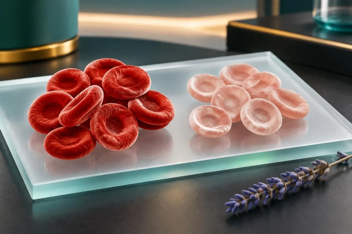
A low MCHC usually means red cells contain less haemoglobin than expected, but the combination of MCHC, MCV, haemoglobin, RDW and iron markers gives the real clinical picture.

Key takeaways
- Low MCHC meaning: Adult MCHC is commonly about 32–36 g/dL; a persistent result below the laboratory range often indicates hypochromia, especially when MCV is also low.
- MCH versus MCHC: MCH measures the average haemoglobin mass in one red cell, usually about 27–33 pg, whereas MCHC measures haemoglobin concentration within packed red cells.
- Most common pattern: Low MCHC plus low MCV and low ferritin strongly supports iron deficiency, but menstrual or gastrointestinal blood loss still needs consideration.
- Thalassaemia clue: Low MCHC with a low MCV, relatively preserved haemoglobin and a normal or high red-cell count can suggest thalassaemia trait rather than simple iron deficiency.
- Iron studies: Ferritin below about 15–30 ng/mL supports depleted iron stores, although inflammation can raise ferritin and conceal iron deficiency.
- Repeat testing: A mildly low MCHC with normal haemoglobin may be rechecked after roughly 4–12 weeks, depending on symptoms, prior results and the suspected cause.
- Urgent context: Chest pain, fainting, breathlessness at rest, black stools or rapidly worsening fatigue need prompt medical assessment regardless of the MCHC value.
- Interpretation limit: MCHC is a calculated CBC index, not a diagnosis; laboratory ranges, hydration, analyser effects and the rest of the CBC can change its meaning.
What does a low MCHC measure?
Low MCHC means the average concentration of haemoglobin inside packed red cells is below the laboratory interval. Adult reference intervals are commonly about 32–36 g/dL, although laboratories may use slightly different limits. Kantesti is an AI blood test analyzer that compares MCHC with haemoglobin, haematocrit, MCV, MCH and RDW rather than treating one flagged number as a diagnosis.
MCHC is calculated from haemoglobin and haematocrit: approximately haemoglobin divided by haematocrit, multiplied by 100. For example, haemoglobin of 12 g/dL and haematocrit of 36% produce an MCHC of about 33.3 g/dL, while haemoglobin of 10 g/dL and haematocrit of 35% produce about 28.6 g/dL.
A low MCHC is called hypochromia on a CBC when the reduced haemoglobin concentration is reflected in the red-cell appearance. Iron deficiency is the commonest explanation, but thalassaemia trait, chronic inflammation, sideroblastic processes and some analytical problems can create a similar result.
A single value just below the interval is less informative than a persistent result or a downward trend. Our blood-test biomarkers guide explains why the same result should be read against the analyser's own range, the patient's age and the rest of the blood count.
| Level | Range | What it means |
|---|---|---|
| Typical adult interval | 32–36 g/dL | Haemoglobin concentration inside red cells is broadly within common adult laboratory limits. |
| Slightly low | 30–31.9 g/dL | May be early iron depletion, a stable individual variant or analytical variation; interpret with MCV and haemoglobin. |
| Clearly low | 27–29.9 g/dL | Hypochromia is more likely, particularly with low MCV, high RDW or low ferritin. |
| Markedly low | <27 g/dL | Needs clinical review, especially when haemoglobin is low or symptoms suggest significant anaemia. |
Why the laboratory range matters
MCHC ranges differ by analyser, population and reporting units; one laboratory may flag below 31 g/dL while another uses 32 g/dL. A result of 31.8 g/dL therefore cannot be classified accurately without the printed reference interval.
The practical question is not whether MCHC is low in isolation, but whether the pattern shows reduced haemoglobin production, smaller cells, active blood loss or a technical discrepancy. A previous CBC from 3–6 months earlier can be more informative than a single borderline result.
How is low MCHC different from low MCH?
Low MCH measures a reduced amount of haemoglobin per red cell, whereas low MCHC measures reduced haemoglobin concentration within the red-cell volume. Typical adult MCH is about 27–33 pg, and typical MCHC is about 32–36 g/dL; they often fall together but are not interchangeable.
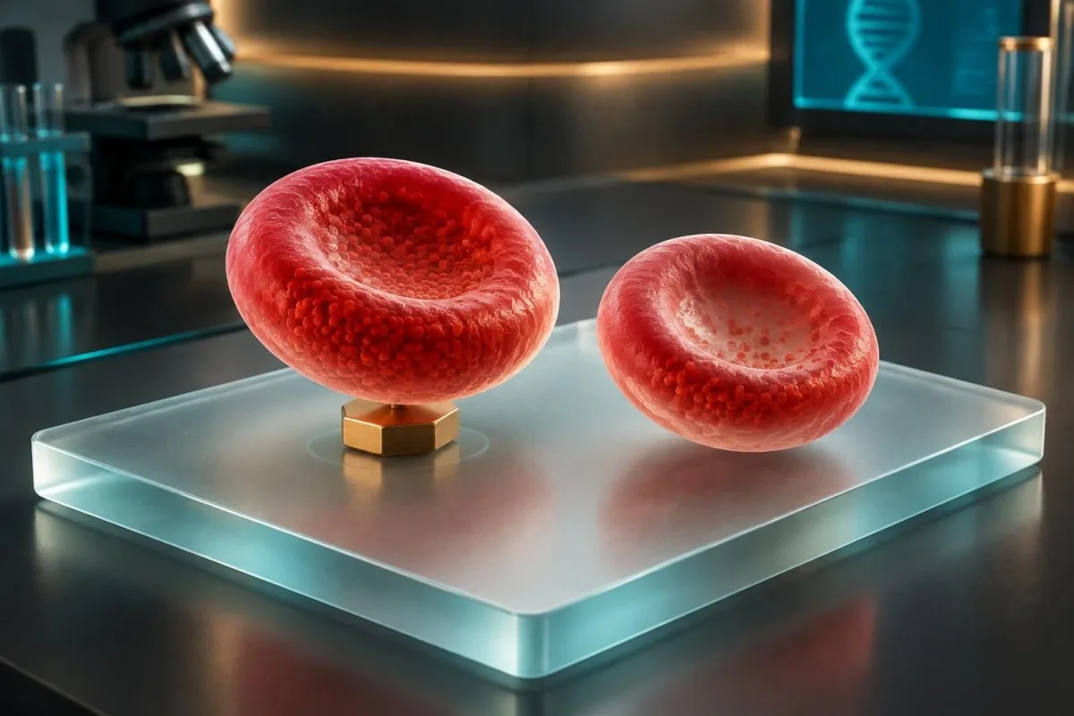
MCH is calculated from haemoglobin and red-cell count, so it answers how much haemoglobin the average cell carries. MCHC uses haemoglobin and haematocrit, so it answers how densely haemoglobin is packed within the measured red-cell volume; this distinction matters when cell size changes.
For example, a person with MCH 25 pg, MCV 72 fL and MCHC 30 g/dL has small, relatively pale cells, a pattern compatible with iron-restricted haemoglobin production. Another person may have MCH 26 pg and MCHC 32 g/dL because the cells are small but not unusually dilute; the interpretation is less straightforward.
MCH and MCHC can disagree because MCV, haematocrit and red-cell count are measured or calculated differently. This is why a low MCH should not automatically trigger iron tablets, particularly when ferritin, RDW and haemoglobin are normal.
A useful calculation check
If haemoglobin is 11.2 g/dL and the red-cell count is 4.4 million/µL, MCH is approximately 25.5 pg. If haematocrit is 34%, MCHC is approximately 32.9 g/dL, showing that a low MCH can coexist with a normal MCHC.
Conversely, a low MCHC with an ordinary MCH may occur when the measured haematocrit is relatively high for the haemoglobin value. Unexpected discordance deserves a review of the original CBC, specimen quality and analyser comments.
What does MCHC mean alongside MCV?
MCHC and MCV together classify the red-cell pattern: low MCHC with low MCV suggests a microcytic, hypochromic process, while low MCHC with normal MCV can represent early iron restriction or a mixed disorder. MCV is commonly about 80–100 fL in adults, but age and laboratory method affect the interval.
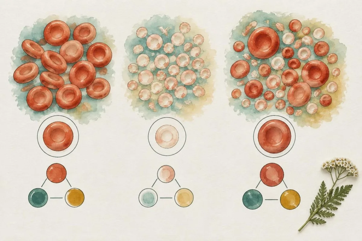
Low MCHC plus MCV below 80 fL is most often associated with iron deficiency or thalassaemia trait. Low MCHC plus normal MCV can occur before cells become small, after recent blood loss, or when a microcytic process is mixed with B12 deficiency, kidney disease or another condition that pushes MCV upward.
RDW adds a time dimension: RDW above roughly 14–15% often indicates uneven cell populations, as occurs when iron deficiency develops progressively. A normal RDW does not exclude iron deficiency, especially early in the process or when the pattern has been stable for years.
Our RDW and MCV interpretation guide shows why a low MCHC with MCV 74 fL and RDW 19% points differently from a low MCHC with MCV 74 fL and RDW 12.5%. Neither pattern identifies the cause alone.
Three practical CBC combinations
Low MCHC, low MCV, high RDW and low haemoglobin is a classic iron-deficiency pattern, although gastrointestinal or menstrual blood loss still requires assessment. Low MCHC, low MCV, normal haemoglobin and a relatively high red-cell count raises thalassaemia trait as a possibility.
Low MCHC, normal MCV and normal haemoglobin may be an early or mild finding that merits iron studies rather than an immediate disease label. Low MCHC with high MCV suggests a mixed process or measurement issue and should prompt a broader CBC review.
When does iron deficiency cause low MCHC?
Iron deficiency causes low MCHC when haemoglobin synthesis becomes iron-limited, often producing low ferritin, low transferrin saturation, rising RDW and eventually low MCV. Ferritin below about 15–30 ng/mL supports depleted stores, but inflammation can raise ferritin and make a seemingly normal result misleading.

Iron deficiency can progress in stages: storage depletion, iron-restricted red-cell production, and established iron-deficiency anaemia. A person may therefore have ferritin 18 ng/mL, transferrin saturation 14%, MCHC 31 g/dL and haemoglobin 12.4 g/dL before haemoglobin falls below the local anaemia threshold.
Low ferritin is relatively specific for depleted iron stores, but the threshold is context-dependent. In inflammation, chronic kidney disease or liver disease, ferritin below 50–100 ng/mL may still be compatible with iron deficiency because ferritin behaves as an acute-phase protein.
The British Society of Gastroenterology guideline recommends confirming iron deficiency and considering the source rather than assuming diet is the explanation (Snook et al., 2021). Our iron studies guide explains how ferritin, serum iron, transferrin and TIBC fit together.
Why diet alone may not explain it
Iron loss, absorption problems and increased requirements can all lower iron stores; a diet change is not always enough to correct the underlying cause. Heavy menstrual bleeding, coeliac disease, bariatric surgery, frequent blood donation and gastrointestinal disease are clinically different pathways.
For a hypothetical 38-year-old with MCHC 29 g/dL, MCV 70 fL and ferritin 8 ng/mL, iron deficiency is strongly supported, but treatment should be paired with a reasoned search for ongoing loss or poor absorption.
Can thalassaemia trait cause low MCHC?
Thalassaemia trait can cause low MCHC and low MCV with mild or no anaemia, often accompanied by a normal or relatively high red-cell count. This pattern differs from iron deficiency, where the red-cell count is often lower and RDW is more frequently increased.

A low MCV below 75 fL with a red-cell count above about 5.0 million/µL can make thalassaemia trait more likely, although these are clues rather than diagnostic cutoffs. The Mentzer index, calculated as MCV divided by red-cell count, is sometimes used; a value below 13 favours thalassaemia trait, but performance varies across populations.
A hypothetical 24-year-old with MCV 66 fL, MCHC 30 g/dL, haemoglobin 12.8 g/dL, red-cell count 5.9 million/µL and ferritin 62 ng/mL has a pattern that is less typical of iron deficiency. Haemoglobin electrophoresis may identify beta-thalassaemia trait through an increased HbA2, while alpha-thalassaemia may require molecular testing.
Iron supplements do not correct thalassaemia trait unless iron deficiency is also present. The haemoglobin electrophoresis guide explains why a normal electrophoresis does not exclude every inherited microcytosis.
Avoiding a common misclassification
Treating every low MCHC and low MCV as iron deficiency can delay recognition of a carrier state and expose a person to unnecessary supplementation. Ferritin, transferrin saturation, family origin, previous CBCs and haemoglobin analysis should be considered together.
Pregnancy changes iron requirements and clinical thresholds, so a carrier state and iron deficiency can coexist. In that situation, a low MCHC may improve with iron while the MCV remains lower than average.
How can inflammation or chronic illness lower MCHC?
Chronic inflammation can lower MCHC by trapping iron in storage cells and reducing iron delivery to the bone marrow, even when total body iron is not severely depleted. This anaemia of inflammation often has normal or mildly low MCV, ferritin that is normal or high, and transferrin or TIBC that is low or normal.

Inflammatory signalling increases hepcidin, a liver-derived regulator that reduces intestinal iron absorption and limits iron release from storage cells. A person with ferritin 120 ng/mL, transferrin saturation 12%, CRP 35 mg/L and MCHC 30.5 g/dL may have functional iron restriction rather than uncomplicated dietary deficiency.
Kidney disease adds a second mechanism: reduced erythropoietin can lower red-cell production, while inflammation may restrict iron availability. Chronic infection, autoimmune disease, cancer and obesity can also alter ferritin interpretation; ferritin above 100 ng/mL does not always exclude iron-restricted erythropoiesis.
Low transferrin can reflect inflammation, liver disease or malnutrition, so it should not be interpreted as a simple measure of iron stores. Our transferrin explanation covers why a low result may be part of an inflammatory pattern.
When inflammation and iron deficiency overlap
Mixed iron deficiency and inflammation can produce ferritin between 30 and 100 ng/mL, low transferrin saturation and a low MCHC. Soluble transferrin receptor, reticulocyte haemoglobin and the response to clinically supervised treatment may help when routine markers disagree.
The evidence is honestly mixed on universal ferritin cutoffs in inflammatory illness. Clinicians often use the entire pattern, including CRP, kidney function, liver tests and recent illness, rather than applying one number mechanically.
Can blood loss explain a low MCHC?
Ongoing blood loss can lower MCHC after iron stores are gradually depleted, with menstrual blood loss and gastrointestinal loss among the most common causes. The CBC may initially show normal MCV and haemoglobin, because newly produced cells become abnormal before the whole circulating red-cell population changes.
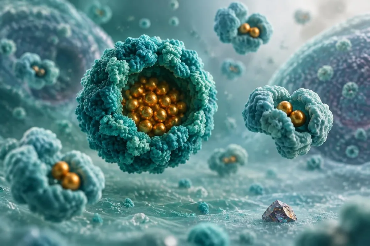
Menstrual blood loss is a frequent explanation in people who menstruate, but the amount is often underestimated. Bleeding lasting more than 7 days, changing protection every 1–2 hours, passing large clots or developing fatigue and breathlessness warrants medical review; our guide to heavy menstrual bleeding describes practical warning signs.
Gastrointestinal loss may be invisible, particularly when stools look normal. A low MCHC with iron deficiency in a man, a postmenopausal woman or anyone with gastrointestinal symptoms generally deserves evaluation for the source; the AGA guideline supports gastrointestinal assessment in appropriate adults with iron-deficiency anaemia (Ko et al., 2020).
A hypothetical 62-year-old with haemoglobin 10.6 g/dL, MCV 76 fL, MCHC 29 g/dL and ferritin 9 ng/mL needs more than dietary advice. The next step is to confirm iron deficiency, review medicines such as NSAIDs and arrange source evaluation through a clinician.
Why the timing of blood loss matters
After sudden blood loss, MCV and MCHC can remain near normal for several days because older circulating cells are still present. Low MCHC develops later if iron replacement cannot match continuing loss or if iron stores were already marginal.
Reticulocytes may rise during marrow recovery, and their haemoglobin content can reveal iron availability earlier than mature-cell indices. A single CBC therefore may not reveal when the process began.
What less common conditions lower MCHC?
Less common low MCHC causes include sideroblastic anaemia, lead exposure, severe copper deficiency and selected marrow disorders, usually identified by the wider CBC, history and specialist testing. These causes are uncommon compared with iron deficiency and thalassaemia trait, but they matter when iron studies do not fit.

Sideroblastic processes impair incorporation of iron into haemoglobin and may produce low MCHC with a low or normal MCV, increased RDW and abnormal iron markers. Ring sideroblasts are identified in bone-marrow examination, not from MCHC alone; alcohol exposure, medicines and acquired marrow disorders can be relevant.
Lead exposure can interfere with haemoglobin synthesis and may be associated with abdominal symptoms, neurological effects or basophilic stippling on a cell sample. Testing is based on exposure history and a blood lead level, not on the degree of MCHC reduction.
Copper deficiency can cause anaemia and neutropenia, especially after gastrointestinal surgery, excess zinc intake or malabsorption. Our article on low copper causes explains why copper, zinc, haemoglobin and white-cell results may need to be reviewed together.
When uncommon causes become more plausible
An unusual cause becomes more plausible when MCHC remains low despite replete iron stores, the CBC shows more than one abnormal cell line, or the patient has a relevant exposure or medication history. Ferritin 180 ng/mL with MCHC 28 g/dL is not proof of sideroblastic anaemia, but it is a reason to widen the assessment.
A specialist may consider a peripheral cell sample, reticulocyte count, B6 status, copper, lead, haemoglobin analysis or marrow testing. These tests should be selected from the pattern rather than ordered indiscriminately.
What if MCHC is low but haemoglobin is normal?
Low MCHC with normal haemoglobin often represents early iron restriction, mild inherited microcytosis, a stable personal pattern or a borderline laboratory variation rather than clinically significant anaemia. The result becomes more concerning when MCV, RDW or ferritin are also abnormal, or when symptoms and trends point in the same direction.
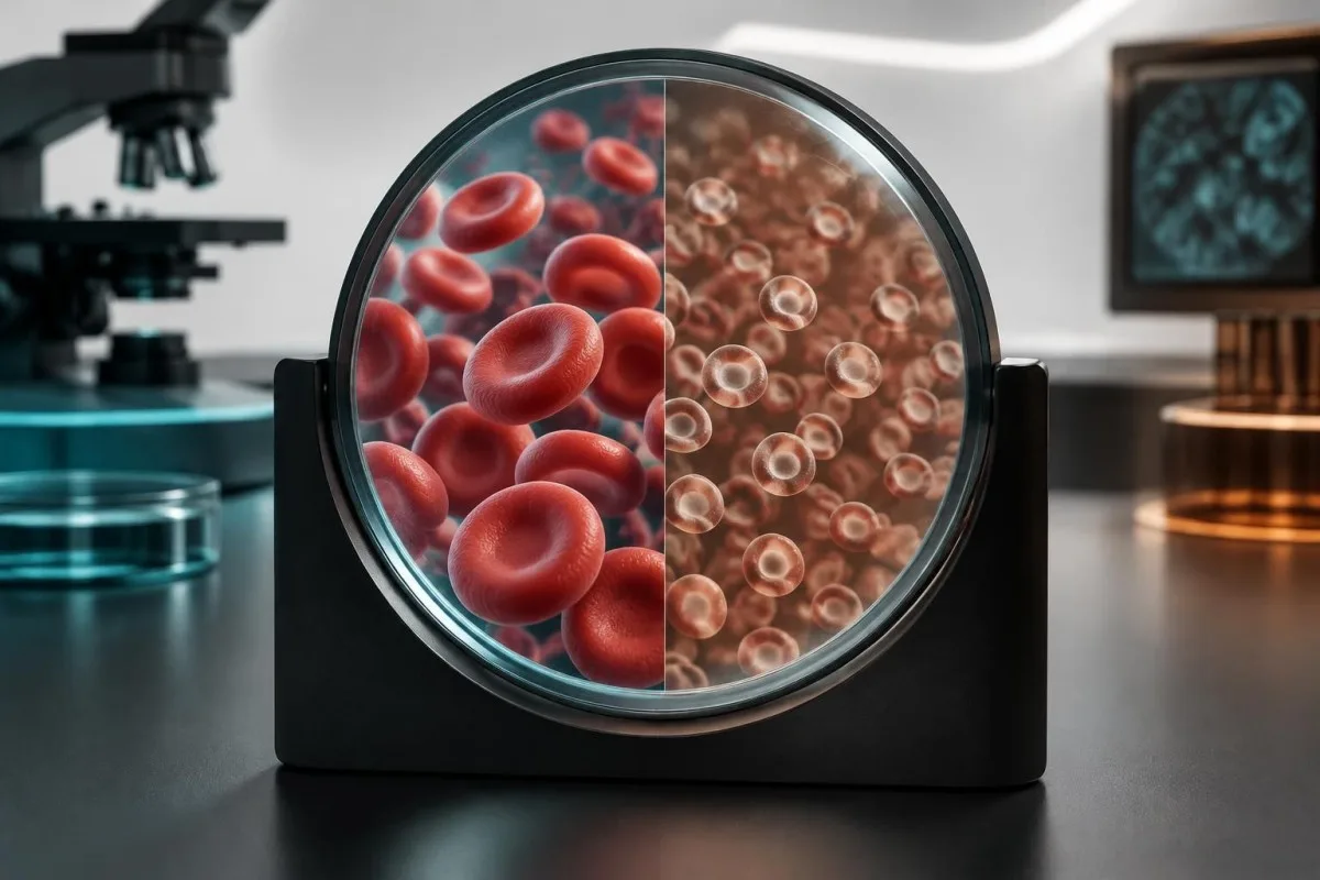
Anaemia is defined by haemoglobin below a population and laboratory threshold, not by MCHC alone. WHO adult thresholds commonly use less than 13 g/dL for men and less than 12 g/dL for non-pregnant women, with adjustments for pregnancy, altitude, smoking and laboratory context.
For example, haemoglobin 13.1 g/dL, MCV 78 fL, MCHC 31 g/dL and RDW 16% may indicate early iron depletion even though haemoglobin is not low. Ferritin and transferrin saturation can clarify whether a preventable deficiency is developing.
A low MCHC with normal MCV, normal RDW and stable haemoglobin over 2 years is less suggestive of an active progressive disorder. Kantesti AI compares current and historical CBC patterns so a borderline result can be viewed as a trend rather than an isolated alert.
Symptoms can precede anaemia
Fatigue, reduced exercise tolerance, restless legs, hair shedding or concentration difficulty can occur with low iron stores before haemoglobin falls. These symptoms are nonspecific, however, and should not be attributed to iron deficiency without appropriate testing.
The low ferritin symptoms guide explains why symptoms and iron stores may change before MCHC becomes markedly abnormal. A normal haemoglobin value does not make every low MCHC result irrelevant.
When are iron studies useful for low MCHC?
Iron studies are useful when low MCHC persists, MCV is low, RDW is rising, haemoglobin is falling, symptoms suggest iron depletion or the cause of anaemia is unclear. The usual panel includes ferritin, serum iron, transferrin or TIBC and transferrin saturation; the exact selection depends on the clinical setting.

Ferritin is the best single marker of iron stores in an otherwise well person, but it rises with inflammation, liver injury and infection. Transferrin saturation is calculated as serum iron divided by TIBC multiplied by 100; a value below about 15–20% commonly indicates restricted iron availability.
A practical example is ferritin 12 ng/mL, serum iron 25 µg/dL, TIBC 450 µg/dL and transferrin saturation 5.6%, together with MCHC 28 g/dL. This combination strongly supports iron deficiency, while ferritin 145 ng/mL, TIBC 220 µg/dL and saturation 12% suggests a different or mixed process.
Testing soon after an acute illness may produce confusing results because serum iron varies during the day and inflammation changes ferritin and transferrin. A clinician may repeat or expand the panel when CRP, kidney function or liver tests suggest that the first result is difficult to interpret.
Tests that can add detail
Reticulocyte haemoglobin, sometimes reported as Ret-He or CHr, reflects iron available to newly produced cells and may respond before mature-cell indices change. A value below the analyser's own interval can support iron-restricted erythropoiesis, but cutoffs vary by instrument.
Soluble transferrin receptor may help distinguish iron deficiency from inflammation because it is less affected by inflammatory signalling, although it is not available in every laboratory. This is one of those areas where context matters more than a number.
How should ferritin and transferrin saturation be read?
Ferritin estimates stored iron, while transferrin saturation estimates how much circulating transport capacity is occupied by iron; both are needed when inflammation or chronic disease may distort one marker. Ferritin below 15 ng/mL is highly suggestive of depleted stores, but a normal result does not reliably exclude deficiency during inflammation.

Ferritin below 15 ng/mL is generally specific for iron deficiency, and values from 15–30 ng/mL often remain clinically suspicious when the CBC is microcytic or symptoms fit. Ferritin above 100 ng/mL usually makes absolute deficiency less likely in a well person, but inflammation can shift that interpretation.
Transferrin saturation below 15% is strongly compatible with iron-restricted erythropoiesis, while values above about 20% are more reassuring when ferritin is adequate. Serum iron alone is weak evidence because meals, supplements and diurnal variation can change it substantially.
A person with ferritin 52 ng/mL, CRP 28 mg/L and transferrin saturation 11% may have iron deficiency masked by inflammation. Conversely, ferritin 52 ng/mL with CRP normal, saturation 24% and stable CBC is less supportive of deficiency; the difference lies in the surrounding data.
When TIBC is low rather than high
TIBC often rises in uncomplicated iron deficiency and falls or remains normal with inflammation, liver disease or poor protein production. A low TIBC with low transferrin saturation therefore points away from a simple isolated deficiency, though mixed disease remains possible.
For additional detail, see our guide to low TIBC results. Do not compare iron-study numbers across laboratories without checking units and assay-specific ranges.
When should a low MCHC be repeated?
Repeat testing is useful when MCHC is only mildly low, the result conflicts with the rest of the CBC, the sample had a quality problem or a recent illness may have distorted the result. The timing is individual, but a repeat CBC in roughly 4–12 weeks is often more informative than repeating it the next day without a clinical reason.
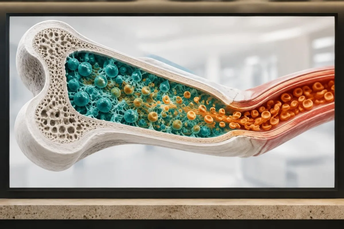
A result of MCHC 31.7 g/dL with haemoglobin 14.2 g/dL, MCV 89 fL and normal RDW may be rechecked after 1–3 months, particularly if the person feels well. A result of 27.5 g/dL with haemoglobin 8.9 g/dL requires evaluation sooner and should not wait for routine surveillance.
Laboratory artefacts can arise from delayed processing, specimen dilution, clumping or calculation discrepancies. Haemolysis, lipaemia and cold-reactive effects can interfere with selected CBC measurements, although the exact direction and size of error depend on the analyser and specimen.
If the laboratory reports a quality flag, the appropriate response may be a fresh sample rather than repeating the same specimen. Kantesti's privacy and data guidance explains how uploaded reports should be handled, but clinical decisions still require the original laboratory and healthcare context.
A sensible repeat-test checklist
Repeat the CBC using the same or a comparable laboratory when possible, and include haemoglobin, haematocrit, RBC count, MCV, MCH, MCHC, RDW and reticulocytes. Document iron supplements, recent infection, pregnancy, blood donation and heavy exercise because each can affect interpretation.
If the initial sample was reported as unsuitable or haemolysed, do not infer that every abnormal value reflects physiology. Our guide to repeat testing after sample problems covers why a redraw can be more reliable.
Which symptoms make low MCHC more concerning?
Low MCHC becomes more clinically concerning when it accompanies breathlessness, chest discomfort, fainting, rapid heartbeat, marked fatigue, neurological symptoms or evidence of ongoing blood loss. The urgency depends mainly on haemoglobin, symptoms, rate of change and suspected cause—not on MCHC alone.
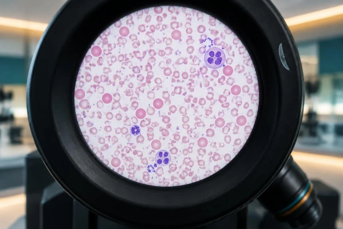
Urgent assessment is appropriate for chest pain, severe breathlessness at rest, fainting, confusion, black tarry stools, vomiting blood or rapidly worsening weakness. A low MCHC with haemoglobin below about 8 g/dL is often clinically significant, but symptoms and cardiovascular disease can make higher values important too.
A gradual fall from haemoglobin 13.5 to 10.8 g/dL over 6 months may matter more than a stable haemoglobin of 10.8 g/dL documented for years. Trend analysis can reveal occult loss, treatment failure or a new production problem before a single report makes the pattern obvious.
For symptoms such as breathlessness, a CBC is only one part of the assessment; heart, lung, thyroid, kidney and clot-related causes can overlap. Our shortness-of-breath blood-test guide discusses why a low MCHC should not become a premature explanation.
Why symptom severity can mislead
Some people tolerate slowly developing anaemia surprisingly well, while others develop palpitations or exertional symptoms with a modest haemoglobin change. Age, pregnancy, cardiopulmonary disease and the speed of decline alter physiological tolerance.
Do not self-treat severe symptoms with over-the-counter iron alone. Iron can cause side effects, obscure the cause of blood loss and be inappropriate in iron-loading disorders.
What is the practical next step after a low MCHC?
The practical next step is to review the complete CBC, compare prior results, assess symptoms and decide whether iron studies, haemoglobin analysis or repeat testing is appropriate. A low MCHC is a clue that needs pattern recognition, not a standalone diagnosis.

Start with the laboratory interval and the severity of the change, then check haemoglobin, haematocrit, RBC count, MCV, MCH, RDW and reticulocytes. Next consider ferritin and transferrin saturation, menstrual or gastrointestinal loss, inflammation, kidney function, diet, medicines and family history.
Kantesti is an AI-powered blood test analysis tool that can organise these related values into a readable pattern, but it cannot examine a person, verify hidden bleeding or replace a clinician's assessment. Uploading the full report is more useful than entering MCHC alone because the relationships among results carry the diagnostic signal.
Kantesti is an AI lab test interpretation service that presents possible explanations and follow-up questions without claiming that a low MCHC proves iron deficiency or thalassaemia. For information about our clinical content and review principles, see the medical content standards.
A clinician-ready summary
Bring the current CBC, older CBCs, iron studies, medication and supplement list, recent illness history and any bleeding symptoms. Mention pregnancy, recent donation, gastrointestinal symptoms, restrictive diets and family history of anaemia because these details can change the next test.
If the pattern is mild and stable, monitoring may be reasonable; if haemoglobin is falling, iron deficiency is confirmed or symptoms are significant, investigation should be more active. The correct plan depends on the complete clinical picture.
Frequently asked questions
What are the most common low MCHC causes?
The most common low MCHC causes are iron deficiency, thalassaemia trait and anaemia associated with chronic inflammation. Adult MCHC is commonly about 32–36 g/dL, and values below this range indicate that red cells contain less concentrated haemoglobin than expected. Low MCHC with low MCV and low ferritin strongly supports iron deficiency, while low MCHC with very low MCV, preserved haemoglobin and a relatively high red-cell count can suggest thalassaemia trait. The CBC pattern and iron studies are more informative than MCHC alone.
What is the difference between low MCH and low MCHC?
Low MCH means the average red cell contains less haemoglobin by mass, usually below about 27 pg, whereas low MCHC means haemoglobin is less concentrated within the red-cell volume, commonly below about 32 g/dL. MCH is calculated using haemoglobin and red-cell count, while MCHC uses haemoglobin and haematocrit. Both may fall in iron deficiency, but they can disagree when red-cell size or haematocrit changes. A complete CBC is needed to interpret either result.
Does low MCHC always mean iron deficiency?
Low MCHC does not always mean iron deficiency because thalassaemia trait, chronic inflammation, sideroblastic processes, lead exposure and laboratory variation can produce a similar result. Ferritin below 15–30 ng/mL supports depleted iron stores, but inflammation can raise ferritin and hide deficiency. Transferrin saturation below about 15–20% adds evidence of restricted iron availability. Symptoms, MCV, RDW, haemoglobin and the cause of any blood loss should also be assessed.
Is low MCHC with normal haemoglobin serious?
Low MCHC with normal haemoglobin is often mild or early, but it can indicate developing iron deficiency or an inherited microcytic pattern. For example, haemoglobin 13.2 g/dL with MCHC 31 g/dL, MCV 77 fL and RDW 17% may justify ferritin testing even though haemoglobin is normal. A stable result with normal MCV and RDW is less concerning than a progressively worsening pattern. Repeat CBC and iron studies are chosen according to symptoms and prior results.
When should iron studies be ordered for low MCHC?
Iron studies are useful when low MCHC persists, MCV is below about 80 fL, RDW is increasing, haemoglobin is falling or symptoms suggest iron depletion. The usual tests are ferritin, serum iron, transferrin or TIBC and transferrin saturation. Ferritin below 15–30 ng/mL strongly supports iron deficiency, while transferrin saturation below 15–20% suggests restricted iron availability. Inflammation, liver disease and kidney disease can make the results harder to interpret.
When should a low MCHC be repeated?
A mildly low MCHC with normal haemoglobin may commonly be repeated after about 4–12 weeks, depending on symptoms, previous results and suspected cause. Immediate or prompt review is more appropriate when haemoglobin is substantially low, the result is changing quickly or there are symptoms such as fainting, chest pain, breathlessness at rest or black stools. The repeat CBC should include MCV, MCH, RDW, RBC count and, when appropriate, reticulocytes. A laboratory quality flag may require a fresh sample sooner.
Get your blood test results explained
Upload your lab report and Kantesti's AI explains each biomarker in plain language within seconds. Used by more than 2 million people worldwide.
References
- Klein, T., Mitchell, S., & Weber, H. (2026). Nipah Virus Blood Test: Early Detection & Diagnosis Guide 2026. Kantesti AI Medical Research. DOI
- Klein, T., Mitchell, S., & Weber, H. (2026). B Negative Blood Type, LDH Blood Test & Reticulocyte Count Guide. Kantesti AI Medical Research. DOI
- Snook J et al. (2021). British Society of Gastroenterology guidelines for the management of iron deficiency anaemia in adults. Gut. DOI PubMed
- Ko CW et al. (2020). AGA Clinical Practice Guidelines on the Gastrointestinal Evaluation of Iron Deficiency Anemia. Gastroenterology. DOI PubMed
- World Health Organization (2011). Haemoglobin concentrations for the diagnosis of anaemia and assessment of severity. WHO Vitamin and Mineral Nutrition Information System.
- 2M+Tests analysed
- 127+Countries
- 100+Languages
About the medical team


E-E-A-T Trust Signals
Experience
Physician-led clinical review of lab interpretation workflows.
Expertise
Laboratory medicine focus on how biomarkers behave in clinical context.
Authoritativeness
Medically reviewed by Dr. Thomas Klein, with contributions from Dr. Sarah Mitchell and Prof. Dr. Hans Weber.
Trustworthiness
Evidence-based interpretation with clear follow-up pathways to reduce alarm.




Medical disclaimer
This article is for educational purposes only and does not constitute medical advice. Always consult a qualified healthcare provider for diagnosis and treatment decisions.
Published · Contact the editorial team
Published by Kantesti Ltd, registered in England & Wales, company no. 17090423, London, United Kingdom.