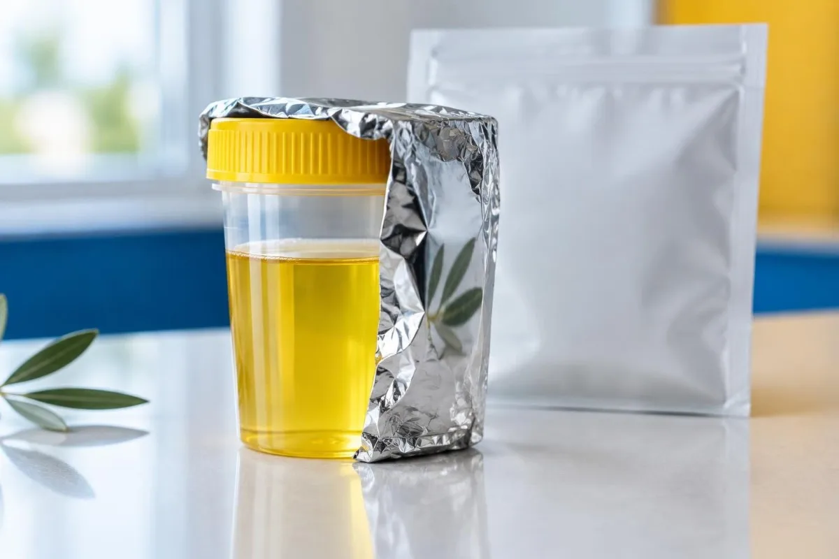
A well-timed, light-protected urine sample can make suspected acute porphyria easier to investigate. Serious symptoms still need immediate assessment.

Key takeaways
- Sample handling: Collect a random urine sample during symptoms when possible, protect it from light promptly, and follow the receiving laboratory’s instructions.
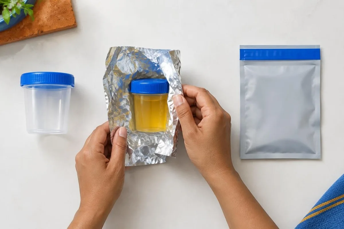
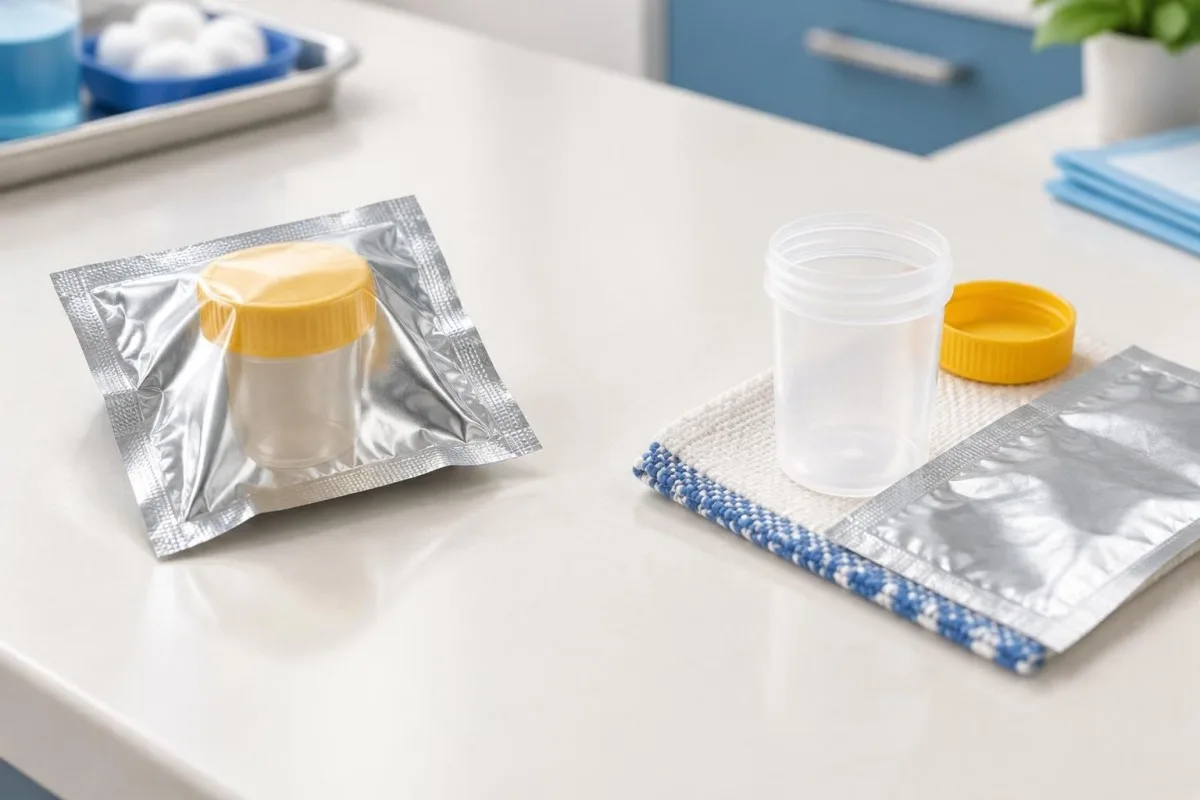
- Light protection: An opaque container or foil around the specimen helps limit light exposure; a clear transport bag alone is not light protection.
- Dilution: Request a quantitative urine PBG result alongside urine creatinine from the same specimen when available.
- Timing: Collect urine before hemin treatment if this can be done promptly, but never delay urgent treatment to obtain a specimen.
- Urgent symptoms: Severe abdominal pain, new weakness, seizures, confusion or breathing difficulty warrant emergency assessment without waiting for a report.
- Interpretation: A substantially raised PBG-to-creatinine ratio during symptoms supports an acute hepatic porphyria workup, but further testing identifies the type.
- Normal results: A reliable normal PBG during ongoing symptoms makes common acute hepatic porphyria attacks less likely; timing, treatment and rare exceptions still matter.
- Collection: A 24-hour urine collection is generally unnecessary for the initial evaluation of a suspected acute attack.
Why does urine porphobilinogen sample handling matter?
Urine porphobilinogen sample handling matters because light exposure, dilution and collection after symptoms or treatment can make a result less representative of a suspected acute porphyria attack. Use a promptly protected random sample and request creatinine alongside PBG; urgent symptoms need assessment before the report arrives.
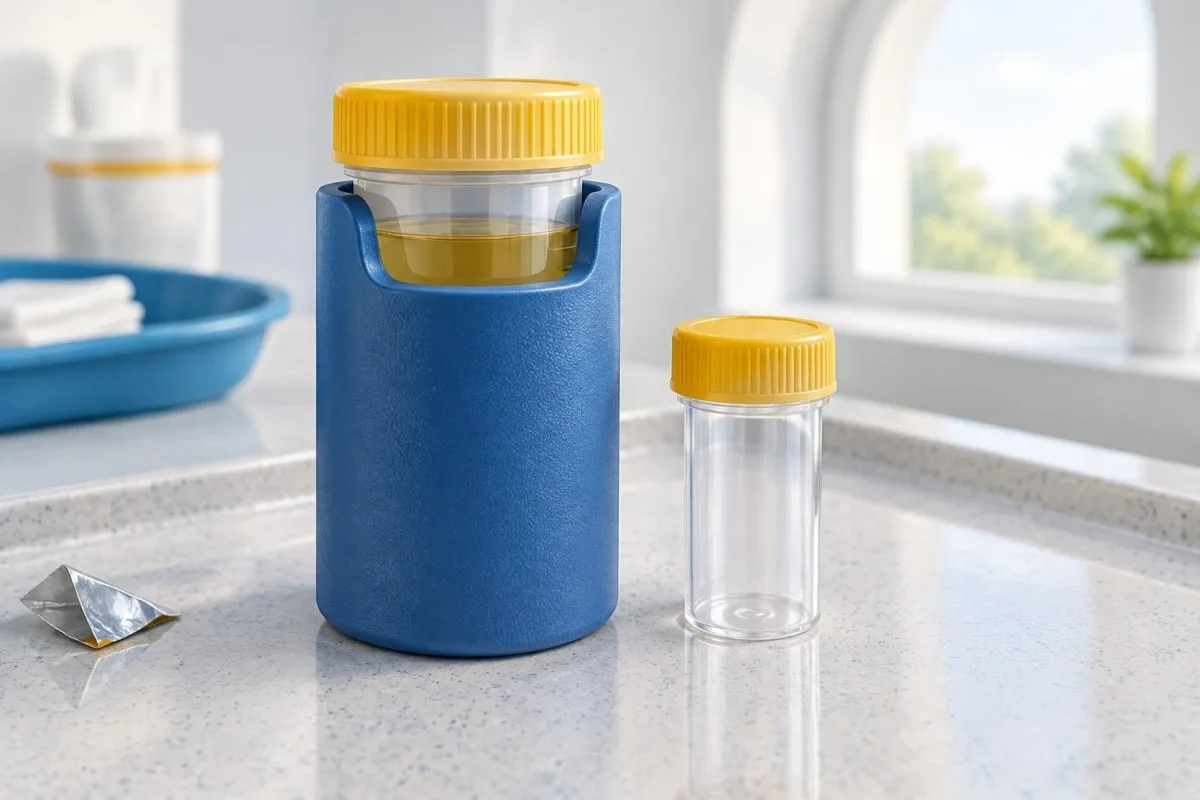
Porphobilinogen, or PBG, is an intermediate in the pathway used to make heme. During attacks of several acute hepatic porphyrias, the body produces excess PBG and related substances that can be detected in urine.
A result reflects both the patient’s biology and what happened to the specimen after collection. Our guide to porphyria blood and urine tests explains why a routine blood panel cannot replace a specific urine PBG test.
Kantesti is an AI blood test analyzer that can help readers understand associated blood results, but it cannot check whether a urine specimen was protected or determine whether a patient is having an acute attack.
Which symptoms should not wait for a PBG report?
Severe or worsening abdominal pain, new muscle weakness, confusion, seizures or breathing difficulty require emergency assessment rather than a wait for urine PBG results. A suspected attack can progress, and other dangerous causes of these symptoms also need evaluation.
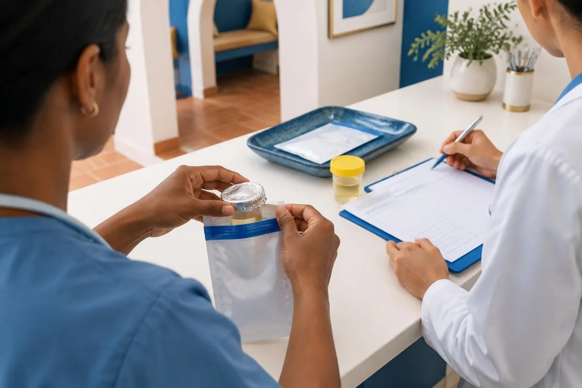
Call emergency services for breathing difficulty, a seizure, rapidly worsening weakness or severe confusion. Severe unexplained abdominal pain, especially with vomiting or new neurological symptoms, also warrants urgent emergency-department assessment.
Tell the clinical team about a personal or family history of porphyria, recent symptoms and medicines. A laboratory can collect urine while clinicians assess breathing, circulation, electrolytes and other possible causes; see our guide to urgent abdominal pain.
Low blood sodium can accompany an acute attack and contribute to neurological symptoms. Its evaluation may include blood and urine tests, as described in our SIADH testing guide.
How does light exposure affect a urine PBG sample?
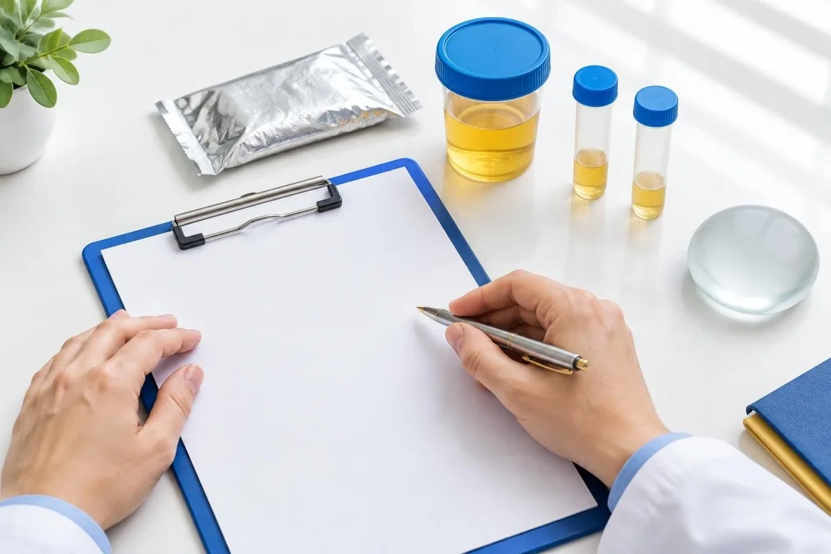
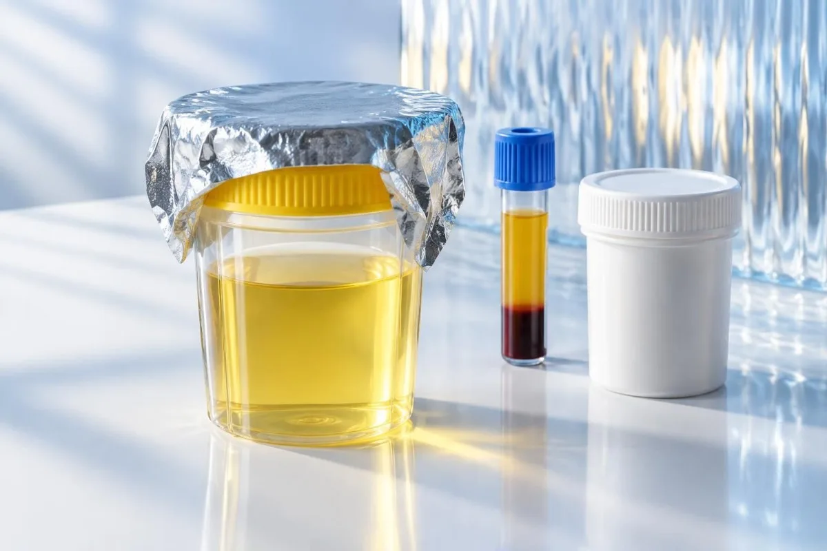
Light can degrade urine PBG and produce a falsely low result, so the sample should be protected from light as soon as practical after collection. The receiving laboratory should specify its accepted container, wrapping and transport procedure.
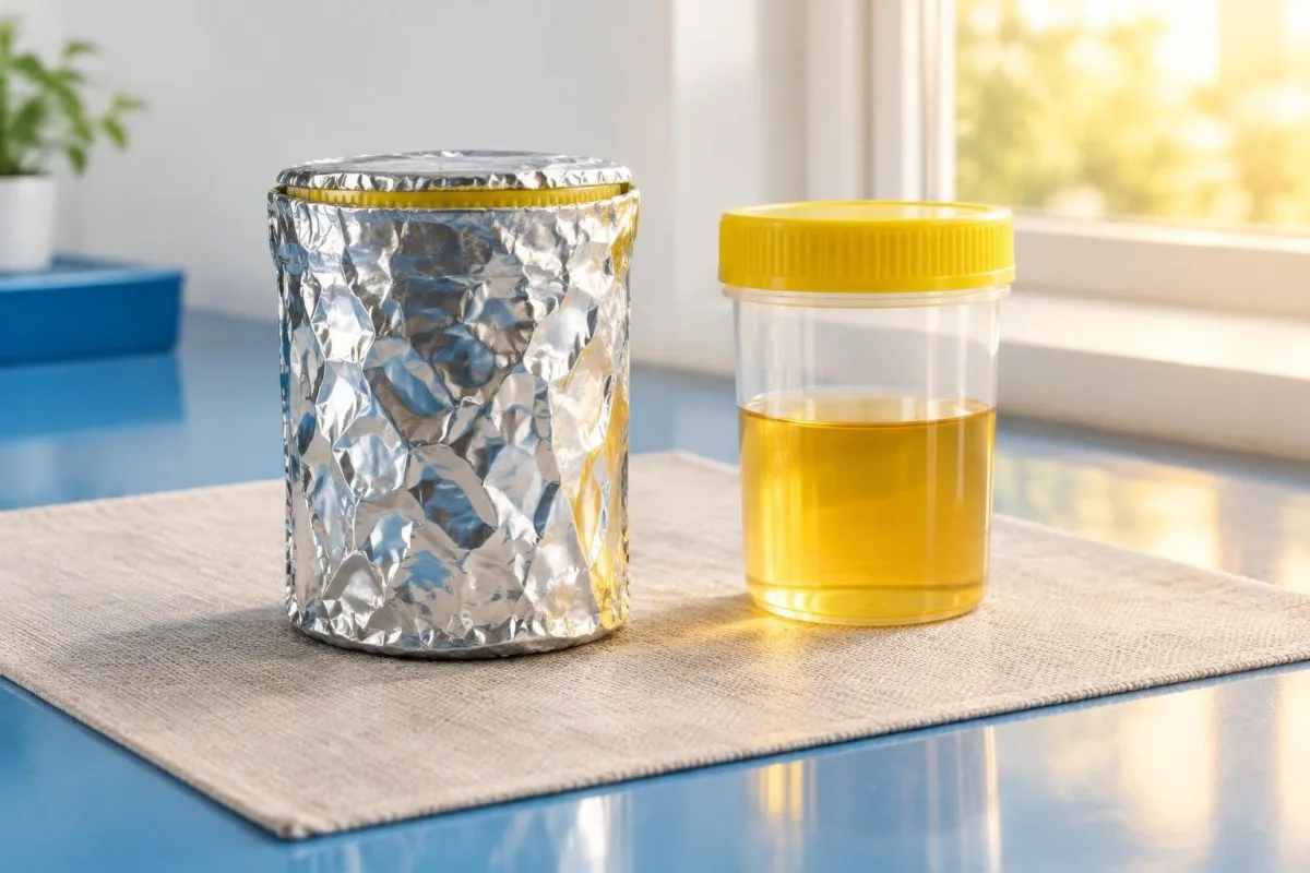
A laboratory may supply an opaque container or instruct staff to wrap the specimen container completely in aluminium foil. Cover the container rather than relying on a transparent specimen bag; the laboratory must still be able to identify the sample correctly.
Brief routine handling is not the same as leaving a clear container on a sunny windowsill for hours. If exposure occurred, report what happened and when—only the testing laboratory can judge whether its validated acceptance criteria were met.
Do not discard a collected sample yourself because you are worried about exposure. Ask the laboratory whether to send it and whether a new, protected specimen should be collected; our article on sample-related laboratory errors explains why reporting handling details matters.
How is a random urine PBG sample collected?
A fresh random, or spot, urine sample is generally appropriate for the first investigation of suspected acute hepatic porphyria; a 24-hour collection is not usually needed. Follow the laboratory’s instructions for container, minimum volume, labelling and transport.
The clinician should request quantitative urine PBG, ideally with urine creatinine measured on the same specimen and often with urine aminolevulinic acid, or ALA. A general urinalysis or urine porphyrins alone is not a substitute for a specific PBG measurement.
In the acute hepatic porphyria recommendations, Balwani and colleagues describe urine PBG testing corrected for creatinine as a first-line diagnostic approach (Balwani et al., 2017). Ask the receiving laboratory whether it needs a separate aliquot for ALA or creatinine.
Unlike a timed urine catecholamine collection, initial PBG testing usually does not require saving every void for a full day. Do not postpone an urgent assessment to begin a 24-hour collection.
Why report random urine PBG with creatinine?
A random urine PBG-to-creatinine ratio helps account for differences in urine concentration, which can otherwise make a dilute specimen appear reassuring. The PBG and creatinine measurements should come from the same urine sample.
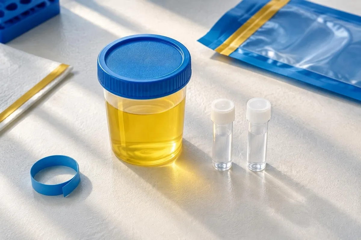
Drinking large amounts of fluid or receiving intravenous fluids can lower the concentration of both urine PBG and urine creatinine. A low PBG concentration per litre may therefore be misleading if the laboratory has not also considered creatinine.
For a hypothetical example, a specimen contains PBG of 2 mg/L and creatinine of 0.1 g/L: the calculated ratio is 20 mg PBG/g creatinine. If both concentrations were reduced by half through dilution, their ratio would still be 20 mg/g; real results must be interpreted with the laboratory’s own units and reference interval.
Creatinine correction is useful, not infallible. Very low urine creatinine, kidney impairment and values close to an assay’s detection limit can complicate interpretation; our urine creatinine guide explains why a ratio needs context.
When should urine PBG be collected?
Collect urine PBG while symptoms are present, preferably before specific treatment if collection can happen promptly. PBG can decline after an attack or treatment, so a sample collected later may be harder to interpret.
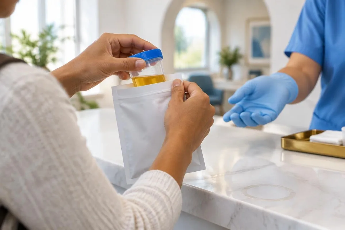
PBG often remains elevated longer after an attack of acute intermittent porphyria than after attacks of hereditary coproporphyria or variegate porphyria. The exact rate of decline varies, so there is no single reliable number of days that applies to every patient.
Anderson’s review of acute hepatic porphyrias discusses the value of prompt first-line biochemical testing and the different courses of PBG elevation across porphyria types (Anderson, 2019). Record when symptoms began, when urine was collected and whether treatment had already started.
If severe symptoms have resolved by the time testing is arranged, discuss the timing with a clinician or porphyria specialist rather than assuming a later normal result answers every question. The same timing principle affects other tests, including diagnostic test windows.
Should urine be collected before hemin or IV fluids?
Collect a protected urine specimen before hemin when feasible, because hemin can lower ALA and PBG, but do not delay necessary treatment or emergency care to obtain it. Fluids can dilute urine, making a same-sample creatinine measurement especially useful.
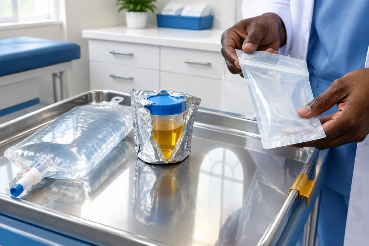
If a patient has already received intravenous fluids, testing can still be useful. Tell the laboratory and treating team about the fluids and request PBG and creatinine together rather than deciding the sample is unusable.
Hemin is a specialist-directed treatment used for significant acute hepatic porphyria attacks. A pre-treatment specimen can preserve evidence of biochemical activity, but treatment decisions depend on the clinical situation and specialist advice.
For example, if urine can be collected immediately in an emergency department, the team may save it before treatment begins. If the patient is deteriorating, care takes priority; our guide to urine sodium interpretation describes another urine result affected by the clinical context.
How should a PBG specimen be stored and transported?
Protect the urine specimen from light and send it promptly under the receiving laboratory’s temperature and transport instructions. Refrigeration or freezing requirements vary by laboratory, method and expected delay.
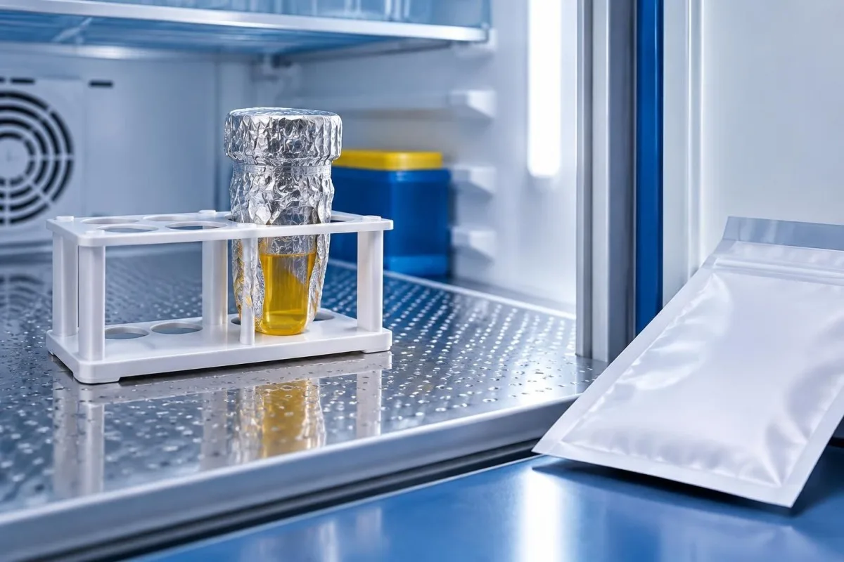
The collection site should ask the testing laboratory about its accepted container, minimum volume, temperature, stability window and whether a frozen aliquot is needed. Do not apply a generic number of hours to every laboratory: validated limits differ.
A patient collecting at home should obtain instructions before collecting and use the supplied packaging. If instructions are unclear, contact the collection service rather than leaving the sample in direct light or choosing a storage method by guesswork.
Record the collection time and any known delay. As with a lactate sample, the interval between collection and processing can affect what a laboratory can confidently report.
What does a high urine PBG result mean?
A markedly elevated quantitative urine PBG, corrected for creatinine and measured during compatible symptoms, strongly supports an acute hepatic porphyria workup. A positive result does not by itself identify which acute hepatic porphyria is present.
In a symptomatic patient, PBG more than 5 times the upper limit of the laboratory’s reference range is a commonly used biochemical threshold supporting an acute attack. The laboratory’s method, units and reference range matter, and a clinician should interpret results below that level in context rather than applying a universal rule.
Mild elevations are less specific than marked elevations and may need repeat measurement, review of kidney function and specialist input. Do not compare a result expressed in mg/g creatinine directly with a reference interval expressed in µmol/mmol creatinine without a valid conversion.
Further testing may include urine, plasma and stool porphyrin patterns followed by appropriate genetic testing. Our article on qualitative and quantitative results explains why a numerical measurement offers different information from a simple positive or negative screen.
Can a normal urine PBG result miss acute porphyria?
A properly collected, quantitative normal PBG during ongoing symptoms makes the common PBG-elevating acute hepatic porphyrias less likely as the cause of those symptoms. A late sample, prior hemin, dilution, handling problems or an uncommon disorder can complicate that conclusion.
A normal PBG does not exclude every porphyria or every cause of neurological and abdominal symptoms. ALA-dehydratase-deficiency porphyria is exceptionally rare and can present with elevated ALA without a corresponding marked PBG increase; lead poisoning can also increase ALA.
If the sample was taken after symptoms settled, was exposed to light for a prolonged period, or was collected after hemin, discuss whether repeat quantitative PBG and ALA during symptoms are appropriate. An urgent clinical assessment should not wait for a repeat sample.
Ask the laboratory whether the result was corrected to urine creatinine and whether the specimen met its handling requirements. Our discussion of heavy-metal testing covers why a suspected alternative diagnosis requires its own appropriately selected tests.
Does urine color or a routine dipstick diagnose porphyria?
Urine color and a routine dipstick cannot diagnose or exclude acute porphyria. Urine may darken after standing in some cases, but normal-looking urine does not rule out an attack.
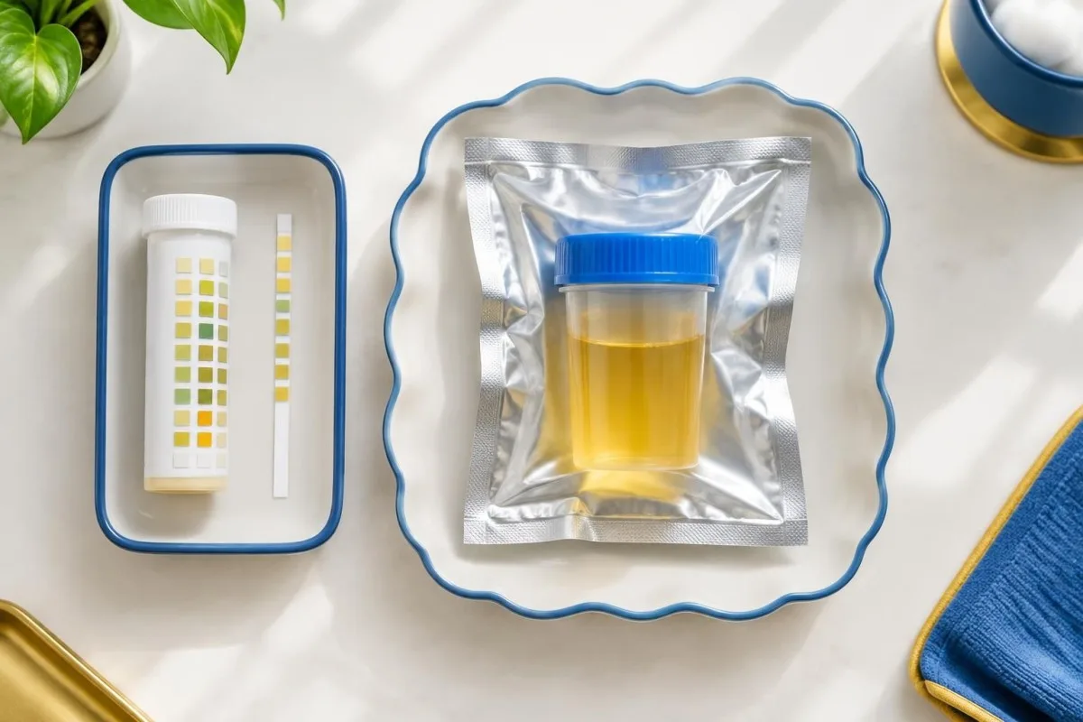
Dark or reddish urine has many possible explanations, including medicines, dehydration and blood. A person should not expose a sample to sunlight to see whether its color changes—that can compromise the requested biochemical test.
A routine dipstick measures selected properties such as blood, protein or glucose; it does not measure quantitative urine PBG. Our urine dipstick guide explains what those common fields do and do not show.
If urine is visibly red or dark, tell a clinician even if porphyria is already being investigated. The separate guide to dark urine causes outlines other reasons for timely assessment.
What happens after an abnormal or uncertain PBG result?
An abnormal or uncertain PBG result calls for a clinician-led review of symptoms, timing, creatinine correction and specimen quality, with porphyria specialist advice when appropriate. Further biochemical testing helps distinguish acute hepatic porphyria types.
The initial result should be reviewed alongside ALA and, when indicated, urine, plasma or stool porphyrin testing. A clinician may request another properly protected spot sample if the first was dilute, collected late or handled outside the laboratory’s requirements.
Genetic testing can help confirm the type and guide family testing after a biochemical diagnosis, but an uncertain genetic variant alone does not prove that a current episode is an acute attack. Our guide to genetic test uncertainty explains how an inconclusive variant differs from a confirmed diagnosis.
Kantesti AI can help readers prepare questions about associated laboratory reports; it cannot select confirmatory porphyria tests or replace a specialist. Our medical validation information describes the limits and oversight of our interpretation work.
What should you ask the laboratory about a PBG sample?
Ask the laboratory which container to use, how to protect it from light, how quickly to deliver it and whether creatinine is measured on the same sample. These details determine whether the laboratory can apply its own validated handling rules.
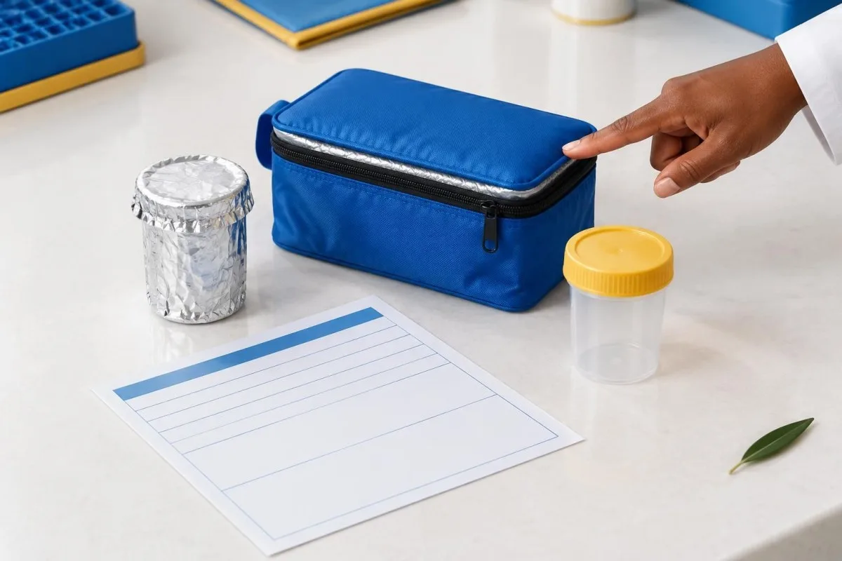
Before collection, confirm whether the request is for quantitative PBG, whether ALA is also required and whether the laboratory reports a creatinine-corrected ratio. Ask about minimum volume, labelling and transport temperature rather than assuming that all centres use the same protocol.
After collection, tell staff if the container was left unwrapped, warmed, delayed or collected after fluids or hemin. Describe the approximate duration and circumstances without trying to calculate how much the result may have changed.
Keep a record of symptom onset and collection time for the clinician. Kantesti is an AI lab test interpretation service that can help organise questions about a report, while our medical content standards explain how we approach health information.
How can you use a PBG report safely in follow-up?
Use a PBG report as one part of a clinician-led assessment, alongside symptoms, urine creatinine, collection timing and the laboratory’s reference interval. Neither an AI explanation nor a single result can establish whether urgent symptoms are safe to monitor at home.

Bring the complete report rather than only a screenshot of the flagged value. The units, reference interval, specimen collection time and comments about sample quality can change the meaning of the result.
Kantesti’s AI blood test interpretation platform can help explain accompanying blood-test terminology and organise questions for a clinician. It should not be used to diagnose porphyria, verify specimen handling or decide whether to delay urgent care.
Our responsible AI guidance explains why clinical judgement remains necessary. For a broader overview of urine findings, see our urinalysis guide.
Frequently asked questions
Does a urine PBG sample need to be protected from light?
Yes. Urine PBG samples should be protected from light promptly because exposure can lower the measured value. A laboratory may recommend an opaque container or wrapping the container in aluminium foil. Follow the laboratory’s instructions and tell staff if the sample was exposed or delayed.
Can drinking too much water affect a urine PBG test?
Yes. Large amounts of fluid can dilute urine and lower the measured PBG concentration per litre. Measuring urine creatinine in the same specimen allows the laboratory to report or assess a PBG-to-creatinine ratio. Do not restrict fluids or delay care without a clinician’s advice.
Do I need a 24-hour urine collection for PBG?
Usually not for the initial assessment of a suspected acute attack. A random urine sample collected during symptoms is generally appropriate for quantitative PBG and creatinine testing. A 24-hour collection can delay investigation and is not a substitute for urgent clinical care.
Can a normal urine PBG result rule out acute porphyria?
A reliable normal PBG result collected during ongoing symptoms makes the common PBG-elevating acute hepatic porphyrias less likely as the cause of that episode. It does not exclude every porphyria, and a sample collected after symptoms resolve or after hemin can be harder to interpret. Dilution and light exposure also matter. A clinician may consider ALA, repeat testing or specialist advice depending on the circumstances.
Should I wait for my urine PBG result if I have severe symptoms?
No. Severe abdominal pain, new weakness, seizures, confusion or breathing difficulty needs urgent assessment, even if a PBG report is pending. Call emergency services for breathing difficulty, seizures or rapidly worsening weakness. A clinical team can collect a protected urine sample while evaluating and treating you.
What does a high PBG-to-creatinine ratio mean?
A markedly elevated PBG-to-creatinine ratio during compatible symptoms supports investigation for acute hepatic porphyria. A result more than 5 times the laboratory’s upper reference limit is a commonly used threshold supporting an acute attack, but the result needs clinical interpretation. Further biochemical and sometimes genetic tests help identify the type. Mild elevations are less specific and may require review or repeat testing.
Get your blood test results explained
Upload your lab report and Kantesti's AI explains each biomarker in plain language within seconds. Used by more than 2 million people worldwide.
References
- Klein, T., Mitchell, S., & Weber, H. (2026). A Pre-Registered, Rubric-Based Automated Technical Benchmark of the Kantesti Blood-Test Interpretation Engine on 100,000 Synthetic Test Cases. Kantesti AI Medical Research. DOI
- Klein, T., Mitchell, S., & Weber, H. (2026). Clinical Validation Framework v2.0 (Medical Validation Page). Kantesti AI Medical Research. DOI
- Balwani M et al. (2017). Acute hepatic porphyrias: Recommendations for evaluation and long-term management. Hepatology.
- Anderson KE (2019). Acute hepatic porphyrias: Current diagnosis & management. Molecular Genetics and Metabolism.
- 2M+Tests analysed
- 127+Countries
- 100+Languages
About the medical team


E-E-A-T Trust Signals
Experience
Physician-led clinical review of lab interpretation workflows.
Expertise
Laboratory medicine focus on how biomarkers behave in clinical context.
Authoritativeness
Medically reviewed by Dr. Thomas Klein, with contributions from Dr. Sarah Mitchell and Prof. Dr. Hans Weber.
Trustworthiness
Evidence-based interpretation with clear follow-up pathways to reduce alarm.

Medical disclaimer
This article is for educational purposes only and does not constitute medical advice. Always consult a qualified healthcare provider for diagnosis and treatment decisions.
Published · Contact the editorial team
Published by Kantesti Ltd, registered in England & Wales, company no. 17090423, London, United Kingdom.