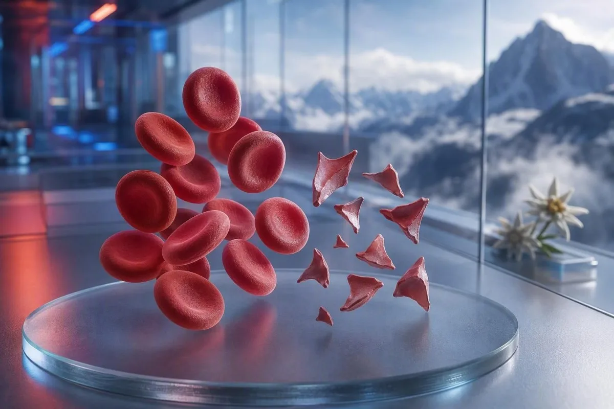
Шистоциттер — бұл фрагменттелген эритроциттер; олар қауіпті ұсақ тамырларда ұюдың белгісі болуы мүмкін, бірақ бір ғана жағындыдағы түсіндірме диагноз болып саналмайды. Шұғылдық фрагменттер пайызына, тромбоциттер санына, гемоглобин динамикасына, бүйрек функциясына, гемолиз маркерлеріне және симптомдарға байланысты.


- Шистоциттер шегі: Жақсы дайындалған ересек адамның перифериялық жағындысында 1%-ден көп шистоцит болса, басқа эритроцит пішінінің өзгерістері болмаса, тромботикалық микроангиопатияға күдік тудырады.
- Шұғыл кластер: Шистоциттер плюс тромбоциттер 150 × 10^9/L-ден төмен, гемоглобиннің төмендеуі, LDH-ның жоғарылауы және гаптоглобиннің төмен болуы сол күні-ақ дәрігердің бағалауын қажет етеді.
- ТТП белгісі: ADAMTS13 белсенділігі 10%-ден төмен болуы иммундық тромботикалық тромбоцитопениялық пурпураны қолдайды; клиникалық күдік жоғары болғанда, растайтын тест нәтижелері келгенге дейін ем басталады.
- Әрбір фрагмент қауіпті емес: Механикалық жүрек қақпақшалары, ауыр гипертензия, диализ контурлары, күйіктер және үлгінің зақымдануы фрагменттелген эритроциттерді тудыруы мүмкін.
- Жағынды — қолмен жасалған дәлел: Автоматтандырылған анализатор шистоциттерді сенімді түрде анықтай алмайды; тәжірибелі морфолог слайдты қарап шығуы тиіс.
- Жүктілік тәуекелді арттырады: 20 аптадан кейін жоғары қан қысымы, несепте ақуыз, немесе тромбоциттер санының төмендеуімен қатар шистоциттер болса, шұғыл түрде акушерлік бағалау қажет.
- Қайта тексеру көмектеседі: Жақында жиналған EDTA үлгісі және қайталама жағынды шоғырлану артефактін шынайы жүріп жатқан эритроциттердің фрагментациясынан ажырата алады.
- Өзінше емделмеңіз: Темір, фолат немесе қоспалар микроангиопатиялық гемолизді емдемейді және шұғыл бағалаудан назарыңызды басқа жаққа бұруы мүмкін.
Фрагменттелген эритроциттер шұғыл медициналық қарауды қажет еткенде
Шистоциттер тромбоциттер саны төмен, анемия, биохимиялық гемолиз, бүйрек зақымдануы, неврологиялық симптомдар, қызба немесе ауыр гипертензиямен бірге пайда болса, сол күні-ақ шұғыл қайта қаралуы керек. Тек жағындыға берілген түсініктеме ғана зиянсыз немесе артефакт болуы мүмкін; қауіпті анықтама — үлгі. 2026 жылғы 7 тамыздағы жағдай бойынша, жаңа шистоциттер плюс тромбоциттер 100 × 10^9/л-ден төмен болса, оны әдеттегі бақылау емес, «қазір-ақ клиницистке хабарласу» нәтижесі ретінде қарастырар едім.
Тромбоциттердің төмендеуі көбіне ең жылдам шұғылдық белгісі болады. Гемоглобин 92 г/л, тромбоциттер 68 × 10^9/л және жаңадан хабарланған шистоциттер — гемоглобин 145 г/л және тромбоциттер 260 × 10^9/л болып тұрақты, 1% фрагменттерге қарағанда әлдеқайда алаңдатарлық. Kantesti — бұл осы кластерді толық қан анализі (complete blood count) бойынша оқитын, морфология туралы түсініктемені оқшау түрде көтермейтін AI қан анализаторы.
Менің 15 жылдық клиникалық тәжірибемде тез айтатын қорытындыларым — нәтижелері бірнеше күн ішінде өзгерген пациенттер: гемоглобиннің төмендеуі, креатининнің көтерілуі және лактатдегидрогеназаның (ЛДГ) көтерілуі. Доктор Томас Клейннің практикалық ережесі қарапайым: осы үлгімен қатар шатасу, жаңа әлсіздік, кеуде ауыруы, ентігу, несептің өте азаюы немесе қатты бас ауыруы болса, бүгін-ақ шұғыл бағалау керек.
Шистоциттер адам өзін нашар сезінсе, қайталап тексеруді күте беруге себеп емес. Қалыпты калий немесе қалыпты лейкоциттер саны тромботикалық микроангиопатия қаупін жоққа шығармайды. Төмен көрсеткішті түсінуге тырысатын пациенттер де біздің нұсқаулықты қарап шығуы мүмкін: төмен эритроциттер санының симптомдары, бірақ жедел симптомдар әрқашан онлайн түсіндіруден жоғары тұрады.
Қазір жедел жәрдем қызметіне қоңырау шалыңыз:
Жаңа бір жақты әлсіздік, естен тану, құрысу, қатты ентігу, кеудеде жаншып тұрған жайсыздық, қара нәжіс немесе шатасудың тез күшеюі клиникаға хабарлама жіберуден гөрі жедел жәрдем қызметін қажет етеді. Бұл симптомдар ағзаның зақымдануын көрсетуі мүмкін, ал шистоциттер пайызын қауіпсіз түрде жоққа шығаратын көрсеткіш болмайды.
Перифериялық жағындыдағы шистоциттердің шын мәнінде нені білдіретіні
Шистоциттер қанайналымдағы жасушалар физикалық түрде «қайырылып» (қырқылып) кеткенде пайда болатын, біркелкі емес эритроцит фрагменттері; көбіне бұл ұсақ тамырларда фибрин жіпшелерімен немесе қалыптан тыс жоғары «қырқу» әсері бар беткейлермен болады. Олар әдетте өткір бұрышты, дулыға тәрізді пішіндерге ие немесе ұсақ үшбұрышты болып келеді және бүтін эритроциттің орталық бозаруын болмайды.
Шынайы шистоциттер — морфологиялық анықтама, стандартты CBC анализаторы шығаратын мән емес. Гематологиядағы стандарттауға арналған Халықаралық кеңес сандық бағалау маңызды болғанда (Zini et al., 2021) оңтайлы жағынды аймағында кемінде 1 000 эритроцитті санауды ұсынады. Бұл деталь неге бір зертхана “сирек фрагменттер” деп, ал екіншісі пайызын хабарлауы мүмкін екенін түсіндіреді.
Классикалық алаңдаушылық — микроангиопатиялық гемолиздік анемия, мұнда эритроциттер қанайналымда тромбоциттерге бай микротромбылармен және фибринмен түйіседі. Дегенмен, ауыр аорталық стеноз, механикалық клапан, экстракорпоральды контурлар және ірі көлемді термиялық жарақаттар басқа механизм арқылы шынайы фрагменттер түзуі мүмкін. Сондықтан сұрақ “Фрагменттер шынайы ма?” емес, “Фрагменттерді не тудырып жатыр?”
Kantesti — бұл толық қан талдауы (CBC) жүктелген нәтижесін қасына қоя алатын және CBC құрамына не кіреді билирубин, ретикулоциттер немесе гаптоглобин сияқты жетіспейтін серіктес нәтижелерді атап көрсететін AI қан талдауы нәтижесі платформасы. Ол слайдтың өзін тексере алмайды, бұл — оқытылған зертханалық маманның жұмысы.
Ересек адамдағы жағындыда шистоцитоздың қаншалықты алаңдатарлық екені
Ересек адамның шеткі жағындысында шистоциттер саны 1%-ден жоғары болса, фрагменттер қызыл жасушалардағы негізгі ауытқу болған кезде тромботикалық микроангиопатияға күмәнді. 1%-ден төмен көрсеткіштер ауруды жоққа шығармайды, әсіресе егер пленка сұйықтық енгізілгеннен кейін, трансфузиядан кейін немесе дамып келе жатқан ауру аясында жасалса.
1% шегі — диагностикалық белгі, ауырлық шкаласы емес. ICSH нұсқаулығы сау ересектер мен мерзімінде туған жаңа туғандар үшін 1%-ден төмен референс аралығын қолданады; шала туған нәрестелерде дәл сол мағынасыздықпен 5%-ге дейін фрагменттер болуы мүмкін (Zini et al., 2021). Ауыр тромбоцитопениямен бірге 2% нәтижесі механикалық жүрек-қантамырды қолдау құрылғысы қойылғаннан кейінгі 4%-ге қарағанда көбірек алаңдатады.
Зертханалар пайыздың орнына “кездейсоқ”, “аз”, немесе “жоғары қуатты өріске 1–2” деп хабарлауы мүмкін. Бұл тіркестер бір-бірімен алмастырылмайды, өйткені өріс өлшемі, жағынды қалыңдығы және есеп беру саясаты әртүрлі. Зертхана ICSH стиліндегі пайыздық есептеуді орындады ма деп сұрау, сипаттамалық тіркесті өзіңіз түрлендіруге тырысқаннан гөрі жиі пайдалырақ.
Жоғары RDW шистоциттерді өлшемейді. RDW жасуша өлшемінің әртүрлілігін көрсетеді және ретикулоцитоз, темір тапшылығы, трансфузия немесе фрагментация кезінде жоғарылауы мүмкін. Біздің егжей-тегжейлі RDW және эритроцит индексі бойынша нұсқаулық неге жоғары RDW өздігінен TTP-ні де, артефактты да қолдамайтынын түсіндіреді.
Нағыз гемолизді қолдайтын қан талдауының үлгісі
Нағыз эритроцит фрагментациясы әдетте жоғары LDH, төмен гаптоглобин, жанама билирубиннің жоғарылауы және анемиямен қатар ретикулоциттік жауап береді. Бірде-бір маркер айқын емес, өйткені бауыр ауруы, қабыну, жақында қан құю және сүйек кемігі патологиясы көріністі бұрмалауы мүмкін.
LDH сезімтал, бірақ спецификалық емес. LDH 600 IU/L-ден жоғары болуы көбіне дұрыс клиникалық жағдайда белсенді гемолизді қолдайды, алайда бұлшықет жарақаты, бауыр зақымдануы, инфекция және қатерлі ісік те оны арттыра алады. Зертханадағы төменгі шектен төмен гаптоглобин, әдетте 0,3 г/Л-ден төмен, плазмаға бөлінген бос гемоглобинді байланыстыратындықтан салмақ қосады.
Ретикулоциттер саны әдетте шеткері (перифериялық) жойылу кезінде артуы тиіс, бірақ фолий тапшылығы, темір тапшылығы, бүйрек ауруы немесе жедел сүйек кемігі тежелуінде төмен болып қалуы мүмкін. Бұл айқын қайшылықты оңай жіберіп алуға болады: науқаста шынайы гемолиз болуы мүмкін, бірақ ретикулоциттік жауап орынсыз төмен. Сол гаптоглобин нәтижесі бойынша нұсқаулық жиі кездесетін интерпретациялық қателіктерді қамтиды.
Kantesti AI бір уақыт нүктесінен алынған гемоглобин, MCV, тромбоциттер, LDH, билирубин, креатинин және ретикулоциттерді салыстыру арқылы фрагменттелген эритроциттер үлгілерін түсіндіреді. 48–72 сағат ішінде гемоглобиннің 20 г/Л-ден астам төмендеуі, бұрынғы салыстырусыз ғана жеңіл ауытқыған мәннен клиникалық тұрғыдан анағұрлым ақпаратты.
Билирубин неге қалыпты болуы мүмкін
Гемолиздің басында, көктамыр ішіне сұйықтық құйылуынан кейін немесе бауыр билирубинді тиімді түрде тазартып жатқанда жанама билирубин қалыпты болуы мүмкін. Сондықтан қалыпты билирубин сенімді фрагменттерді, төмен гаптоглобинді және төмендеп бара жатқан гемоглобинді жоққа шығармауы тиіс.
Неге ТТП гематологиялық шұғыл жағдай ретінде емделеді
Тромботикалық тромбоцитопениялық пурпура, немесе TTP, тромбоцитопениямен бірге шистоциттер және айқынырақ балама жоқ болғанда микроангиопатиялық гемолиздік анемия болғанда күдіктенеді. Емделмеген иммундық TTP тез үдеуі мүмкін, сондықтан клиницистер ADAMTS13 нәтижелері қайтып келгенше плазма алмастыруды және иммундық емді бастауы ықтимал.
ADAMTS13 белсенділігі 10%-ден төмен TTP-ны қатты қолдайды, бірақ нәтиже бірнеше сағат немесе күн алуы мүмкін. ISTH диагностикалық нұсқаулығы PLASMIC сияқты клиникалық құралдармен алдын ала ықтималдықты бағалауды, сонымен бірге плазмамен емдеуге дейін үлгі алуды ұсынады (Zheng et al., 2020). Ұпай — әрекетке көпір, мамандық пайымның орнына жүрмейді.
Қызба, неврологиялық белгілер, бүйрек жеткіліксіздігі, тромбоцитопения және гемолиздік анемиядан тұратын таныс “пентада” көптеген нақты науқастарда толық болмайды. Джордж және Нестер тромботикалық микроангиопатияны бес міндетті қорап емес, қабаттасатын себептері бар синдром ретінде сипаттады (George & Nester, 2014). Тәжірибеде бес белгінің бәрін күту — қауіпті ескі әдет.
Гемолиз кезінде тромбоциттер 30 × 10^9/Л-ден төмен болса, гематологиямен әсіресе шұғыл түрде талқылау қажет. Плазмалық тромбоцит құю әдетте күдікті TTP кезінде өмірге қауіпті қан кету болмаса немесе инвазивті процедураны күтуге болмайтын жағдай болмаса жүргізілмейді. Қатысты тексерулер үшін біздің коагуляция тестіне арналған нұсқаулық PT және aPTT TTP кезінде неге қалыпты болуы мүмкін екенін түсіндіреді.
Дәрігерлер шистоциттердің басқа да қауіпті себептерін қарастырады
Шистоциттер сонымен қатар диссеминацияланған тамырішілік ұюда, Шига-токсиндік гемолиздік уремиялық синдромда, ауыр артериялық гипертензияда, жүктілікке байланысты микроангиопатияда, кейбір дәрілерде және трансплантацияға байланысты тромботикалық микроангиопатияда да кездеседі. Оған байланысты тромбоциттік, коагуляциялық, бүйректік және клиникалық айғақтар әдетте осы жағдайларды ажыратады.
DIC көбіне фрагменттерді ұзаққа созылған PT, ұзаққа созылған aPTT, фибриногеннің төмендеуі немесе төмендеп бара жатуымен және D-димердің айқын жоғарылауымен біріктіреді. TTP-де көбіне күнделікті коагуляция көрсеткіштері салыстырмалы түрде қалыптырақ болады. Ауру науқаста фибриноген 1,5 г/Л-ден төмен болса, тұтынулық коагулопатияға қатысты алаңдаушылықты күшейтеді, бірақ фибриноген бастапқыда қалыпты болып көрінуі мүмкін, өйткені ол жедел қабыну кезінде артады.
Ауыр артериялық гипертензия қысым микроқантамырларға зақым келтіргенде эритроциттерді «кесіп» тастауы мүмкін; бас ауыруы, көрудің өзгеруі, кеуде ауыруы немесе бүйрек дисфункциясы бар 180/120 мм сын.бағ. деңгейінде немесе одан жоғары көрсеткіш — төтенше жағдай. Сондықтан қан қысымын өлшеу нәтижесі жағынды (мазок) есебінің қасына жазылуы тиіс. Науқастар екіншілік артериялық гипертензияға зертханалық белгілер жедел қауіп жойылғаннан кейін.
Диареядан кейін несеп шығарудың азаюы, анемия және тромбоцитопения гемолиздік-уремиялық синдромға күдік тудырады. Нәжісті тексеру Шига токсинін анықтауға көмектесуі мүмкін, бірақ алдымен сұйықтық жағдайын, креатининді, калийді және неврологиялық бағалауды ескеру шұғыл көмекті бағыттайды. Жоғары лактат немесе тұрақсыз өмірлік көрсеткіштер күту-байқау тәсілінен гөрі сепсис пен ДИК-ті бағалауды талап етеді; мыналарды қараңыз сепсис маркерлерінің үлгілеріне арналған бөлек нұсқаулығымыз лактаттың, прокальцитониннің, CRP-ның және CBC-ның ауысымдары неге бір-бірімен бәсекелес тесттер ретінде емес, бірге түсіндірілетінін түсіндіреді..
Фрагменттелген қызыл қан жасушаларының механикалық себептері
Механикалық бұзылу TTP-сыз да шынайы шистоциттер түзуі мүмкін, әсіресе протезделген жүрек қақпақшаларында, ауыр аорталық стенозда, қарыншалық көмекші құрылғыларда, диализ контурларында немесе экстракорпоральды мембраналық оксигенацияда. Бұл себептер бәрібір медициналық қарауды қажет етеді, бірақ ем иммундық TTP-ге емес, механикалық көзге бағытталады.
Механикалық-қақпақшаға тән үлгіде көбіне созылмалы жеңіл LDH жоғарылауы және тромбоциттер тұрақты болғанда төмен гаптоглобин болады. Жаңа анемия, ентігудің күшеюі, қою несеп немесе қақпақша дыбысының өзгеруі паравальвулярлық ағуды немесе құрылғы дисфункциясын меңзеуі мүмкін және кардиологияға шұғыл қаралуға түрткі болуы тиіс. Жақпада (мазокта) белгі бар; эхокардиография көзін анықтайды.
Спортшылар кейде марафон фрагменттерді түсіндіре ала ма деп сұрайды. Аяқтың жерге соғылуынан болатын гемолиз ұзақ қашықтыққа жүгіруден кейін уақытша бос гемоглобин мен төмен гаптоглобинге әкелуі мүмкін, бірақ сенімді шистоцитозды жаттығумен жай ғана байланыстыруға болмайды — әсіресе тромбоциттер төмендеп жатса. Оны неғұрлым кең марафон жүгірушісінің зертханалық нұсқаулығымен салыстырыңыз.
Ауыр күйіктер және ірі тамырлық хирургия физикалық және эндотелийлік зақым арқылы фрагменттелген жасушалар түзуі мүмкін. Осындай жағдайларда әр 6–24 сағат сайын зертханалық динамика бір ғана жақпадан гөрі көбірек ақпарат беруі мүмкін. Қоздырғыш оқиғадан кейін CBC-нің тұрақты болуы жұбататын белгі; гемоглобиннің үздіксіз төмендеуі — жоқ.
Жинау және слайд артефакттері шистоциттерді қалай ұқсатып көрсетеді
Үлгіні нашар жинау, жақпаны дайындауды кешіктіру, антикоагулянтты түтіктердің жеткіліксіз толтырылуы және жақпа жиегінің зақымдануы қанайналым деңгейіндегі гемолизді көрсетпей-ақ шистоциттерге ұқсас пішіндер тудыруы мүмкін. Таза жиналған және моноқабатта (monolayer) қаралған қайталама үлгі — клиникалық көрініс тыныш болған кезде әдеттегі бірінші тексеріс.
Кренирленген жасушалар, жақпа жиегінің бұрмалануы, кептіру артефактісі және бір-біріне қабаттасқан жасушалар жиі кездесетін жалған ұқсастықтар. Олар көбіне біркелкі емес, диффузды түрде немесе қауырсындалған шетінде ғана көрінеді, ал идеал оқылатын аймақта біркелкі болмайды. Тәжірибелі морфолог нағыз фрагменттің айқын геометриясын слайд дайындау кезінде ғана бұрмаланған жасушадан ажыратады.
Қиын жиналған үлгі түтікті гемолиздеуі және калийді немесе LDH-ті жалған көтеруі мүмкін, бірақ ол автоматты түрде нағыз айналымдағы шистоциттерді тудырмайды. Егер нәтиже гемолиз индексін немесе “үлгі бұзылған” деген түсіндірмені қамтыса, клиницистер әдетте ауруды анықтамас бұрын қайта үлгі алады. Біздің мақаламызда калийді алу кезіндегі қателер туралы нұсқаулығымызды оқыңыз бірдей преаналитикалық мәселе неге бірнеше нәтижені бір мезетте жаңылыстыруы мүмкін екені түсіндіріледі.
EDTA-ға тәуелді тромбоциттердің түйірленуі тромбоциттер санын жалған төмендетіп, үрей тудыратындай көрінетін комбинацияны «жасап» беруі мүмкін. Дилюцияға түзетілген цитратты түтікпен алынған тромбоциттер саны және жаңа жағынды мұны анықтай алады. Нюанс маңызды: артефакт шынайы аурумен қатар болуы мүмкін, сондықтан қайталама тест симптомдары бар адамға күтімді ешқашан кешіктірмеуі тиіс.
Неге анализатордың ескертулері мен зертханалық тұжырымдар жаңылыстыруы мүмкін
Автоматты қан анализаторлары қалыптан тыс эритроцит популяцияларын белгілеуі мүмкін, бірақ олар тромботикалық микроангиопатияны жоққа шығаратындай сенімділікпен шистоциттерді өздігінен тәуелсіз диагноз қоя алмайды. Клиникалық сұрақ гемолиз немесе TTP болса, қолмен перифериялық қан жағындысын қайта қарау әлі де қажет.
“Бөлшектенген қызыл жасушалар бар” деген ескертпе санын, жағынды сапасын және қатар жүретін морфологияны туралы сұрақтар туғызуы тиіс. Темір тапшылығынан, мегалобластты анемиядан немесе тұқым қуалайтын эритроцит бұзылыстарынан болатын айқын анизопойкилоцитоз 1% ережесін азырақ нақты етеді. Бөлшектер диагностикалық тұрғыдан ең пайдалысы — олар көптеген қатысы жоқ пішіндердің арасында емес, қалыптан тыс морфологияның басым бөлігі болған кезде.
Kantesti — зертханалық талдау нәтижелерін түсіндіру қызметі; ол «тромбоциттер саны төмен» сияқты тромбоциттер түйіршіктері бар деген ескертпемен қатар келетін сәйкессіз комбинацияларды белгілеуге арналған. Ол зертхананың микроскопиялық қарап шығуын немесе қайталама жағынды сұрай алатын клиницисті алмастырмайды. Сапаны тексеру жөніндегі біздің тәсіліміз мынада сипатталған: медициналық валидацияға шолу.
Дельта-тексеру ағымдағы нәтижені бұрынғы нәтижемен салыстырады және кенеттен орынсыз өзгерісті анықтай алады. Гемоглобиннің 138 г/л-ден 87 г/л-ге 24 сағат ішінде төмендеуі клиникалық тұрғыдан маңызды, бірақ көктамырішілік сұйықтықтардан сұйылту немесе таңбасы қате қойылған үлгі әлі де мүмкін. Туралы көбірек оқыңыз: зертханалық көрсеткіштердің кенет өзгерістері.
Қан талдауының нәтижесінде шистоциттер айтылса, не істеу керек
Шистоциттер алғаш рет хабарланған болса, сол күні талдауды тағайындаған клиницистке немесе қызметке хабарласыңыз; ал неврологиялық симптомдар, ентігу, кеуде ауыруы, қою несеп, қатты бас ауыруы, қызба немесе несеп шығарудың азаюы болса, дереу шұғыл медициналық көмекке жүгініңіз. Жағынды туралы ескертпені өздігінен диагноз ретінде түсіндірмеңіз.
Нақты есепті қолда ұстаңыз: гемоглобин, тромбоциттер, креатинин, билирубин, LDH және кез келген гемолиз индексі. Сондай-ақ жақында болған жүктілікті, диареялық ауруды, қан қысымының өте жоғары көрсеткіштерін, жаңа дәрілерді, қақпақшаға жасалған операцияны, диализді, трансфузияны және аутоиммундық ауруды да ескеріңіз. Бұл мәліметтер морфология өрісіндегі бір ғана сөзден гөрі дифференциалды диагнозды көбірек өзгертеді.
Мен пациенттердің темірді себеп анықталмай тұрып бастау арқылы бағалы уақыт жоғалтатынын көремін, өйткені олар “анемия” деген сөзді бірден оқиды. Темір тапшылығы қатар болуы мүмкін, бірақ ол бөлшектермен жүретін тромбоциттердің жылдам төмендеуін түсіндірмейді. Доктор Томас Кляйн тек ерекшеленген сандарды көшіріп жазудың орнына толық есептің фотосын түсіруді ұсынады; көшіріп жазу көбіне үлгісін үйлестіретін зертханалық түсіндірмені түсіріп қалуы мүмкін.
Тромбоциттер өте төмен болса немесе қан кету болса, клиницист басқа нұсқау бермейінше аспиринді, ибупрофенді және тағайындалмаған антикоагулянттарды қолданбаңыз. Тағайындалған антикоагулянтты шұғыл медициналық нұсқаусыз тоқтатпаңыз. Шаршауы немесе басы айналуы бар адамдар біздің бас айналу кезіндегі қан талдаулары шұғыл себептер жоққа шығарылғаннан кейін сұрақтар дайындау үшін пайдалана алады.
Жүктілік кезіндегі, жаңа туғандардағы және балалардағы шистоциттер
Жүктілік кезінде шистоциттер, егер олар гипертензиямен, протеинуриямен, бауыр ферменттерінің жоғарылауымен, тромбоциттердің төмендігімен, бас ауыруымен, көру симптомдарымен немесе құрсақтың жоғарғы бөлігіндегі ауырумен бірге пайда болса, шұғыл бағалауды қажет етеді. HELLP синдромы, преэклампсия, TTP, атипиялық HUS және ДИК бір-бірімен қабаттасуы мүмкін, ал босануға дейінгі/босанудан кейінгі уақыт көмектеседі, бірақ диагнозды түпкілікті шешпейді.
HELLP синдромы классикалық түрде гемолиз, бауыр ферменттерінің жоғарылауы және тромбоциттердің төмендігі арқылы анықталады; ол көбіне жүктіліктің 20 аптасынан кейін немесе босанудан көп ұзамай байқалады. Тромбоциттер саны 100 × 10^9/л-ден төмен немесе AST 70 IU/л-ден жоғары болса, алаңдаушылықты қолдауы мүмкін, бірақ жергілікті акушерлік топтар бүкіл клиникалық көріністі түсіндіреді. Осы комбинациямен кез келген жоспарлы антенаталдық қабылдауды күтпеңіз.
Мерзіміне жетіп туған нәрестелерде 1% шистоциттер аз болуы мүмкін, ал шала туған нәрестелерде 5%-ке дейін болуы мүмкін, өйткені неонаталдық эритроцит морфологиясы өзгеше (Zini et al., 2021). Алайда диареясы бар балада бозару, несептің азаюы және тромбоцитопения болса, бөлшектер HUS-қа меңзеуі мүмкін және дереу педиатриялық бағалауды қажет етеді.
Тромбоциттер мен гемоглобин үшін анықтамалық аралықтар жасқа және жүктілік кезеңіне қарай айтарлықтай өзгереді. Жалпыға арналған қосымша “қалыпты” деп белгілеген нәтиже контекстте клиникалық тұрғыдан әлі де алаңдатуы мүмкін. Біздің жүктілік кезіндегі тромбоциттер диапазондары туралы ресурстарымыз және сол күнгі жүктілікке қатысты зертханалық белгілер пайдалы фондық ақпарат беріңіз, бірақ ол жүктілік кезіндегі триаждың орнына жүрмейді.
Дәрігерлер нағыз шистоциттерді анықтау үшін тағайындайтын тесттер
Микроангиопатиялық гемолизге күдік болғанда жүргізілетін алғашқы зерттеулер жиынтығына әдетте қайталама CBC және жағынды, ретикулоциттер саны, LDH, гаптоглобин, билирубин фракциялары, креатинин, несеп талдауы, PT, aPTT, фибриноген және D-димер кіреді. Егер TTP күдік болса, плазма алмасуға дейін ADAMTS13 белсенділігі мен ингибиторға тестілеу алынуы тиіс, бірақ емді күтуге болмайды.
Тікелей антиглобулиндік тест (DAT), сондай-ақ Кумбс тестілеуі деп аталады, иммундық гемолизді иммундық емес фрагментациядан ажыратуға көмектеседі. Оң DAT екінші үдерісті жоққа шығармайды, бірақ классикалық TTP әдетте DAT-теріс болады. Несеп микроскопиясы бүйрек микроқантамырлық зақым белсенді болғанда ақуыздық немесе жасушалық цилиндрлерді анықтай алады; біздің түсіндірмемізді қараңыз гранулалы зәр цилиндрлері.
Нәжістегі шигатоксинге тестілеу үйлесімді асқазан-ішек ауруынан кейін қарастырылады, ал комплементке тесттер, дәрілік шолулар және генетикалық немесе функционалдық зерттеулер атипиялық HUS үшін қолданылуы мүмкін. Тапсырыс ел мен ауруханаға қарай өзгереді. 48 сағат ішінде креатининнің 26,5 мкмоль/л-ге артуы жедел бүйрек зақымдануының бір критерийіне сәйкес келеді және ол күрделі санға айналмай тұрып-ақ назар аударуды қажет етеді.
Маңызды анемия кезінде ретикулоциттер өндірілу индексі 2-ден төмен болса, шикі ретикулоцит пайыздық көрсеткіші жоғары көрінсе де, сүйек кемігінің жеткіліксіз жауап беруін көрсетеді. Бұл есеп анемия кезіндегі эритроциттердің төмен массасын түзетеді. ретикулоцит және гематология маркері жөніндегі нұсқаулық пайыздардың неліктен жаңылыстыруы мүмкін екенін түсіндіруге көмектеседі.
Жағындыны қайта алу қашан орынды және қашан емес
Жағындыны бірнеше сағат ішінде 24 сағатқа дейін қайталау орынды, егер фрагменттер аз болса, пациент өзін жақсы сезінсе, тромбоциттер мен гемоглобин тұрақты болса және үлгінің сапасына күмән болса. Тромбоцитопения, белсенді гемолиз, ағзалық зақымдану немесе алаңдататын симптомдар болған кезде қайталауды қауіпсіз кешіктіру емес.
Жақсы қайталама үлгі дұрыс толтырылған EDTA түтігіне дереу жиналып, пленкаға тез дайындалуы тиіс. Тапсырыс нақты түрде күдікті шистоциттерді және тромбоциттердің түйірленуін (clumping) қарауды сұрауы керек, егер екеуі де ықтимал болса. Зертхана екі пленканы қатар салыстыра алады, бұл автоматтандырылған CBC-ні жай ғана қайта жүргізуден әлдеқайда пайдалы.
Бағыт маңызды. Үш күн ішінде тромбоциттер саны 210-нан 165-ке, одан 98 × 10^9/л-ге дейін төмендесе, алғашқы екі көрсеткіш жергілікті анықтамалық интервал ішінде түсуі мүмкін болса да, бұл алаңдатарлық. Kantesti AI осы траекторияларды айқындау үшін бойлық контекстті қолданады; бұл принцип біздің қан талдауының трендін талдау жөніндегі нұсқаулығымызды пайдаланып INR тарихыңызбен даталарды салыстырыңыз.
Құю (трансфузия) бастапқы морфологияны уақытша сұйылтып немесе бүркемелеуі мүмкін. Екінші үлгі алынғанға дейін эритроциттер алған болсаңыз, командаға күнін және қанша бірлік (unit) болғанын айтыңыз. Біздің ауруханадан шыққаннан кейінгі мәндердің өзгеруі туралы нұсқаулық емнен кейінгі үрдістерге неге басқа бастапқы деңгей (baseline) қажет екенін түсіндіреді.
Шистоцит ауруына ұқсас, бірақ ондай емес болып көрінетін жағдайлар
Темір тапшылығы, витамин B12 тапшылығы, бауыр ауруы, тұқым қуалайтын эритроциттік бұзылыстар және ауыр инфекция тромботикалық микроангиопатияның тән фрагментация үлгісінсіз-ақ қалыптан тыс эритроцит пішіндерін тудыруы мүмкін. Жағындындағы толық морфология және клиникалық уақыт желісі фрагменттерді асыра бағалаудың алдын алады.
Витамин B12-нің ауыр тапшылығы анемиямен, LDH жоғарылауымен, тромбоциттердің төмендеуімен және кейде фрагменттермен жүретін псевдо-тромботикалық микроангиопатияны туғызуы мүмкін. Ол көбіне айқын макроцитоз береді, ретикулоциттік жауап төмен болады және LDH көрсеткіштері кейде 2 500 IU/л-ден жоғары—бұл белгілер автоматты түрде TTP емін емес, B12 және метилмалон қышқылын тестілеуді бастауға тиіс. Біздің B12 мен фолат жөніндегі нұсқаулықта қамтылған осы маңызды еліктеушіні түсіндіреді.
Темір тапшылығы әдетте микроцитозды, гипохромияны, қарындаш тәрізді жасушаларды және RDW-ның жоғарылауын туғызады; ауыр жағдайларда фрагменттер туралы хабарлануы мүмкін, бірақ олар сирек негізгі белгі болады. Ферритиннің 15 мкг/л-ден төмен болуы, әйтпесе асқынбаған ересек адамда, темір қорларының сарқылуын қатты қолдайды, алайда қабыну ферритинді көтеру арқылы тапшылықты бүркемелеуі мүмкін.
Менің тәжірибемде ең жұбататын нәтиже тек “шистоциттер жоқ” дегеннің өзі емес, тұрақты тромбоциттер, бүйрек функциясының тұрақтылығы және биохимиялық гемолиздің болмауымен бірге келетін, қисынды балама түсіндірме болып табылады. Егер қорытындылар сәйкес келмей тұрса, гематолог соңғы есептің мәтінін ғана емес, нақты слайдты қарап шығуы мүмкін.
Жағындыдағы шұғыл жағдайды жіберіп алмастан АИ интерпретациясын қолдану
AI қан анализінің нәтижелерін ұйымдастыруға және қауіпті комбинацияларды анықтауға көмектесе алады, бірақ CBC PDF ішінен шистоцит диагнозын растай алмайды және шұғыл клиникалық бағалауды алмастыра алмайды. Перифериялық жағынды — визуалды микроскопия, ал бастапқы зертханалық слайд — ақиқаттың қайнар көзі болып қала береді.
Kantesti — AI-мен қуатталған қан талдауы құралы, ол есептер бойынша байланысты нәтижелерді, соның ішінде тромбоциттер траекторияларын, гемоглобин өзгерістерін, креатининді, билирубинді және LDH-ты картаға түсіреді. Бұл әрбір ауытқыған маркерді бөлек оқудан туындайтын жиі кездесетін қателікті азайта алады. Ол шистоцит деп аталатын пішіннің шынайы фрагмент пе, жағынды жиегінің артефакті ме, әлде есеп беру конвенциясы ма — соны көре алмайды.
Жүктеу сапасы маңызды, өйткені бірлік немесе ондықтың түсіп қалуы нәтиженің мағынасын кері айналдыра алады. Есепті бөліспес бұрын пациент идентификаторларын, жинау күнін, бірліктерді, анықтамалық диапазондарды және нәтижеде “estimated” (бағаланған), “pending” (күтіліп тұр), немесе “sample haemolysed” (үлгі гемолизденген) деп жазылған-жазылмағанын тексеріңіз. Біздің AI есеп беру дәлдігі бойынша чек-листті қараңыз. соңғы адамдық тексеріс үшін пайдалы.
Kantesti-тің клиникалық әдістері біздің Медициналық консультативтік кеңес, тарапынан енгізілген ұсыныстармен қаралады, ал біздің технология AI түсіндіру технологиясы бойынша нұсқаулықта Kantesti-тің нейрожелісі іргелес биомаркерлерді, тренд бағытын және ішкі сәйкестікті қалай өлшейтіні түсіндіріледі. Менің тәжірибемде бұл әсіресе. сипатталған. Доктор Томас Клейн ретінде мен клиницист жаңа фрагменттері бар, тромбоциттері 100 × 10^9/L-ден төмен және симптомдары бар пациент үшін зертханаға тікелей қоңырау шалуын әлі де қалаймын — өйткені дәл сол кезде жылдамдық пен клиникалық тексеріс ең маңызды болады.
Жиі қойылатын сұрақтар
Який рівень шистоцитів є небезпечним?
Ересек адамның шеткі қан жағындысын мұқият қайта қарап, 1%-ден астам шистоциттердің болуы, фрагменттелген жасушалар негізгі эритроциттік ауытқу болған кезде, тромботикалық микроангиопатияға күмән тудырады. Шистоциттер пайызы кез келген деңгейде, егер ол тромбоциттер саны 150 × 10^9/л-ден төмен болғанда, гемоглобиннің төмендеуінде, LDH деңгейінің жоғарылауында, гаптоглобиннің төмендеуінде, бүйрек зақымдануында немесе неврологиялық симптомдармен бірге кездессе, шұғыл болып саналады. 1%-ден төмен нәтиже дамып келе жатқан жағдайды сенімді түрде жоққа шығармайды. Клиникалық көрініс қауіптілікті тек пайызға қарағанда көбірек анықтайды.
Шистоциттер нашар қан алу (қан тапсыру) салдарынан пайда болуы мүмкін бе?
Қиын жинақ, дайындықтың кешігуі, жеткіліксіз толтырылған ЭДТА түтігі немесе нашар дайындалған жағынды жасуша пішіндерін шистоциттерге ұқсатып көрсетуі мүмкін, дегенмен бұл нағыз айналымдағы фрагментацияны тудырмайды. Зертханалар мұны жаңа жиналған үлгімен және оңтайлы жағынды аймағын қолмен қарап шығумен нақтылай алады. Гемолизденген үлгі сондай-ақ калий мен ЛДГ көрсеткіштерін жалған түрде арттыруы мүмкін, бұл есепті одан да алаңдатарлық етіп көрсетуі ықтимал. Қайта тексеру симптомдар, тромбоциттердің төмендігі немесе анемия болған жағдайда шұғыл бағалауды кідіртуі тиіс емес.
Шистоциттер әрқашан ТТП-ны білдіре ме?
Шистоциттер әрдайым тромботикалық тромбоцитопениялық пурпураны немесе TTP-ны білдірмейді. Олар ДИК-те, ауыр артериялық гипертензияда, Шига-токсиндік HUS-та, жүктілікке байланысты бұзылыстарда, механикалық жүрек қақпақшаларында, диализ контурларында және ірі тіндік зақымдануда кездесуі мүмкін. Шистоциттер тромбоцитопениямен және иммундық емес гемолиздік анемиямен қатар болғанда, әсіресе ADAMTS13 белсенділігі 10%-тен төмен болғанда, TTP жоғары басымдықтағы мәселе ретінде қарастырылады. ADAMTS13 тестілеуі уақыт алатындықтан, күдікті TTP нәтижесі түскенге дейін шұғыл мамандық бағалауды қажет етеді.
Які аналізи крові підвищені при гемолізі зі шистоцитами?
Нағыз фрагментациялық гемолиз көбіне LDH және тікелей емес билирубинді арттырады, гаптоглобинді төмендетеді және гемоглобин төмендеген кезде ретикулоциттер санының жоғарылауын туғызады. Белсенді гемолиз кезінде LDH 600 ХБ/л-ден асып кетуі мүмкін, бірақ ол спецификалық емес, өйткені бұлшықет, бауыр және басқа тіндердің зақымдануы да оны арттырады. Бауырдың өндіруі жеткілікті болғанда гаптоглобиннің шамамен 0,3 г/л-ден төмен болуы тамырішілік гемолизді қолдайды. Креатинин, тромбоциттер саны, ПТ, аПТТ, фибриноген және D-димер негізгі себепті анықтауға көмектеседі.
Жүктілік кезінде шистоциттер қалыпты ма?
Шистоциттер жүктіліктің қалыпты, күнделікті табысы болып саналмайды, егер олар гипертензиямен, протеинуриямен, бауыр ферменттерінің жоғарылауымен, тромбоциттердің төмендеуімен, бас ауыруымен, көрудің өзгерістерімен немесе құрсақтың жоғарғы бөлігіндегі ауырсынумен қатар пайда болса. Бұл біріктірілім HELLP синдромын, преэклампсияны, TTP, атипиялық HUS немесе DIC-ті көрсетуі мүмкін және шұғыл акушерлік бағалауды қажет етеді. Осы жағдайдағы тромбоциттер санының 100 × 10^9/л-ден төмен болуы әсіресе алаңдатарлық. Жүктілікке тән анықтамалық диапазондар гемолиз үлгісін елемеуге қауіпсіз деп есептеуге негіз бермейді.
Механикалық жүрек қақпақшасы шистоциттер тудыруы мүмкін бе?
Механикалық жүрек қақпақшалары және ауыр аорталық стеноз шынайы шистоциттерге әкелуі мүмкін, өйткені жоғары ығысу (shear) әсеріндегі қан ағыны эритроциттерді физикалық түрде фрагментациялайды. Бұл көбіне жеңіл созылмалы гемолизді туғызады: LDH деңгейі жоғарылайды және гаптоглобин төмендейді, ал тромбоциттер саны тұрақты болып қалады. Жаңа анемия, қою түсті несеп, ентігу немесе қақпақша қызметінің өзгеруі жүрекке қайта қарауды қажет етеді, өйткені ағып кету немесе құрылғы ақауы себеп болуы мүмкін. Бұл анықтаманы жүрек тарихын ескермей автоматты түрде TTP деп белгілеуге болмайды.
Бүгін AI арқылы қан талдауы нәтижесін алыңыз
Kantesti-ті лезде әрі дәл зертханалық талдау үшін сенетін бүкіл әлем бойынша 2 миллионнан астам пайдаланушыға қосылыңыз. Қан анализінің нәтижелерін ж.үктеп, бірнеше секунд ішінде 15,000+ биомаркерлерінің жан-жақты түсіндірмесін алыңыз.
📚 Сілтеме жасалған ғылыми жарияланымдар
Кляйн, Т., Митчелл, С., & Вебер, Х. (2026). Kantesti Қан талдауын түсіндіру қозғалтқышының 100 000 синтетикалық тест жағдайындағы алдын ала тіркелген, рубрикаға негізделген автоматтандырылған техникалық бенчмарк. Kantesti AI медициналық зерттеулер.
Кляйн, Т., Митчелл, С., & Вебер, Х. (2026). Клиникалық валидациялау негіздемесі v2.0 (Медициналық валидация беті). Kantesti AI медициналық зерттеулер.
📖 Сыртқы медициналық сілтемелер
Чжэн С.Л. және басқалар (2020). Тромботикалық тромбоцитопениялық пурпура диагностикасы бойынша ISTH нұсқаулықтары. Thrombosis and Haemostasis журналы.
⚕️ Медициналық ескерту
Бұл мақала тек білім беру мақсатында ғана және медициналық кеңес болып табылмайды. Диагноз және ем туралы шешімдер үшін әрдайым білікті медицина қызметкеріне жүгініңіз.
E-E-A-T сенім сигналдары
Тәжірибе
Дәрігер басқаратын зертханалық талдауды түсіндіру жұмыс ағындарын клиникалық шолу.
Сараптама
Биомаркерлердің клиникалық контексте қалай әрекет ететініне зертханалық медицина фокусы.
Билік
Доктор Томас Клейн жазған, доктор Сара Митчелл және профессор доктор Ханс Вебер шолған.
Сенімділік
Дабыл қаупін азайту үшін айқын бақылау жолдарымен дәлелге негізделген түсіндіру.