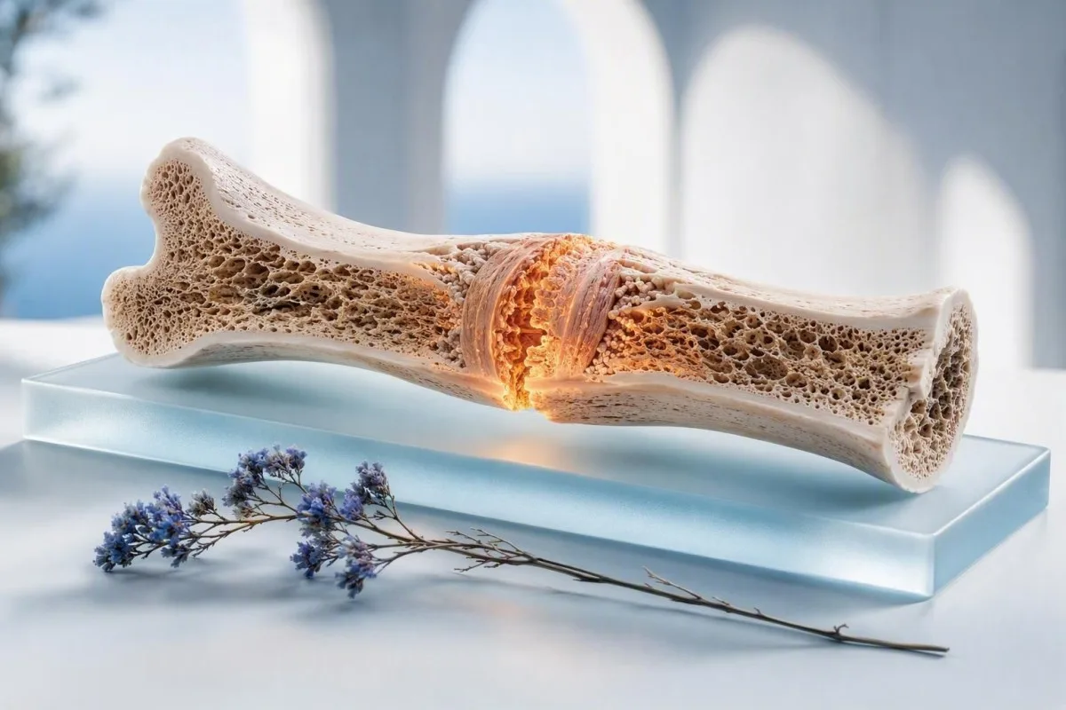
A broken bone can raise alkaline phosphatase as new bone forms, usually beginning after the first week and settling over several weeks. The rest of the liver panel, the degree of elevation, and your symptoms determine whether that explanation is enough.


- Expected timing: Total ALP may begin rising 7-10 days after a significant fracture, often peaks around 3-6 weeks, and can remain above baseline for 6-12 weeks.
- Not every fracture raises ALP: A single uncomplicated wrist or ankle fracture may produce no detectable total-ALP change because the signal is small and liver ALP is included in the same test.
- Typical adult range: Many laboratories use an adult total ALP reference interval near 30-120 U/L, but the upper limit can differ by 20-30 U/L between laboratories.
- Bone pattern: High ALP with normal GGT, bilirubin, ALT, and AST makes bone healing or another bone source more likely, although it does not prove it.
- Liver pattern: High ALP plus elevated GGT or direct bilirubin deserves evaluation for cholestasis, medication effects, or a bile-duct problem rather than being attributed to a fracture.
- Useful recheck: If you feel well and other liver tests are normal, repeating ALP with GGT in 4-8 weeks often provides more clinical value than an immediate extensive work-up.
- Urgent symptoms: Jaundice, dark urine, pale stool, fever, worsening right-upper-abdominal pain, or severe itch need prompt medical assessment regardless of a recent fracture.
- Bone-health clues: Persistent elevation with low phosphate, low 25-hydroxyvitamin D, high PTH, diffuse bone pain, or recurrent low-impact fractures warrants a metabolic bone evaluation.
Can a broken bone make alkaline phosphatase high?
Yes—bone healing can raise alkaline phosphatase (ALP), most often from about 1 week after a substantial fracture through the next 1-3 months. In an otherwise well adult with normal bilirubin, GGT, ALT, and AST, a modest isolated rise is commonly compatible with repair rather than liver disease. The number alone cannot confirm that; the trend and companion tests do the real work.
ALP is an enzyme released partly by osteoblasts, the cells that lay down new bone matrix. After a fracture, osteoblast activity accelerates around the developing callus, so bone-derived ALP can enter circulation. In my clinical experience, an ALP of 135-180 U/L after a major tibial, femoral, or pelvic fracture is often less alarming than the same value without an obvious skeletal reason.
The practical catch is that total ALP combines bone and liver fractions. A small fracture can heal perfectly without changing a total ALP result, while a large or multiple fracture can create a clearer rise. Kantesti is an AI blood test analyzer that places ALP beside GGT, bilirubin, calcium, phosphate, and prior results rather than treating one flagged value as a diagnosis.
There is no universally accepted “safe fracture ceiling” such as 200 U/L. Laboratory methods differ, bone size differs, and a value 1.2 times the upper limit of normal means something very different from a persistent value 4 times the upper limit. For perspective on a mild result outside the reference interval, see our guide to borderline ALP results.
What does a high ALP result measure?
Adult total ALP commonly runs about 30-120 U/L, but each laboratory’s printed reference interval is the result that applies to you. ALP comes mainly from liver bile-duct cells and bone-forming cells; placenta and intestine contribute in particular physiological settings.
ALP testing measures enzyme activity, usually in U/L or IU/L, not an amount of enzyme protein. A result of 150 U/L can be only 1.25 times a laboratory upper limit of 120 U/L, whereas that same 150 U/L is nearly 1.9 times an upper limit of 80 U/L. This is why I ask patients for the laboratory range printed beside the result before interpreting the flag.
Children and teenagers often have ALP values several times adult ranges because growth plates are active. Pregnancy can also elevate ALP in the third trimester through placental production. Adults with a healing fracture have a different physiology altogether, and a bone-specific ALP assay or fractionation can help when the distinction remains unclear; our explanation of ALP isoenzymes shows how this works.
A result should never be graded as “critical” by a fixed total-ALP number alone. The level, prior baseline, symptoms, and liver-test pattern determine urgency. A 90 U/L increase from a personal baseline of 55 U/L is sometimes more informative than a stable 150 U/L recorded on several previous panels.
When does ALP rise after a fracture and when does it fall?
After a significant fracture, ALP usually starts to rise after 7-10 days, reaches its broad peak around weeks 3-6, and trends down over 6-12 weeks. The timeline is approximate, not a laboratory rule, and surgical fixation, delayed union, age, and fracture size can shift it.
The first few days after injury are dominated by local inflammatory and repair signals, not mature mineral deposition. ALP rises later because osteoblasts become more active as soft callus is converted toward mineralized hard callus. Older observational work in femoral fractures described an increase after the first week, with a maximum in the first month and gradual normalization afterward; that pattern is useful, but it is not precise enough to date a fracture from a blood test.
ALP may peak later than pain improvement or cast removal. I have seen patients walking comfortably at 6 weeks while their ALP is still slightly above range, particularly after long-bone injuries. A falling value on repeat testing is reassuring only when it is compared in the same laboratory, as discussed in our article on real versus apparent lab change.
A rising ALP beyond 12-16 weeks is not automatically a sign of failed healing. It does, however, make me review the radiology report, vitamin D status, phosphate, renal function, weight-bearing plan, and liver markers rather than simply assuming “the fracture is still doing it.”
Which fractures are most likely to raise ALP?
Large bones, multiple fractures, high-energy injuries, and fractures with abundant callus formation are more likely to raise total ALP than a single small fracture. A normal ALP does not mean a fracture is not healing, and a high result does not measure how strong the repair is.
Femoral shaft, tibial shaft, pelvic, and vertebral fractures involve more remodeling surface than a small finger fracture. Multiple injuries can add further osteoblastic activity. In older adults, however, low baseline bone turnover and vitamin D deficiency may blur this expected rise, so I would not use ALP as a stand-alone healing test.
Surgery can complicate the story. Intramedullary fixation, bone grafting, and revision procedures may restart local bone formation, while post-operative antibiotics, analgesics, and reduced food intake can affect liver-associated tests. A post-discharge panel needs its clinical dates attached; our guide to lab changes after hospital care explains why.
Children are a special case because physiological ALP from growth can be high before any injury. Pediatric laboratories use age- and puberty-specific ranges, often far above adult limits. An adult reference interval should never be applied to a 12-year-old with a fracture.
How do doctors tell bone ALP from liver or bile-duct ALP?
A high ALP with normal GGT, normal bilirubin, and stable ALT/AST favors a bone source, while high ALP plus high GGT or bilirubin favors a hepatobiliary source. Neither pattern is perfect, but this paired interpretation prevents many unnecessary scans and missed bile-duct problems.
GGT is present in bile-duct and liver tissue but is not typically increased by bone formation. Thus, an ALP of 165 U/L with GGT 22 U/L, normal bilirubin, and a healing fracture points more toward bone than an ALP of 165 U/L with GGT 180 U/L. Some laboratories also use 5′-nucleotidase, which supports a liver source when elevated.
The American College of Gastroenterology recommends confirming an ALP elevation with GGT when the source is unclear, then pursuing cholestatic causes if the pattern persists (Kwo et al., 2017). Kantesti AI interprets high ALP after fracture by checking this pattern, the local upper limits, and the direction of change across dates.
Bone-specific ALP is useful when GGT is normal but uncertainty remains—for example, in someone with both a recent fracture and chronic liver disease. It is not ordered in every case because many results become clear with a repeat liver panel and careful history. Our focused review of high ALP with normal GGT covers the exceptions.
Which liver and bile-duct patterns should not be blamed on a fracture?
ALP elevation accompanied by jaundice, direct bilirubin elevation, dark urine, pale stool, or a raised GGT requires liver and bile-duct evaluation even if you recently broke a bone. Fracture healing does not cause bile to back up.
Clinicians often calculate an R ratio: ALT divided by its upper limit, then divided by ALP divided by its upper limit. An R ratio of 2 or lower indicates a cholestatic pattern, 2-5 a mixed pattern, and above 5 a hepatocellular pattern. The formula is a classification tool, not a diagnosis, but it helps decide whether ultrasound, medication review, or specialist referral is sensible.
EASL cholestasis guidance identifies ultrasound as a common first imaging step when cholestatic liver tests persist or symptoms suggest blockage (EASL, 2009). Gallstones, biliary strictures, primary biliary cholangitis, fatty liver with another process, and medicine-related injury can all raise ALP. A fracture does not protect someone from these unrelated conditions.
New itch without rash can be a surprisingly useful clue when ALP and GGT are both elevated. So can urine that looks tea-colored or stool that is putty-colored; review our practical discussion of cholestatic blood-test patterns and seek clinical advice rather than waiting for the cast to come off.
When can persistent ALP signal another bone disorder?
Persistent ALP elevation with normal GGT can reflect vitamin D deficiency with osteomalacia, hyperparathyroidism, Paget disease, healing stress injury, or less commonly bone involvement from cancer. The accompanying calcium, phosphate, PTH, 25-hydroxyvitamin D, symptoms, and imaging decide which possibility fits.
Osteomalacia commonly produces a pattern of raised ALP with low or low-normal phosphate, low 25-hydroxyvitamin D, and secondary PTH elevation; calcium may still be normal. A 25-hydroxyvitamin D result below 25 nmol/L (10 ng/mL) is severe deficiency in many clinical frameworks, but the treatment dose should reflect age, kidney function, malabsorption, and calcium level.
Paget disease becomes more likely with a clearly elevated ALP in an older adult, focal deep bone pain, skull enlargement, hearing change, or characteristic radiographic findings. It is uncommon, and an isolated 10-20% ALP rise after a known fracture is not the usual presentation. High PTH with normal calcium has several explanations too, as outlined in our PTH and normal calcium guide.
Kidney disease changes mineral metabolism and can obscure the simple picture. Reduced eGFR, phosphate abnormalities, and persistently high PTH deserve coordinated interpretation, particularly when a fracture followed minimal trauma. This is one area where a single “normal calcium” result can be falsely reassuring.
Which symptoms make high ALP after a fracture urgent?
Seek same-day medical assessment for high ALP with yellow eyes or skin, fever, severe right-upper-abdominal pain, repeated vomiting, confusion, or rapidly worsening itch. These symptoms suggest a possible liver, bile-duct, or systemic problem that a healing fracture cannot explain.
Jaundice usually becomes clinically visible when bilirubin is roughly above 2-3 mg/dL (34-51 µmol/L), although skin tone and lighting alter what people notice. Dark urine plus pale stool is more specific for conjugated bilirubin reaching urine and failing to reach the gut. These are not symptoms to monitor casually for a week.
A second urgency track concerns the fracture itself: fever of 38.0°C or higher, escalating local pain after initial improvement, new drainage, loss of circulation or sensation, or shortness of breath needs direct clinical assessment. ALP does not diagnose an orthopedic complication, infection, pulmonary embolism, or compartment syndrome. I advise patients to keep the fracture symptoms and laboratory question separate in their minds.
If the principal symptom is yellowing or dark urine, a home urine test may show bilirubin but cannot identify the cause. Our article on positive urine bilirubin explains what a clinician commonly checks next.
What tests should be repeated after a fracture-related ALP rise?
For a modest isolated ALP rise after fracture, repeat ALP with GGT, bilirubin, ALT, AST, calcium, phosphate, creatinine, and sometimes 25-hydroxyvitamin D in about 4-8 weeks. The best interval changes if symptoms occur, the ALP is more than 2-3 times the upper limit, or other tests are abnormal.
Repeat testing is most interpretable when performed at the same laboratory, ideally without a major acute illness or new medication started in the previous few days. Fasting is usually not needed for ALP, though it may be requested when the same draw includes triglycerides or glucose. Write down the fracture date, operation date, supplements, and alcohol intake; those details make a panel far more clinically useful.
If GGT is raised, I would broaden the review to medication exposures, alcohol, viral risk, metabolic risk, autoimmune clues, and biliary symptoms. A clinician may add ultrasound or targeted antibody tests depending on the pattern. See what a liver panel includes before assuming ALP is the only liver-related result.
As Dr. Thomas Klein, I want patients to use interpretation tools as preparation, not as a replacement for diagnosis. Kantesti is an AI lab test interpretation service that can organize these values into a time-based summary, while our clinical standards and limits are described on the medical validation page.
Could medicines, alcohol, or illness raise ALP at the same time?
Yes—anticonvulsants, some antibiotics, anabolic drugs, herbal products, alcohol-associated liver disease, and acute illness can raise ALP or GGT while a fracture is healing. A temporally convenient fracture should not stop a medication review.
Antiseizure medicines such as phenytoin and phenobarbital can increase GGT through enzyme induction and may affect bone metabolism with long-term use. Amoxicillin-clavulanate is a well-recognized cause of delayed cholestatic drug injury, sometimes appearing days to weeks after the medicine ends. The timing, not merely the medication list, matters.
Over-the-counter products deserve equal attention. Concentrated green tea extracts, bodybuilding mixtures, and multi-ingredient “liver support” products can create liver-test confusion, while high-dose vitamin D can disturb calcium balance. Stopping a prescribed drug without medical advice is risky; instead, bring all containers or photographs to the review.
Rapid weight loss can transiently change liver enzymes and increase gallstone risk, creating a separate reason for abnormal tests. Our discussion of ALT after weight loss helps explain why AST and ALT may not move in parallel with ALP.
Does a rising ALP prove that a fracture is healing?
No—ALP is not a validated stand-alone test for fracture union, delayed union, or non-union. It reflects osteoblastic activity somewhere in the body, while serial examination and imaging remain the standard ways to assess whether a fracture is bridging and stable.
A rising ALP may occur during active callus formation, but the same result can appear with vitamin D deficiency, liver disease, or another bone process. Conversely, an older adult with a low-turnover state may have a normal total ALP despite genuine healing. In orthopedic follow-up, pain with loading, fracture-site tenderness, alignment, and imaging carry more decision-making weight.
Delayed union is usually considered when expected progression on imaging and symptoms stalls over months, but definitions vary by bone, injury pattern, and surgical approach. I become more concerned if pain is not improving, mechanical stability worsens, or radiographs show little bridging over sequential visits—not simply because ALP has not normalized by week 8.
Nutrition, smoking, diabetes, vascular status, infection risk, and medication exposure can all slow repair. If progress stalls, a targeted slow-healing laboratory review may identify anemia, glucose dysregulation, low protein intake, or mineral abnormalities worth correcting.
Should you take vitamin D or calcium because ALP is high?
Do not treat a high ALP number with calcium or high-dose vitamin D unless deficiency or another indication has been established. A healing fracture needs adequate protein, vitamin D, calcium, and overall energy intake, but excess supplementation can create kidney stones, hypercalcemia, or misleading lab results.
Most adults need about 1,000 mg of calcium daily from food plus supplements combined; women over 50 and men over 70 are often advised to target 1,200 mg daily, depending on national guidance and dietary intake. Protein requirements during recovery are individualized, although 1.0-1.2 g/kg/day is commonly used for older adults without contraindications. These are nutrition targets, not an ALP-lowering prescription.
For vitamin D, clinicians usually measure 25-hydroxyvitamin D, not 1,25-dihydroxyvitamin D, when assessing stores. A common maintenance dose is 800-1,000 IU daily, but confirmed deficiency may need a different supervised regimen. The evidence is mixed on whether routine supplementation accelerates union in people who are already replete.
Avoid starting several products at once before a repeat panel. It becomes impossible to know whether a changing calcium, phosphate, or liver result reflects healing, the supplement, or an unrelated illness. Our comparison of vitamin D3 and D2 explains why the form can matter.
How should you track ALP without overreacting to one test?
Track ALP as a dated trend with the laboratory’s upper limit, GGT, bilirubin, fracture date, and new medicines—not as a sequence of isolated red flags. A 15 U/L movement can be analytical and biological variation, while a consistent multi-test rise has more clinical meaning.
Use the actual ratio to the upper limit of normal when comparing laboratories. For example, 140 U/L with a local upper limit of 120 U/L equals 1.17 times upper limit, while 140 U/L with an upper limit of 90 U/L equals 1.56 times upper limit. That distinction can change whether a clinician watches, repeats, or investigates.
A useful personal record includes fracture location, date of injury, procedure dates, cast removal, mobility changes, alcohol changes, new antibiotics, and supplements. Kantesti is an AI biomarker interpretation platform that compares serial laboratory reports and can flag when ALP rises alongside liver markers rather than alone. Our article on an AI blood comparison workflow shows the practical data points to retain.
Privacy matters when records include radiology reports and results from several clinics. Kantesti uses GDPR-aligned handling, but no automated report can know whether you have jaundice, a painful abdomen, or a new prescription unless you add that context and speak with a qualified clinician.
What should you ask your clinician about high ALP after a fracture?
Ask whether the elevation is isolated, whether GGT and bilirubin support a liver source, how many times above your laboratory’s upper limit it is, and when to repeat it. Those four questions usually separate a sensible monitoring plan from a need for prompt investigation.
Bring the complete report rather than only the highlighted ALP line. Ask: “Was my ALP high before the fracture?” and “Would bone-specific ALP, GGT, or an ultrasound actually change the plan?” A prior normal GGT and a new modest ALP rise 4 weeks after a tibial fracture is a very different scenario from unexplained ALP elevation that predates injury.
Ask about metabolic bone testing if the fracture occurred after a low-impact fall, if you have had two adult fragility fractures, or if there is persistent diffuse bone pain. Calcium, phosphate, PTH, vitamin D, kidney function, thyroid testing, and bone-density assessment are chosen individually. There is no value in ordering every possible marker without a clinical question.
As of July 31, 2026, my advice remains straightforward: symptoms and the laboratory pattern outrank a single explanation that happens to be convenient. Our doctors’ oversight principles are available through the Medical Advisory Board, and Dr. Thomas Klein’s clinical approach is to escalate early when cholestatic symptoms appear, then avoid needless testing when the trend is clearly settling.
Frequently Asked Questions
How high can alkaline phosphatase get after a fracture?
A fracture can cause a mild to moderate total ALP rise, often beginning 7-10 days after injury and peaking around 3-6 weeks, but there is no validated universal upper number for normal healing. In many adult laboratories, a result up to about 1.5 times the local upper limit with normal GGT, bilirubin, ALT, and AST can be compatible with bone repair. Values above 3 times the laboratory upper limit, a rising trend after 12-16 weeks, or abnormal liver-associated tests deserve clinical review. The fracture type, number of fractures, age, and baseline ALP all affect the result.
Can a broken bone cause high ALP and normal GGT?
Yes, a healing broken bone can raise ALP while GGT remains normal because bone-forming osteoblasts contribute ALP but do not usually increase GGT. This pattern is most convincing when bilirubin, ALT, and AST are also normal and the ALP rise occurs 1-6 weeks after a substantial fracture. A normal GGT does not prove the source is bone, because vitamin D deficiency, Paget disease, and other bone conditions can create the same pattern. Repeat ALP and GGT in 4-8 weeks is often reasonable if the person feels well and the clinician agrees.
How long does alkaline phosphatase stay elevated after a broken bone?
ALP commonly remains elevated for 6-12 weeks after a significant fracture, although some people return to baseline sooner and others take longer. The enzyme rise often starts after day 7 and reaches a broad peak around weeks 3-6 as mineralized callus develops. An ALP result still slightly high at week 8 can fit uncomplicated recovery if it is declining and GGT, bilirubin, ALT, and AST are normal. A persistent or rising elevation beyond 12-16 weeks should prompt a review of healing progress, liver tests, vitamin D, phosphate, and medications.
Does high alkaline phosphatase mean my fracture is healing?
High ALP can be consistent with active bone formation, but it does not prove that a fracture is healing normally. Total ALP includes liver and bone fractions, and a normal total ALP can occur in a fracture that is healing appropriately. Orthopedic clinicians assess union through symptoms, examination, stability, and serial imaging rather than using ALP alone. New pain, poor function, or limited radiographic progress over several months needs orthopedic review regardless of the ALP result.
When should I worry about high ALP after a fracture?
You should seek prompt medical advice for high ALP after a fracture if you also have yellow eyes or skin, dark urine, pale stool, fever of 38.0°C or higher, severe right-upper-abdominal pain, repeated vomiting, or substantial generalized itch. These symptoms may indicate cholestasis or another liver and bile-duct problem rather than bone healing. A result more than 3 times the laboratory upper limit, a raised GGT or bilirubin, or a continued rise on repeat testing also warrants timely evaluation. In an otherwise well person with a modest isolated rise, a clinician may instead repeat ALP with GGT and a liver panel in 4-8 weeks.
What tests help distinguish bone ALP from liver ALP?
GGT, bilirubin, ALT, AST, 5′-nucleotidase, and sometimes bone-specific ALP help distinguish bone from liver-related ALP elevation. High ALP with normal GGT and normal bilirubin favors a bone source, particularly 1-12 weeks after a fracture. High ALP plus elevated GGT or direct bilirubin favors a hepatobiliary source and may lead to medication review and abdominal ultrasound. Calcium, phosphate, PTH, 25-hydroxyvitamin D, and creatinine are useful when a metabolic bone cause is possible.
Get AI-Powered Blood Test Analysis Today
Join over 2 million users worldwide who trust Kantesti for instant, accurate lab test analysis. Upload your blood test results and receive comprehensive interpretation of 15,000+ biomarkers in seconds.
📚 Referenced Research Publications
Klein, T., Mitchell, S., & Weber, H. (2026). Kantesti LTD. (2026). A Pre-Registered, Rubric-Based Automated Technical Benchmark of the Kantesti Blood-Test Interpretation Engine on 100,000 Synthetic Test Cases. Figshare. https://doi.org/10.6084/m9.figshare.32095435. Kantesti AI Medical Research.
Klein, T., Mitchell, S., & Weber, H. (2026). Kantesti LTD. (2026). Clinical Validation Framework v2.0 (Medical Validation Page). Zenodo. https://doi.org/10.5281/zenodo.17993721. Kantesti AI Medical Research.
📖 External Medical References
⚕️ Medical Disclaimer
This article is for educational purposes only and does not constitute medical advice. Always consult a qualified healthcare provider for diagnosis and treatment decisions.
E-E-A-T Trust Signals
Experience
Physician-led clinical review of lab interpretation workflows.
Expertise
Laboratory medicine focus on how biomarkers behave in clinical context.
Authoritativeness
Written by Dr. Thomas Klein with review by Dr. Sarah Mitchell and Prof. Dr. Hans Weber.
Trustworthiness
Evidence-based interpretation with clear follow-up pathways to reduce alarm.