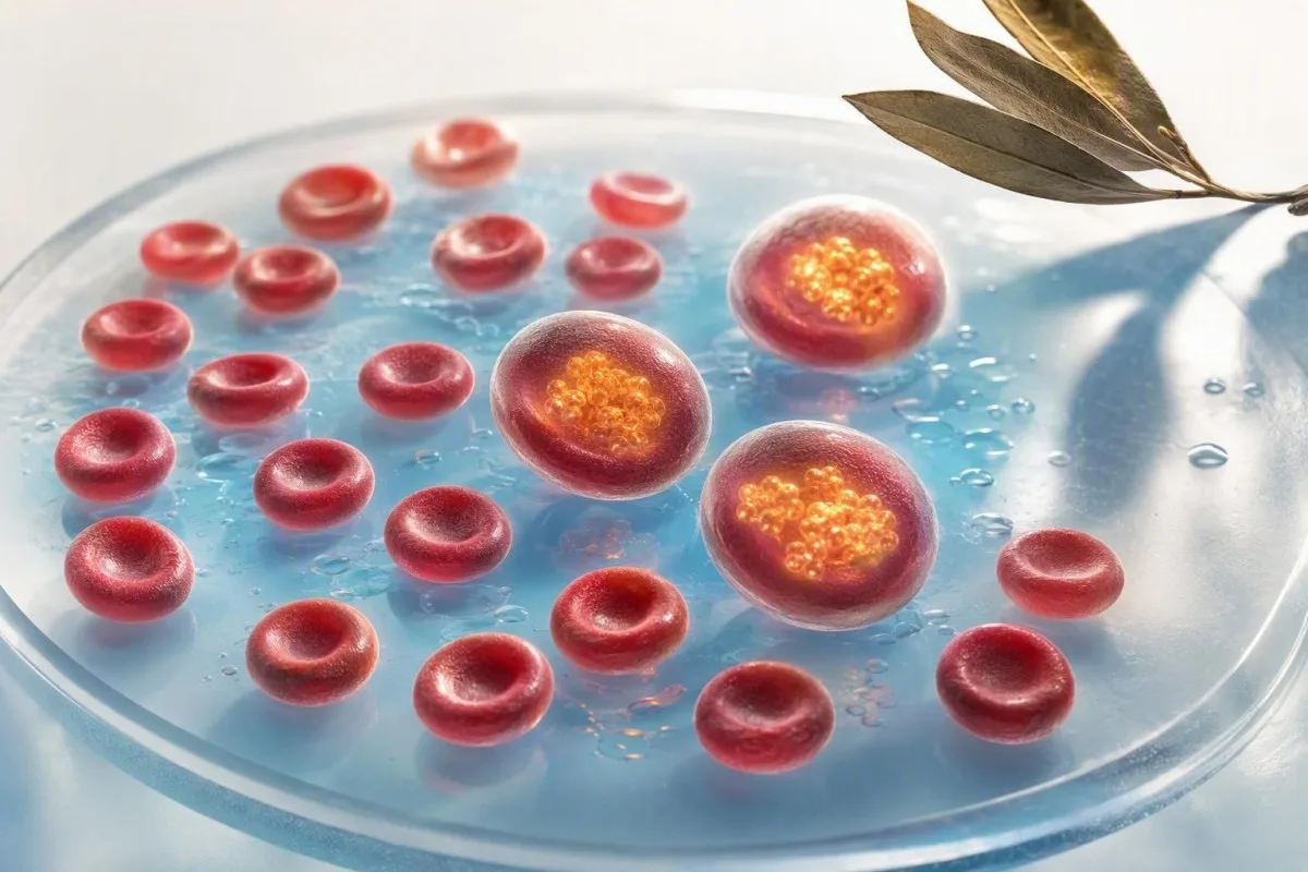
A low red-cell count with normal hemoglobin usually does not mean anemia. It often means each circulating cell is slightly larger or carries more hemoglobin, though hydration, pregnancy, medicines, and a changing CBC pattern can also explain it.


- RBC count below a lab range with hemoglobin of 12.0 g/dL or higher in most non-pregnant women, or 13.0 g/dL or higher in most men, is not anemia by itself.
- MCV above 100 fL is the commonest reason for low RBC normal hemoglobin because fewer, larger cellular elements can carry the same total hemoglobin.
- MCH often rises with macrocytosis; hemoglobin is approximately the RBC count multiplied by MCH, after unit conversion.
- Pregnancy dilution can lower RBC count and hematocrit before hemoglobin crosses the usual anemia thresholds of 11.0 g/dL in the first and third trimesters.
- Vitamin B12 testing may need methylmalonic acid when B12 is borderline at roughly 200-350 pg/mL and MCV is rising.
- Alcohol, hypothyroidism, liver disease, and medicines can raise MCV even when hemoglobin remains normal.
- A repeat CBC in 4-12 weeks is often more informative than treating one isolated low RBC result, provided there are no red-flag symptoms or other low cell lines.
- Urgent review is sensible for shortness of breath at rest, chest pain, fainting, black stools, jaundice, or a rapidly falling hemoglobin.
Why a Low RBC Count Can Coexist With Normal Hemoglobin
What does low RBC mean when hemoglobin is normal? Most often, it means the red cellular elements are larger than average, so a lower number of cells still carries an adequate total amount of hemoglobin. A lone low RBC count is therefore not synonymous with anemia, which is defined primarily by hemoglobin concentration and clinical context.
The apparent mismatch becomes much less mysterious when you remember the arithmetic: hemoglobin concentration reflects total hemoglobin, whereas RBC count reflects how many cellular elements occupy a litre of blood. A person with 3.75 × 10¹²/L cells and an MCH of 32 pg can have about the same hemoglobin as someone with 4.50 × 10¹²/L cells and an MCH of 27 pg.
In my clinical practice, I commonly see this after an otherwise healthy patient has a CBC for fatigue or an annual examination: RBC 3.8, hemoglobin 13.1 g/dL, MCV 101 fL. Dr Thomas Klein's practical rule is simple—read the hemoglobin, MCV, MCH, hematocrit, RDW, white cells, and platelets together, rather than allowing one red flag on a report to define the story.
Kantesti is an AI blood test analyzer that interprets a low RBC normal hemoglobin pattern by comparing cell number, size, hemoglobin content, and prior results rather than labeling the result as anemia. For a fuller explanation of the calculation behind this mismatch, see our guide to RBC count versus hemoglobin.
RBC Count Versus Hemoglobin: The Three CBC Numbers to Compare
RBC count, hemoglobin, and hematocrit measure different properties of the same circulating cells. RBC counts are reported as cells per litre, hemoglobin as grams per decilitre or grams per litre, and hematocrit as the fraction of blood volume occupied by cellular elements.
Many adult laboratories use an RBC reference interval of roughly 3.8-5.2 × 10¹²/L for women and 4.3-5.7 × 10¹²/L for men, but the printed range from the reporting laboratory always takes priority. Hemoglobin commonly runs about 12.0-15.5 g/dL in adult women and 13.5-17.5 g/dL in adult men; these ranges vary with altitude, smoking status, assay method, and local population.
Hematocrit is closely linked to cell size: a useful approximation is hematocrit (%) = RBC × MCV ÷ 10 when RBC is expressed in millions per microlitre. This explains why a low RBC count can sit beside a normal hematocrit if MCV is high, and why what is included in a CBC matters more than any single result.
Kantesti AI checks whether the numerical relationship makes physiological sense before highlighting a discrepancy. A result can be technically outside the reference range yet be unimportant for that individual—especially when it has been stable for years and the rest of the full blood count is unremarkable.
Macrocytosis Is the Commonest Explanation for This Pattern
Macrocytosis means the average red cellular element is larger than 100 fL, and it is the leading explanation for a low RBC count with normal hemoglobin. Larger cells reduce the cell count needed to deliver the same overall hemoglobin concentration.
An MCV of 101-105 fL with normal hemoglobin is often mild macrocytosis, not an emergency. The useful distinction is whether it is new, progressive, or accompanied by high RDW, because RDW above about 14.5% suggests the cells vary more in size and may point to an evolving nutritional deficit, recovery phase, or mixed process.
Aslinia, Mazza, and Yale's clinical review describes alcohol use, vitamin B12 or folate deficiency, medications, liver disease, hypothyroidism, and marrow disorders among established macrocytosis causes (Aslinia et al., 2006). Their useful clinical point still holds: MCV alone does not identify the cause, and a peripheral cell sample slide can sometimes clarify whether there are oval macrocytes, target-like forms, or immature cells.
High MCH is expected when cells are larger, because MCH measures hemoglobin per cell, not hemoglobin concentration inside the cell. Our article on high MCH and macrocytosis and the RDW, MCV, and MCHC guide can help you identify whether this is a uniform-size pattern or a mixed-cell-size pattern.
Why MCHC often stays normal
MCHC usually remains near 32-36 g/dL in uncomplicated macrocytosis because it measures hemoglobin concentration within the packed cells, not cell size. A high MCH with normal MCHC is therefore coherent; it says each cell is bigger and contains more hemoglobin, not that each cell is unusually densely packed.
Could Early Vitamin B12 or Folate Deficiency Be Present?
Early vitamin B12 or folate deficiency can cause macrocytosis before hemoglobin falls, but not every MCV above 100 fL is a vitamin problem. The likelihood rises when MCV is climbing, RDW is high, diet is restrictive, or symptoms include tongue soreness, pins and needles, memory change, or persistent fatigue.
A serum B12 below 200 pg/mL (148 pmol/L) strongly supports deficiency in many laboratories, but a result from 200-350 pg/mL can be indeterminate. Methylmalonic acid rises when intracellular B12 is inadequate, although reduced kidney function can raise it too; homocysteine can rise with either B12 or folate deficiency.
O'Leary and Samman reviewed how B12 deficiency may cause neurological symptoms without anemia, which is why I do not dismiss numbness or gait change simply because hemoglobin is 13.0 g/dL (O'Leary & Samman, 2010). Folate deficiency can also increase MCV, but taking folic acid alone can improve the CBC while allowing B12-related nerve injury to continue.
Testing often starts with B12, folate, reticulocyte count, thyroid-stimulating hormone, liver markers, and a medication review; the exact order depends on the person. If B12 is low or borderline, pernicious anemia testing may include intrinsic-factor antibodies, particularly in people over 50 or those with autoimmune thyroid disease.
Alcohol, Liver, Thyroid, and Medication Clues in Macrocytosis
Alcohol exposure, hypothyroidism, liver dysfunction, and several medicines can produce macrocytosis with normal hemoglobin. These causes are common enough that a careful history often provides more value than an immediate broad battery of rare-disease tests.
Regular alcohol intake can raise MCV even without folate deficiency or obvious liver enzyme elevation; MCV may take 2-4 months to normalize after abstinence because circulating cellular elements survive for about 120 days. A raised GGT or AST predominance can strengthen that clue, although neither test proves alcohol is the cause; our GGT interpretation guide explains the limits.
Hypothyroidism can slow marrow maturation and create a mild macrocytosis, sometimes with constipation, cold intolerance, elevated LDL cholesterol, or a TSH above the reporting range. Checking TSH is particularly reasonable when MCV is persistently above 100 fL and no nutritional or alcohol explanation is evident; thyroid test abbreviations are useful when a report uses UK terminology.
Medications worth discussing include hydroxyurea, methotrexate, some antiseizure medicines, antiretrovirals, and chemotherapy; metformin and proton-pump inhibitors matter indirectly because prolonged use can impair B12 status. Do not stop a prescribed medicine because of an MCV of 102 fL—your prescriber needs to weigh the CBC finding against the reason the medicine was prescribed.
Hydration, Posture, and Lab Variation Can Shift a Borderline RBC Count
A marginally low RBC count can be a measurement and plasma-volume issue rather than a change in red-cell production. Drinking a large volume of fluid, receiving intravenous fluids, recent endurance exercise, and even posture before sampling can dilute circulating cellular elements.
Hemoglobin and hematocrit often fall modestly after acute fluid loading because the denominator—plasma volume—has expanded, while the total cell mass has not changed. In a well person, an RBC change of a few tenths of × 10¹²/L without a parallel hemoglobin trend is usually less persuasive than a sustained change documented on two or three comparable draws.
Pre-analytical issues can also matter: an underfilled EDTA tube, a sample delayed in transport, or cold agglutinins can distort automated indices. Cold agglutinins, for example, can make analyzers report a falsely low RBC count and falsely high MCV or MCH, so the laboratory may warm and rerun the sample if the pattern is striking.
For a retest, use the same laboratory if possible, avoid unusually heavy exercise for 24-48 hours, and arrive normally hydrated rather than deliberately over-drinking. Our guide to real blood-test changes between visits explains why a flagged result is not automatically a meaningful biological change.
Pregnancy Can Lower RBC Count Before It Represents Anemia
Pregnancy normally expands plasma volume more quickly than red-cell mass, so RBC count and hematocrit commonly fall even when hemoglobin remains acceptable. This is physiological hemodilution, and it is most apparent from the late first trimester into the second trimester.
ACOG uses hemoglobin thresholds of below 11.0 g/dL in the first and third trimesters and below 10.5 g/dL in the second trimester to define anemia in pregnancy (ACOG, 2021). An RBC count just below the non-pregnant reference range with hemoglobin 11.4 g/dL at 24 weeks is therefore often physiological, though it still deserves routine antenatal monitoring.
The pattern changes when MCV is low, RDW is rising, ferritin is low, or hemoglobin drops between visits; those features raise concern for iron deficiency rather than simple dilution. Ferritin below 30 ng/mL is commonly used in pregnancy to support iron deficiency, but ferritin rises during infection or tissue response, so it is not a perfect stand-alone test.
Kantesti AI is an AI lab test interpretation service that places CBC results beside trimester-specific context, rather than comparing every pregnant person with non-pregnant adult intervals. If you are expecting, our review of same-day pregnancy lab red flags distinguishes a routine CBC question from symptoms that need prompt maternity assessment.
Endurance Training Can Produce a Similar Dilution Pattern
Endurance athletes may show a low RBC count, hematocrit, and sometimes hemoglobin because training expands plasma volume. This “sports anemia” is generally a dilution effect, but true iron deficiency and low energy availability must not be overlooked.
A runner training 60-90 km weekly may have RBC 3.9 × 10¹²/L, hemoglobin 13.2 g/dL, and a normal reticulocyte count without impaired oxygen delivery. The same values in an athlete who has falling performance, heavy menstrual bleeding, a ferritin of 12 ng/mL, or recurrent bone stress injuries deserve a different interpretation.
Distance events add other confounders: foot-strike hemolysis, sweat-related fluid shifts, inflammation after racing, and temporary rises in CK or creatinine. Testing 24-72 hours after a marathon can therefore produce a CBC that does not reflect the athlete's baseline; stable training-week results are more useful.
I ask athletes about diet, gastrointestinal symptoms, menstrual loss, altitude exposure, and supplements before suggesting iron. The endurance athlete blood-test guide covers the cluster of ferritin, vitamin D, thyroid, and energy-availability clues that can coexist with an apparently benign low RBC count.
Reticulocytes Explain Some Temporary RBC and MCV Mismatches
A high reticulocyte count can transiently increase MCV because reticulocytes are larger than mature red cellular elements. This may happen during recovery from recent blood loss, hemolysis, or successful treatment of iron, B12, or folate deficiency.
Reticulocytes are newly released cellular elements that normally comprise roughly 0.5-2.5% of circulating cells, although the absolute reticulocyte count is more useful when anemia exists. A high reticulocyte count with normal hemoglobin may be a reassuring recovery signal, but it can also indicate ongoing cell destruction or recent occult loss.
The combination that merits more attention is low RBC or falling hemoglobin plus high reticulocytes, elevated indirect bilirubin, elevated LDH, and low haptoglobin. That cluster supports hemolysis, while a low reticulocyte response in true anemia suggests reduced marrow production; neither conclusion should come from the reticulocyte percentage alone.
A cell sample slide is particularly helpful after an unexplained change because it can show polychromasia or morphology that an automated analyzer cannot fully classify. For the practical distinction between inadequate production and a marrow response, read our reticulocyte and hematology markers guide.
When a Low RBC Pattern Needs More Than Routine Follow-Up
Low RBC with normal hemoglobin needs earlier medical review when macrocytosis is persistent, MCV exceeds 110 fL, or white cells and platelets are also low. The concern is not the isolated RBC number; it is a multi-line CBC change or a progressive pattern that may signal marrow, liver, thyroid, or systemic disease.
A 72-year-old with MCV 113 fL, RBC 3.7 × 10¹²/L, platelets 128 × 10⁹/L, and new neutropenia needs a timely clinician-led work-up even if hemoglobin remains 13.0 g/dL. In contrast, a 29-year-old with MCV 101 fL, isolated RBC 3.9 × 10¹²/L, stable results, normal B12, and no symptoms can often be assessed non-urgently.
Possible specialist tests include repeat CBC with manual differential, cell sample slide review, reticulocytes, liver and thyroid studies, B12 markers, and occasionally bone-marrow assessment. Myelodysplastic syndromes become more likely with age and multi-line cytopenias, but mild isolated macrocytosis is far more often explained by alcohol, medication, thyroid disease, or nutrition.
Kantesti flags a low RBC result more prominently when it appears with falling platelets, low neutrophils, rising MCV, or a clear adverse trend. Our blood cancer testing pathway explains why a CBC is a starting signal rather than a diagnosis of cancer.
A Sensible Follow-Up Plan for Low RBC Normal Hemoglobin
The best next step for a stable, mild low RBC normal hemoglobin result is usually a repeat CBC plus targeted tests, not blind treatment. Timing depends on the numbers, symptoms, pregnancy status, and whether the finding is new; many stable cases can be rechecked in 4-12 weeks.
Start by comparing the current CBC with at least one older result and checking MCV, MCH, RDW, hematocrit, white cells, platelets, and flags from the analyzer. As of July 31, 2026, this trend-first approach remains more clinically reliable than reacting to a single value 0.1 × 10¹²/L below a laboratory range.
If macrocytosis persists, commonly useful follow-up tests are B12, folate, reticulocytes, TSH, liver markers, and a cell sample slide; iron studies are most useful when MCV is low, ferritin is low, or blood loss is plausible. The iron studies reference guide explains why serum iron alone fluctuates too much to diagnose deficiency.
At Kantesti, we recommend keeping the PDF report, collection date, fasting status, recent exercise, alcohol intake, menstrual or pregnancy context, and medications next to each result. Our clinical validation approach describes why pattern checks and clinician review matter when an AI interpretation identifies a CBC mismatch.
Diet and Supplements: Avoid Treating the Wrong CBC Pattern
Do not start iron solely because the RBC count is low when hemoglobin is normal and MCV is high. Iron deficiency usually produces smaller cells and may lower MCV below 80 fL, whereas macrocytosis points toward a different set of causes.
Vitamin B12 occurs naturally in animal-derived foods and fortified products, while folate is abundant in legumes, leafy greens, citrus, and fortified grains. A strictly plant-based diet without reliable B12 supplementation can lead to deficiency over time because body stores may delay symptoms for several years; food history is clinically relevant, not a moral judgment.
Typical preventive B12 supplements range from 25-100 mcg daily or 1,000 mcg several times weekly, but confirmed deficiency is treated differently and should be guided by a clinician. Folate supplements are often 400 mcg daily for people planning pregnancy, yet high-dose folic acid should not substitute for checking B12 when macrocytosis or neurological symptoms are present.
Kantesti is an AI biomarker interpretation platform used across 127 countries to connect nutrient-related CBC patterns with the laboratory's own units and ranges. For food-focused context, see folate-rich foods and MCV clues before buying multiple supplements at once.
Seven Questions That Make a CBC Follow-Up More Productive
The most useful question is not “How do I raise my RBC count?” but “What do my MCV, MCH, RDW, and prior CBCs show?” Asking for the pattern shifts the conversation from a red-flag marker to the cause most likely to matter.
Ask whether the low RBC is new, whether hemoglobin or hematocrit has changed, and whether MCV is above 100 fL or below 80 fL. Ask whether white cells or platelets are also outside range, because that single detail can determine whether a routine repeat is enough or whether the clinician wants a faster review.
Ask whether your medicines, alcohol pattern, thyroid status, kidney function, diet, menstrual loss, pregnancy, or recent illness could explain the findings. It is also reasonable to ask whether a repeat sample should be drawn before extensive testing; a blood-test second-opinion checklist can help organize the discussion.
As Dr Thomas Klein, I encourage patients to bring the actual report rather than a screenshot of one red number; reference intervals and analyzer comments matter. Our Medical Advisory Board supports physician oversight of educational interpretations, particularly when CBC patterns are persistent or mixed.
Symptoms That Change the Urgency of a Low RBC Result
A low RBC count with normal hemoglobin rarely requires emergency care by itself, but symptoms and a changing trend can make the situation urgent. New chest pain, breathlessness at rest, fainting, black stools, yellow skin or eyes, rapidly worsening weakness, or heavy active bleeding need prompt clinical assessment regardless of one CBC value.
Anemia symptoms correlate more closely with the speed and depth of hemoglobin decline than with RBC count alone. Someone whose hemoglobin has fallen from 14.0 to 9.5 g/dL over days needs urgent evaluation even if their RBC count was previously normal, while a stable RBC of 3.9 × 10¹²/L and hemoglobin 13.2 g/dL often does not explain severe breathlessness.
Contact a clinician soon for persistent fatigue with numbness, balance difficulty, mouth soreness, unexplained weight loss, recurrent infections, easy bruising, or a CBC showing more than one low cell type. Those findings do not establish a dangerous diagnosis, but they justify not waiting months for a repeat.
Kantesti AI can organize CBC trends and identify questions for a clinician, but it cannot replace examination, history, or urgent care when symptoms are concerning. Our AI blood-test technology guide explains how pattern analysis is designed to support—rather than override—medical judgment.
Frequently Asked Questions
Can you have a low RBC count but not be anemic?
Yes. A low RBC count with normal hemoglobin usually means you are not anemic by standard CBC definitions, because anemia is assessed mainly by hemoglobin concentration. In many adult laboratories, hemoglobin of at least 12.0 g/dL for women and 13.0 g/dL for men is within the usual non-anemic range, although the reporting laboratory and personal context matter. Macrocytosis, where MCV is above 100 fL, is a common reason because fewer larger cells can carry a normal total amount of hemoglobin. A clinician should review MCV, MCH, RDW, symptoms, and prior CBCs before deciding whether follow-up is needed.
What does low RBC and high MCV mean?
Low RBC with high MCV most often means macrocytosis: the average red cellular element is larger than usual, generally above 100 fL. Common causes include alcohol exposure, vitamin B12 deficiency, folate deficiency, hypothyroidism, liver disease, and certain medications. A normal hemoglobin level means the pattern is not anemia at present, but a rising MCV or high RDW can be an early clue that deserves targeted testing. Persistent MCV above 110 fL, or macrocytosis with low platelets or white cells, needs more timely clinician review.
Can dehydration cause a low RBC count?
Dehydration usually concentrates the blood and tends to raise, rather than lower, measured RBC count, hemoglobin, and hematocrit. Overhydration, intravenous fluids, endurance training, or pregnancy-related plasma expansion can lower these concentrations by dilution without reducing total red-cell mass. A small RBC shift of 0.1-0.3 × 10¹²/L can also reflect ordinary biological or laboratory variation. Repeating the CBC when normally hydrated and clinically well is often the clearest way to settle a borderline result.
Is low RBC with normal hemoglobin common in pregnancy?
Yes. Pregnancy commonly lowers RBC count and hematocrit because plasma volume expands more than red-cell mass, particularly during the second trimester. ACOG defines anemia in pregnancy as hemoglobin below 11.0 g/dL in the first and third trimesters and below 10.5 g/dL in the second trimester. A low RBC count with hemoglobin above these thresholds may be physiological, but ferritin, MCV, RDW, symptoms, and the direction of change still help identify iron deficiency or another cause. Pregnant people with dizziness, shortness of breath at rest, palpitations, or heavy bleeding should contact their maternity team promptly.
Should I take iron for a low red blood cell count?
Do not take iron solely because RBC count is low, especially if hemoglobin is normal and MCV is above 100 fL. Iron deficiency more often produces microcytosis, with MCV below about 80 fL, low ferritin, and eventually falling hemoglobin. Taking iron when it is not needed can cause nausea, constipation, and misleading follow-up results, while it does not address B12 deficiency, thyroid disease, alcohol effects, or medication-related macrocytosis. Ferritin, transferrin saturation, CBC indices, diet, and possible blood loss should guide an iron decision.
When should low RBC with normal hemoglobin be repeated?
A stable, mild low RBC result with normal hemoglobin is commonly repeated in about 4-12 weeks, depending on symptoms, MCV, pregnancy status, and the rest of the CBC. Repeat testing should happen sooner when hemoglobin is falling, MCV is above 110 fL, white cells or platelets are low, or there are symptoms such as jaundice, numbness, black stools, or shortness of breath. Using the same laboratory and avoiding unusually hard exercise for 24-48 hours can make the comparison cleaner. Your clinician may add B12, folate, reticulocytes, TSH, liver tests, or a cell sample slide at the same time.
Get AI-Powered Blood Test Analysis Today
Join over 2 million users worldwide who trust Kantesti for instant, accurate lab test analysis. Upload your blood test results and receive comprehensive interpretation of 15,000+ biomarkers in seconds.
📚 Referenced Research Publications
Klein, T., Mitchell, S., & Weber, H. (2026). AI Blood Test Analyzer: 2.5M Tests Analyzed | Global Health Report 2026. Kantesti AI Medical Research.
Klein, T., Mitchell, S., & Weber, H. (2026). RDW Blood Test: Complete Guide to RDW-CV, MCV & MCHC. Kantesti AI Medical Research.
📖 External Medical References
⚕️ Medical Disclaimer
This article is for educational purposes only and does not constitute medical advice. Always consult a qualified healthcare provider for diagnosis and treatment decisions.
E-E-A-T Trust Signals
Experience
Physician-led clinical review of lab interpretation workflows.
Expertise
Laboratory medicine focus on how biomarkers behave in clinical context.
Authoritativeness
Written by Dr. Thomas Klein with review by Dr. Sarah Mitchell and Prof. Dr. Hans Weber.
Trustworthiness
Evidence-based interpretation with clear follow-up pathways to reduce alarm.