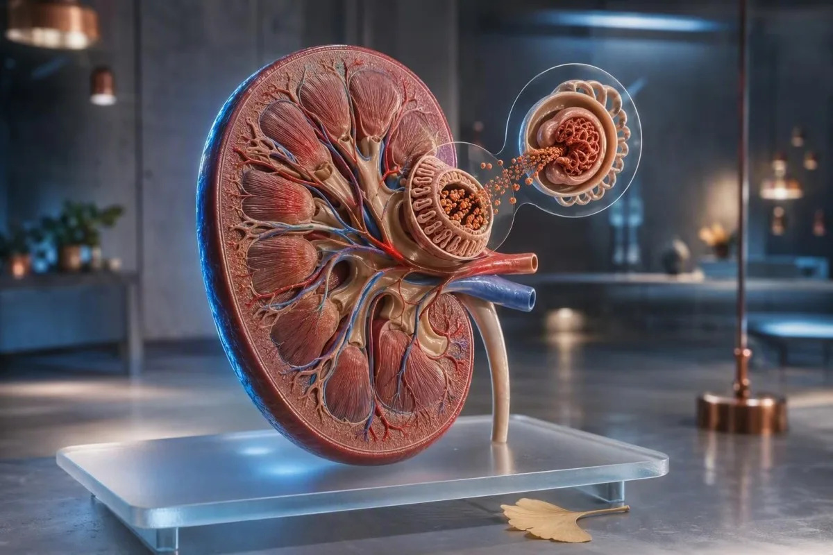
Muutama kupla nopean virtsasuihkun jälkeen on yleensä fysiikkaa. Vaahto, joka toistuvasti peittää kulhon, kestää huuhtelun jälkeen tai esiintyy turvotuksen kanssa, ansaitsee virtsan albumiinitestin arvuuttelun sijaan.


- Lyhyet kuplat jotka katoavat muutamassa sekunnissa heijastavat yleensä virtsan nopeutta, wc-puhdistusainejäämiä tai väkevää virtsaa munuaisvaurion sijaan.
- Pysyvä vaahtuisuuden virtsa on huolestuttavampi, kun tiheä valkoinen kerros pysyy 1–2 minuuttia toistuvasti.
- Virtsan ACR alle 3 mg/mmol (30 mg/g) on normaalia tai lievästi lisääntynyttä albumiinin eritystä useimmilla aikuisilla.
- Albuminuria 3–30 mg/mmol (30–300 mg/g) tulisi vahvistaa toistetuilla aamunäytteillä vähintään 3 kuukauden ajan.
- ACR yli 30 mg/mmol (300 mg/g) on vaikea lisääntynyt albuminuria ja vaatii nopeaa kliinistä arviointia.
- Hälytysmerkit sisältävät kasvojen tai jalkojen turvotusta, näkyvää verta, hengenahdistusta, vähentynyttä virtsaneritystä, kuumetta tai raskautta päänsäryn ja korkean verenpaineen kanssa.
- Näytteenottoon liittyvät seikat: Vältä raskasta liikuntaa 24 tuntia ennen näytteenottoa, käytä puhdasta keskivirtsanäytettä heti aamulla ja kerro lääkärille kuukautisista tai virtsatieinfektiosta.
- Verikokeet auttavat: kreatiniini, eGFR, seerumin albumiini, glukoosi ja HbA1c selvittävät, heijastavatko virtsan proteiinit munuaissairautta, diabetesta vai systeemistä sairautta.
Kuinka erottaa vaarattomat kuplat pysyvästä vaahtuisesta virtsasta
Vaahtoavan virtsan syyt vaihtelevat voimakkaasta suihkusta, joka osuu veteen, munuaisfilttereiden läpi tihkuvaan albumiiniin. Vaarattomat kuplat ovat suuria, kirkkaita ja häviävät nopeasti; huolestuttava vaahto on hienompaa, valkoista, toistuvaa ja kestää noin 1–2 minuuttia, vaikka astia olisi puhdas.
Pintajännitys selittää useimmat yksittäiset kuplat. Voimakas aamuvirtsasuihku, kuivuminen, väkevöityneet liuenneet aineet tai pesuainejäämät voivat vangita ilmaa pintajännitykseen ilman proteiinin menetystä; tämä on erityisen yleistä pitkän lennon, kuumeen tai kovan treenin jälkeen.
Kuviolla on enemmän merkitystä kuin yksittäisellä havainnolla. 15 vuoden kliinisessä käytännössäni pyydän potilaita huomaamaan vaahtoa kolmena eri päivänä käyttäen puhdasta, valkaisematonta astiaa; toistettava hienovaahtokerros on parempi syy testaukseen kuin dramaattisen näköinen episodi liikunnan jälkeen.
Vaahto itsessään ei voi diagnosoida proteinuriaa, koska fosfaatit, sappisuolat, siemenneste ja virtsateiden tulehdus voivat myös muuttaa pintajännitystä. Jos virtsa on vaahtoavan sijaan sameaa, erotusdiagnostiikka siirtyy kohti kiteitä, soluja tai infektiota; oppaamme samean virtsan vihjeet auttaa erottamaan nämä kuviot.
Yksinkertainen kotihavainnointisääntö
Älä valokuvaa WC-kulhoa tai yritä kotitekoisia “ravistustestejä”. Huomaa sen sijaan, esiintyykö vaahtoa puhtaassa astiassa vähintään 3 päivänä 7 päivästä, kestääkö se yli minuutin, ja esiintyykö turvotusta tai verenpaineen muutoksia. Nämä yksityiskohdat tekevät laboratoriotilauksesta paljon hyödyllisemmän.
Miksi proteiini virtsassa voi luoda kestävää vaahtoa
Vaahtoava virtsaproteiini on useimmiten albumiinia, negatiivisesti varautunutta proteiinia, joka normaalisti pysyy verenkierrossa. Kun munuaisten suodatin muuttuu läpäisevämmäksi, albumiini alentaa pintajännitystä ja voi muodostaa pieniä, stabiileja kuplia.
Terve aikuinen erittää yleensä alle 30 mg albumiinia päivässä, vaikka rutiininomainen liuskatesti ei ehkä havaitse matalatasoista albumiinin menetystä. Herkempi virtsan albumiini-kreatiniinisuhde eli ACR korjaa albumiinin virtsan konsentraation mukaan ja on ensisijainen seulontamenetelmä.
Albumiini ei ole ainoa merkittävä proteiini. Plasmasoluhäiriöiden kevyet ketjut, tiettyjen lääkkeiden jälkeiset tubulaariset proteiinit ja ohimenevät proteiinit kuumeen aikana voivat päästä suodattumaan liuskatestistä, joka on suunniteltu pääasiassa albumiinille; siksi negatiivinen liuskatesti ei ratkaise jokaista jatkuvan vaahdon tapausta.
Tohtori Thomas Klein tarkastelee virtsalöydöksiä yhdessä seerumin kreatiniinin, eGFR:n, albumiinin ja glukoosin kanssa sen sijaan, että hän kohtelee visuaalista oiretta diagnoosina. Meidän seerumin proteiiniohje selittää, miksi matala seerumin albumiinitulos ja kohonnut virtsan ACR yhdessä painavat enemmän kuin kumpikaan yksin.
Yleisiä munuaisiin liittymättömiä vaahtuisen virtsan syitä
Useimmilla ohimenevästi vaahtoavilla virtsan oireilla on muu kuin munuaissairaudesta johtuva selitys: väkevä virtsa, nopea virtsaaminen, viimeaikainen siemensyöksy, saippuajäämät, kuume tai rasittava liikunta. Nämä syyt yleensä korjaantuvat 24–48 tunnin kuluessa normaalin nesteytyksen ja toipumisen jälkeen.
Kuivuminen tekee virtsasta tummempaa ja väkevämpää, joten kuplia voi ilmestyä tiheämpinä, vaikka albumiinin eritys olisi normaalia. Tavoittele vaaleankeltaista virtsaa seuraavan päivän aikana sen sijaan, että juoisit liikaa nesteitä; useiden litrien juominen nopeasti voi itsessään alentaa natriumpitoisuutta alttiilla henkilöillä.
Liikunta voi ohimenevästi nostaa virtsan albumiinia. 10 kilometrin juoksukilpailu, korkeatehoinen voimaharjoitus tai pitkäkestoinen altistus kuumuudelle voi tuottaa proteiinia näytteessä, joka on otettu välittömästi sen jälkeen, minkä vuoksi laboratoriot suosittelevat yleensä voimakkaan liikunnan välttämistä 24 tuntia ennen ACR-testausta.
Siemenneste virtsaputkessa siemensyöksyn jälkeen voi tuottaa ohimeneviä kuplia ja heikosti positiivisen proteiiniliuskan. Näkyvästi vaaleanpunainen tai teevärinen virtsa muuttaa kysymyksen täysin ja se tulisi arvioida tämän oppaan avulla verta virtsassa, erityisesti 45 vuoden iän jälkeen tai jos henkilö on altistunut tupakoinnille.
Milloin pysyvä vaahto viittaa kliinisesti merkittävään proteiinin menetykseen
Jatkuva vaahtoaminen vaatii virtsan proteiinitestauksen, kun sitä esiintyy toistuvasti ja erityisesti yhdistettynä diabetekseen, korkeaan verenpaineeseen, turvotukseen tai alentuneeseen eGFR:ään. Albuminuria voi olla varhainen munuaisvaroitus, vaikka kreatiniini pysyisikin laboratoriomittausalueella.
KDIGO määrittelee ACR:n alle 3 mg/mmol tai 30 mg/g A1:ksi; 3–30 mg/mmol A2:ksi; ja yli 30 mg/mmol A3-albuminuriaksi. Luokat ennustavat munuais- ja sydän- ja verisuoniriskiä tarkimmin yhdistettynä eGFR:ään, eivätkä niitä käytetä yksinään (KDIGO, 2024).
Diabetes ja hypertensio ovat yleisimpiä albuminurian kroonisia aiheuttajia maailmanlaajuisesti, mutta ne eivät ole ainoita. Glomerulonefriitti, lihavuuteen liittyvä glomerulopatia, lupus, pre-eklampsia, uniapnea ja tietyt tulehduskipulääkkeet voivat myös edistää; jatkuvat löydökset vaativat kliinikon selvittämään mekanismin.
Kantesti on Tekoälyinen verikoeanalysaattori joka sijoittaa kreatiniinin, eGFR:n, seerumin albumiinin, glukoosin ja HbA1c:n yhteen pitkittäiseen kontekstiin. Se ei voi mitata virtsan ACR:ää veripaneelista, mutta se voi auttaa tunnistamaan aineenvaihdunnallisen tai munuaismallin, joka tekee virtsatuloksesta kiireellisemmän.
Mitkä virtsan proteiinitestit vastaavat parhaiten kysymykseen
Aamuvirtsan kertanäyte-ACR on paras ensisijainen testi epäillylle albumiinin menetykselle useimmilla aikuisilla. Proteiini-kreatiniinisuhde (PCR) on hyödyllinen, kun epäillään muuta kuin albumiiniproteiinia tai kun kokonaisproteiinin määrä on määritettävä.
ACR ilmoitetaan mg/mmol Isossa-Britanniassa ja monissa muissa maissa tai mg/g Yhdysvalloissa. ACR 3 mg/mmol vastaa noin 30 mg/g; tämä muunnos on tärkeä, koska potilaat joskus sekoittavat eri yksiköt suureksi tulosmuutokseksi.
Dipstick-proteiinitulos 'trace' tai 1+ on seulontavihje, ei diagnoosi. Emäksinen virtsa, väkevä virtsa ja kontaminaatio voivat aiheuttaa harhaanjohtavia positiivisia tuloksia, kun taas laimea virtsa ja ei-albumiiniproteiinit voivat antaa vääristä varmuutta; kvantitatiivinen testaus ratkaisee tämän epävarmuuden.
24 tunnin keräystä käytetään nyt valikoiduissa tilanteissa, kuten erittäin suuressa proteiinin menetyksessä, raskauden arvioinnissa tai ristiriitaisissa kertanäytteissä. Keräys on helppo alimitoittaa, joten lukekaa käytännön ohjeemme 24 tunnin virtsankeräyksen opas ennen sen aloittamista.
Kuinka kerätä virtsan proteiinitestaus tarkasti
Tarkka virtsan proteiinitestaus alkaa puhtaalla aamuvirtsan keskivirta-näytteellä, joka kerätään 24 tunnin jälkeen ilman raskasta liikuntaa. Ensimmäinen virtsa heräämisen jälkeen on suositeltava, koska asento, nesteytys ja päivittäinen aktiivisuus vaikuttavat vähemmän albumiinin eritykseen.
Pese kädet, käytä mukana toimitettua steriiliä astiaa, aloita virtsaaminen wc-pönttöön ja ota sitten keskivaihe koskematta astian sisäpuoleen tai kanteen. Toimita näyte nopeasti; jos viivästys on väistämätön, noudata laboratorion jäähdytysohjeita sen sijaan, että jättäisit sen lämpimään autoon.
Siirrä ei-kiireellistä testausta oireellisen virtsatieinfektion, kuumeen, ripulin, kuukautisten aikana tai 24 tunnin sisällä voimakkaan liikunnan jälkeen. Nämä tilat voivat väliaikaisesti lisätä albumiinin eritystä, kun taas kuukautiskontaminaatio voi myös aiheuttaa näennäistä verta ja proteiinia; leukosyyttiesteraasin tuloksiin voi selventää mahdollista infektio-mallia.
Ortostaattinen proteinuria ansaitsee maininnan, erityisesti teini-ikäisillä ja nuorilla aikuisilla. Proteiini, joka ilmenee päivävirtsassa, mutta puuttuu aamuvirtsanäytteestä, on usein hyvänlaatuinen, vaikka kliinikon tulisi vahvistaa malli olettamisen sijaan.
Kuinka tulkita ACR- ja proteiini-kreatiniini-tulokset
ACR alle 3 mg/mmol on yleensä rauhoittava, kun taas vahvistettu ACR 3 mg/mmol tai korkeampi vaatii riskipohjaista seurantaa. Yksi kohonnut näyte ei vahvista kroonista munuaissairautta, koska liikunta, infektio ja glukoosipiikit voivat aiheuttaa lyhytkestoista albuminuriaa.
Diabetesta sairastamattomilla aikuisilla uudelleen kohonnut ACR toistetaan yleensä aamuvirtsanäytteellä viikkojen kuluessa. Krooninen munuaissairaus vaatii epänormaaliutta vähintään 3 kuukauden ajan, ellei ole selkeää akuuttia munuaisvauriota tai merkittävästi kohonnutta proteiinia, joka vaatii nopeampaa tarkistusta (KDIGO, 2024).
ACR yli 70 mg/mmol, noin 700 mg/g, edellyttää yleensä kiireellistä erikoislääkärikeskustelua, vaikka eGFR olisikin säilynyt, erityisesti jos virtsassa on verta. Nefroottisen alueen proteinuria on yleensä PCR yli 300 mg/mmol tai proteiinin erittyminen yli 3,5 g/päivä, johon liittyy usein turvotusta ja matalaa seerumin albumiinia.
Kantesti on AI-laboratoriotestien tulkintapalvelu joka voi järjestää munuaisverimerkinnät eri päivämäärien yli ja merkitä laskevan eGFR-trendin. Trendin tulkinta tukee, mutta ei koskaan korvaa, kliinikon virtsamikroskopiaa, ACR-vahvistusta ja lääkityksen tarkistusta.
Verikokeet, jotka tekevät vaahtuisen virtsan tuloksista merkityksellisiä
Kreatiniini, eGFR, seerumin albumiini, glukoosi, HbA1c, elektrolyytit ja lipidit auttavat selittämään, heijastaako virtsan proteiini munuaisten suodatusvauriota vai laajempaa aineenvaihduntahäiriötä. Normaali kreatiniini ei sulje pois varhaista albuminuriaa.
eGFR lasketaan kreatiniinista ja se laskee monilla diabeetikoilla lievempää albuminavuotoa myöhemmin. eGFR alle 60 mL/min/1.73 m² 3 kuukauden tai pidemmän ajan ajan täyttää yhden kroonisen munuaissairauden kriteerin, mutta arvoja on tulkittava iän, lihasmassan ja äskettäisen sairauden mukaan.
Alhainen seerumin albumiini alle noin 35 g/L yhdessä runsaan virtsaproteiinin ja uusien nilkkaturvotusten kanssa herättää huolta merkittävästä munuaisten proteiinitappiosta. Päinvastoin, korkea albumiini tumman virtsan kanssa heijastaa useammin dehydraatiota; meidän eGFR-arvon ohjetta kuivumisen jälkeen selittää, miksi uusintatestaus toipumisen jälkeen voi olla järkevää.
American Diabetes Association suosittelee vähintään vuosittaisia UACR- ja eGFR-testejä tyypin 2 diabeteksessa ja tyypin 1 diabeteksessa, joka on kestänyt 5 vuotta tai enemmän (ADA Professional Practice Committee, 2024). HbA1c kertoo keskimääräisestä glykemiasta, mutta äkillinen glukoosin nousu voi silti ohimenevästi lisätä virtsan albumiinia ennen kuin HbA1c muuttuu.
Lääkkeet, lisäravinteet ja sairaudet, jotka voivat vaikuttaa virtsan proteiiniin
Tulehduskipulääkkeet, litium, jotkin syöpähoidot ja vakavat infektiot voivat muuttaa munuaisten suodatusta tai tubulaarista proteiininkäsittelyä. Älä lopeta määrättyä lääkitystä vain siksi, että virtsa näyttää vaahtoiselta; pyydä virtsa- ja munuaisten verikokeiden tarkistusta.
Tulehduskipulääkkeet, kuten ibuprofeeni ja naprokseeni, voivat vähentää munuaisten verenkiertoa, erityisesti dehydraation, sydämen vajaatoiminnan tai olemassa olevan CKD:n aikana. Riski kasvaa, kun NSAID yhdistetään diureetin ja ACE-estäjän tai ARB:n kanssa, yhdistelmä, jota lääkärit joskus kutsuvat nimellä “triple whammy”.”
SGLT2-lääkkeet voivat aiheuttaa pienen varhaisen eGFR-laskun, mutta yleensä vähentävät albuminuriaa ajan myötä asianmukaisesti valituilla potilailla. Tämä odotettu hemodynaaminen muutos eroaa etenevästä vauriosta; katso eGFR-muutokset SGLT2-hoidolla käytännön eroista.
Suuret annokset C-vitamiinia voivat häiritä joitakin virtsan dipstick-testejä, kun taas kreatiini voi vaikeuttaa kreatiniinin tulkintaa vahingoittamatta välttämättä munuaisia. Tuo tapaamiseen täydellinen luettelo reseptilääkkeistä, käsikauppalääkkeistä, proteiinijauheista ja yrttivalmisteista – nimet ja annokset ovat tärkeitä.
Diabetes ja verenpaine: tuottoisimmat riskitarkistukset
Diabetes ja verenpainetauti tekevät jatkuvasta vaahtoisuudesta kliinisesti merkittävämpää, koska molemmat voivat aiheuttaa albuminuriaa vuosia ennen oireiden ilmenemistä. Verenpaine 140/90 mmHg tai enemmän ja ACR 3 mg/mmol tai enemmän tulisi käynnistää jäsennelty ensisijainen hoitotarkistus.
Monilla CKD- ja albuminuriapotilailla lääkärit pyrkivät standardoituun systoliseen verenpaineeseen alle 120 mmHg:iin, kun se on siedetty, vaikka tavoitteet on individualisoitava iän, kaatumisriskin, raskauden ja mittausmenetelmän mukaan. Väärin tehdyt kotimittaukset eivät saa yksinään johtaa lääkityksen muutoksiin.
Albuminuria on myös verisuoniriskin merkki. Syy siihen, että olemme huolissamme ACR:n noususta ja korkeasta verenpaineesta, on se, että yhdessä ne viittaavat endoteelin ja munuaisten glomerulusten stressiin, kun taas jompikumpi löydös kerran akuutin sairauden aikana voi olla ohimenevä; meidän verenpainelääkitysopas hahmottelee laajemman työselostuksen.
Kantesti on AI-biomarkkerien tulkinta-alusta käytetään 127+ maassa glukoosin, HbA1c:n, munuaisten markkereiden ja lipiditrendien vertailuun. Jatkuvalle vaahtoiselle virtsalle relevantti seuraava testi on edelleen laboratoriovirtsan ACR, mieluiten tarkistettuna saman kliinisen tiimin kanssa, joka hoitaa verenpainetta tai diabetesta.
Vaahtuisen virtsan raskauden aikana ja lapsilla noudatetaan eri sääntöjä
Vaahtoinen virtsa raskauden aikana vaatii samanaikaista arviointia, jos se esiintyy päänsäryn, näkömuutosten, ylävatsakivun, äkillisen turvotuksen tai kohonneen verenpaineen kanssa. Proteiinin erittyminen yli 20 viikon jälkeen voi olla osa pre-eklampsiaa, mutta pelkkä ulkonäkö ei koskaan diagnosoi.
Raskauden aikana proteiini-kreatiniinisuhde 30 mg/mmol tai enemmän, tai 300 mg proteiinia 24 tunnissa, tukee merkittävää proteiinuriaa asianmukaisessa kliinisessä tilanteessa. Normaalin näköinen virtsanäyte ei sulje pois pre-eklampsiaa, jos verenpaine on 140/90 mmHg tai korkeampi ja siihen liittyy huolestuttavia oireita.
Lapsilla on usein ohimenevää proteiinuriaa kuumeen, urheilun tai dehydraation jälkeen. Jatkuva proteiini kolmessa näytteessä, korkea verenpaine, turvotus tai veri virtsassa vaatii lastenlääkärin arvioinnin; ensimmäisen aamun testaus on erityisen hyödyllistä postuuraalisen proteiinurian poissulkemisessa nuorilla.
Raskaus muuttaa suodatusta, joten kreatiniiniarvot, jotka näyttävät “normaaleilta” ei-raskaana olevilla aikuisilla, eivät välttämättä ole rauhoittavia. Oppaamme raskauden GFR-arvot antaa kontekstia, mutta kiireelliset oireet tulee ohjata suoraan äitiyspoliklinikkaan eikä verkkointerpretatioon.
Varoitusmerkit: milloin vaahtuisen virtsan hoito vaatii kiireellistä lääketieteellistä apua
Hakeudu kiireellisesti lääkärin arviointiin, jos vaahtoiselle virtsalle ilmenee hengenahdistusta, nopeasti lisääntyvää jalka- tai kasvojen turvotusta, merkittävästi vähentynyttä virtsaneritystä, näkyvää verta, kuumetta kylkikivun kanssa tai raskauden varoitusmerkkejä. Nämä yhdistelmät voivat viitata merkittävään munuaisiin, sydämeen, infektioon tai raskauteen liittyvään sairauteen.
Uusi kasvojen turvotus herätessä, kengät kiristävät illalla ja painonnousu yli 2 kg 2-3 päivässä voivat heijastaa nesteen kertymistä. Kun nämä oireet liittyvät jatkuvaan vaahtoon, lääkärit tarkistavat nopeasti virtsan proteiinin, kreatiniinin, albumiinin, verenpaineen ja joskus rintakehän löydökset.
Teen värinen virtsa intensiivisen liikunnan jälkeen, johon liittyy voimakasta lihaskipua, ei ole tyypillinen hyvänlaatuinen vaahto; se voi viitata myoglobiinin vapautumiseen ja vaatii kiireellistä veri- ja virtsatutkimusta. Sama koskee koksin väristä virtsaa, jonka määrä on vähentynyt, mikä voi heijastaa munuaisten suodattimien verenvuotoa tai lihasvauriota.
Tohtori Thomas Klein neuvoo potilaita olemaan odottamatta kotona tehtyä liuskatestiä, kun hälytysmerkkejä on läsnä. Liikuntaan liittyvistä väsimuutoksista artikkelimme juoksijan hematuriasta selittää hyvänlaatuisen tilanteen ja piirteet, jotka eivät ole hyvänlaatuisia.
Mitä lääkärit tekevät positiivisen virtsan proteiinituloksen jälkeen
Positiivisen ACR- tai PCR-tuloksen jälkeen lääkärit yleensä vahvistavat tuloksen, mittaavat verenpaineen ja eGFR:n, tutkivat virtsan sedimentin ja tarkistavat diabeteksen, lääkitykset, autoimmuunioireet ja sukuhistorian. Seuraava vaihe riippuu määrästä, pysyvyydestä ja mukana olevasta verestä tai vähentyneestä suodatuksesta.
Virtsamikroskopia voi tunnistaa punasoluja, valkosoluja ja hyytymiä, joita pelkkä proteiinimäärä ei voi selittää. Punsoluhyytymät tai dysmorfiset punasolut viittaavat munuaisten suodattimen lähteeseen ja nopeuttavat nefrologin lähetettä; yksinkertaisia hyaliinihyytymiä voi esiintyä kuivumisen tai liikunnan jälkeen, kuten käsitellään hyaliinihyytymiä koskevassa oppaassamme.
Jos albuminuria vahvistetaan, hoito kohdistuu yleensä ajuriin: verenpaineen hallintaan, glukoosin hallintaan, painonhallinnan ja tupakoinnin lopettamisen tukeen sekä munuaisia suojaaviin lääkkeisiin, jos se on tarpeen. Munuaisten ultraääni, immunologiset testit, seerumin elektroforeesi tai munuaisbiopsia ovat valikoivia testejä – eivät automaattisia seurauksia yhdestä 1+ liuskatestistä.
Kantesti:n kliinistä metodologiaa tarkastellaan strukturoitujen turvallisuusstandardien mukaisesti, ja lukijat voivat tarkastella lääketieteellinen validointilähestymistapamme. Tehtävämme on tehdä olemassa olevista verituloksista helpommin keskusteltavia, ei diagnosoida munuaisten suodattimen häiriötä kuvan tai yhden laboratoriotuloksen perusteella.
Käytännön seitsemän päivän suunnitelma pysyvään vaahtoon
Jatkuvalle vaahtoavalle virtsalle ilman hälytysmerkkejä järjestetään perusterveydenhuollon virtsan ACR ja virtsatutkimus 1-2 viikon kuluessa, mieluiten käyttäen aamunäytettä. Kirjaa ylös verenpaine, nesteytys, liikunta, lääkitykset ja kestääkö vaahto yli 1 minuutin seitsemän päivän ajan.
Päivä 1-3: juo normaalisti, vältä intensiivistä liikuntaa ja tarkkaile vain puhtaassa WC-kulhossa ilman lisättyä pesuainetta. Älä yritä “huuhtoa pois” ongelmaa liiallisella vedellä; tavoitteena on edustava näyte, ei keinotekoisesti laimennettu.
Päivä 4-7: pyydä virtsatutkimus ja ACR, ja kysy, ovatko kreatiniini, eGFR, kalium, seerumin albumiini, glukoosi ja HbA1c sopivia olosuhteisiisi. Tuo aiemmat tulokset mukanaan, koska muutos ACR 2:sta 8 mg/mmol:iin merkitsee enemmän kuin kumpikaan luku ilman kontekstia; pitkittäisellä tulosseurannalla voi auttaa järjestämään päivämääriä.
Syyskuun 4. päivästä 2026 alkaen järkevä viesti pysyy rauhoittavana mutta tiukkana: useimmat erilliset kuplat ovat vaarattomia, kun taas toistuva hieno vaahto ansaitsee kvantitatiivisen virtsatestin. Jos tarvitset apua kysymysten valmistelussa lääkärille, lääketieteellistä neuvontatiimiämme määrittää koulutussisältömme taustalla olevat kliiniset standardit.
Usein kysytyt kysymykset
Onko vaahtoava virtsa aina merkki proteiinista virtsassa?
Ei, vaahtoava virtsa ei aina ole merkki proteiinin menetyksestä. Nopea virtsasuihku, nestehukka, wc-puhdistusainejäämät, kuume, liikunta ja siemenneste voivat aiheuttaa kuplia, jotka katoavat muutamassa sekunnissa. Jatkuva, hieno valkoinen vaahto, joka toistuu useita päiviä, viittaa vahvemmin proteinuriaan ja se tulisi tarkistaa virtsan albumiini-kreatiniini-suhteella (ACR). ACR 3 mg/mmol, joka vastaa 30 mg/g, tai korkeampi arvo katsotaan riittävän epänormaaliksi vaatimaan kliinistä kontekstia ja usein toistuvaa testausta.
Kuinka kauan virtsassa oleva vaahto voi kestää, ennen kuin minun pitäisi huolestua?
Vaahto, joka jää tiiviinä kerroksena noin 1–2 minuutiksi toistuvasti, on huolestuttavampi kuin suuret kuplat, jotka katoavat nopeasti. Kesto ei ole diagnostinen testi, koska wc-kemikaalit ja virtsan pitoisuus voivat muuttaa kuplien pysyvyyttä. Jos kuvio toistuu 3 päivänä tai useammin, järjestä virtsan analyysi ja virtsan ACR 1–2 viikon kuluessa. Hakeudu päivystykseen nopeammin, jos vaahtoa ilmenee yhdessä turvotuksen, näkyvän veren, hengenahdistuksen, kuumeen tai virtsanerityksen vähenemisen kanssa.
Mitä virtsan proteiinitestiä minun pitäisi pyytää?
Paras ensitesti useimmille aikuisille on aamuvirtsan albumiini-kreatiniinisuhde, jota kutsutaan nimellä ACR tai UACR. ACR alle 3 mg/mmol, eli 30 mg/g, on yleensä normaali tai lievästi lisääntynyt, kun taas 3–30 mg/mmol osoittaa kohtalaisesti lisääntynyttä albuminuriaa. Proteiini-kreatiniinisuhde voidaan lisätä, kun epäillään kokonaisproteiinin tai ei-albumiiniproteiinin menetystä. Rutiininomainen liuskatesti on hyödyllinen seulontatesti, mutta se on vähemmän tarkka kuin ACR vähäisen albumiinin menetyksen havaitsemisessa.
Voiko nestehukka aiheuttaa vaahtoavaa virtsaa?
Kyllä, kuivuminen voi tehdä virtsasta väkevämpää ja aiheuttaa enemmän näkyviä kuplia tai vaahtoa ilman jatkuvaa proteiinikatoa munuaisista. Se liittyy usein tummempaan kellertävään virtsaan, janoon, altistumiseen kuumuudelle, oksenteluun, ripuliin tai pitkään juomatta olemiseen. Palaa normaaliin nesteiden nauttimiseen 24–48 tuntia ennen ei-kiireellistä testiä, mutta vältä liiallista juomista pelkästään tuloksen laimentamiseksi. Jos vaahto säilyy nesteytyksen jälkeen, pyydä virtsan ACR-testausta.
Voiko liikunta aiheuttaa proteiinia ja vaahtoa virtsassa?
Voimakas liikunta voi aiheuttaa väliaikaista albuminuriaa ja vaahtoavaa virtsaa, erityisesti juoksun, raskaan vastusharjoittelun, kestävyyskilpailujen tai kuumassa tapahtuvaan harjoittelun jälkeen. Vaikutus yleensä häviää 24–48 tunnissa, minkä vuoksi ACR-näyte tulisi mieluiten ottaa 24 tunnin kevyen harjoittelun jälkeen. Pitkittynyttä proteinuriaa aamuvirtsanäytteessä toipumisen jälkeen ei todennäköisesti selitä pelkkä liikunta. Tumma tai kolanvärinen virtsa, johon liittyy voimakasta lihaskipua, vaatii kiireellistä lääketieteellistä arviota.
Mikä ACR-taso on vaarallinen?
ACR yli 30 mg/mmol tai 300 mg/g on merkittävästi lisääntynyt albuminuria, joka vaatii välitöntä lääketieteellistä arviointia, erityisesti jos eGFR on alentunut tai virtsassa on verta. ACR yli 70 mg/mmol, noin 700 mg/g, käynnistää yleensä kiireellisen erikoislääkärin konsultaation, vaikka kreatiniiniarvo olisi normaali. Riski määräytyy ACR:n, eGFR:n, verenpaineen, diabeteksen tilan, oireiden ja toistettujen mittausten yhdistelmän perusteella. Yksittäinen kohonnut ACR tulisi yleensä varmistaa, ellei kliininen tilanne ole kiireellinen.
Hanki tekoälypohjainen verikoeanalyysi tänään
Liity yli 2 miljoonan käyttäjän joukkoon maailmanlaajuisesti, jotka luottavat Kantesti:hen saadakseen välittömän ja tarkan laboratoriotestianalyysin. Lataa verikoetuloksesi ja saat kattavan tulkinnan 15,000+-biomarkkereista sekunneissa.
📚 Viitatut tutkimusjulkaisut
Klein, T., Mitchell, S., & Weber, H. (2026). Kliinisen validoinnin viitekehys v2.0 (lääketieteellisen validoinnin sivu). Kantesti AI Medical Research.
Klein, T., Mitchell, S., & Weber, H. (2026). Tekoälyverikoeanalysoija: 2,5M analysoitua testiä | Maailman terveysraportti 2026. Kantesti AI Medical Research.
📖 Ulkoiset lääketieteelliset lähteet
⚕️ Lääketieteellinen vastuuvapauslauseke
Tämä artikkeli on tarkoitettu vain koulutustarkoituksiin eikä se muodosta lääketieteellistä neuvontaa. Ota aina yhteyttä pätevään terveydenhuollon ammattilaiseen diagnoosi- ja hoitopäätöksiä varten.
E-E-A-T-luottamussignaalit
Kokea
Lääkärin johtama kliininen arviointi laboratoriotulkinnan työnkuluista.
Asiantuntemus
Laboratoriolääketiede keskittyy siihen, miten biomarkkerit käyttäytyvät kliinisessä kontekstissa.
Auktoriteetti
Kirjoittanut tohtori Thomas Klein, tarkistanut tohtori Sarah Mitchell ja professori tohtori Hans Weber.
Luotettavuus
Näyttöön perustuva tulkinta selkeillä jatkopoluilla, jotka vähentävät hälytystä.