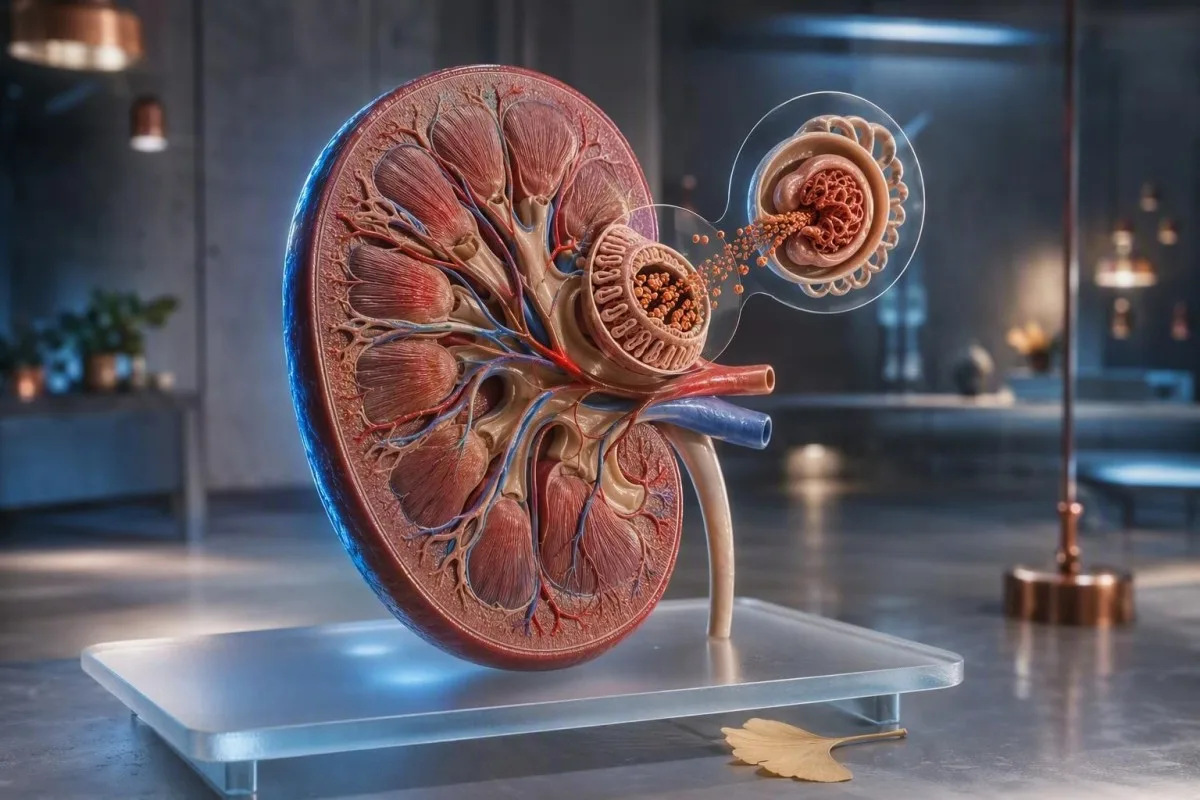
A gyors vizeletürítés utáni néhány buborék általában fizikai jelenség. Az az iszapos hab, amely ismételten beborítja a csészét, az öblítés után is megmarad, vagy duzzanattal jár együtt, vizeletfehérje-tesztet érdemel a találgatás helyett.


- Rövid buborékok amelyek másodperceken belül eltűnnek, általában a vizelet sebességét, a WC-tisztítószer maradványait vagy a koncentrált vizeletet tükrözik, nem pedig vesebetegséget.
- Tartós habos vizelet aggasztóbb, ha sűrű fehér réteg marad 1-2 percig ismétlődő alkalmakkor.
- Vizelet ACR 3 mg/mmol (30 mg/g) alatti érték normális vagy enyhén emelkedett albumin kiválasztást jelent a legtöbb felnőttnél.
- Albuminuria A 3-30 mg/mmol (30-300 mg/g) közötti értékeket legalább 3 hónapon keresztül ismételt kora reggeli mintákkal kell megerősíteni.
- ACR 30 mg/mmol felett (300 mg/g) súlyosan emelkedett albuminuria, és azonnali klinikai értékelést igényel.
- Vörös zászlók arc- vagy lábduzzanat, látható vér, légszomj, csökkent vizeletmennyiség, láz, vagy terhesség fejfájással és magas vérnyomással.
- A mintavétel szempontjai: kerülje a megerőltető testmozgást 24 órán keresztül, használjon tiszta középsugár vizeletmintát kora reggel, és tájékoztassa a kezelőorvost a menstruációról vagy húgyúti fertőzésről.
- Vérvizsgálatok segítenek: a kreatinin, eGFR, szérum albumin, glükóz és HbA1c tisztázzák, hogy a vizeletfehérje vesebetegséget, cukorbetegséget vagy szisztémás betegséget tükröz-e.
Hogyan különböztethetjük meg az ártalmatlan buborékokat a tartós habos vizelettől
Habos vizelet okai a gyors áramlástól a vese szűrőjén átfolyó albuminig terjednek. Az ártalmatlan buborékok nagyok, átlátszóak és gyorsan eltűnnek; a gondot okozó hab finomabb, fehér, ismétlődő, és körülbelül 1-2 percig megmarad tiszta tál ellenére is.
A felületi feszültség magyarázza a legtöbb egyszeri buborékot. Erős első reggeli vizeletsugár, kiszáradás, koncentrált oldott anyagok, vagy maradék tisztítószer levegőt zárhat be a víz felszínén fehérjeveszteség nélkül; ez különösen gyakori hosszan tartó repülőút, láz vagy megerőltető edzés után.
A minta többet számít, mint egyetlen megfigyelés. 15 éves klinikai gyakorlatom során arra kérem a betegeket, hogy három külön napon figyeljenek meg habot tiszta, fehérítetlen tálban; az ismételhető finom habréteg jobb ok a vizsgálatra, mint egy edzés utáni drámai megjelenésű epizód.
A hab önmagában nem diagnosztizálhatja a proteinuria-t, mert a foszfátok, epesavak, ondó és a húgyúti gyulladás is megváltoztathatja a felületi feszültséget. Ha a vizelet habos helyett zavaros, a különbségtételben a kristályok, sejtek vagy fertőzés játszik szerepet; útmutatónk a zavaros vizelet jelzéseihez segít ezeket a mintákat elkülöníteni.
Egy egyszerű otthoni megfigyelési szabály
Ne fényképezze le a WC-csészét, és ne végezzen otthoni “rázkódtató teszteket”. Ehelyett jegyezze fel, hogy a hab legalább 7 napból 3 napon jelentkezik-e tiszta tálban, tovább tart-e 1 percnél, és jelen van-e duzzanat vagy vérnyomásváltozás. Ezek a részletek sokkal hasznosabbá teszik a laboratóriumi kérést.
Miért okozhat stabil habot a fehérje a vizeletben
Habos vizeletfehérje leggyakrabban albumin, egy negatív töltésű fehérje, amely normálisan a véráramban marad. Amikor a glomeruláris szűrő áteresztőbbé válik, az albumin csökkenti a felületi feszültséget és stabil kis buborékokat hozhat létre.
Egy egészséges felnőtt általában naponta kevesebb mint 30 mg albumint választ ki, bár egy rutinszerű tesztcsík nem képes kimutatni az alacsony szintű albuminvesztést. Az érzékenyebb vizelet albumin-kreatinin arány, vagy ACR, korrigálja az albumint a vizelet koncentrációjára, és az elsődleges szűréshez ajánlott.
Az albumin nem az egyetlen fontos fehérje. A plazmasejt-rendellenességek könnyű láncai, bizonyos gyógyszerek utáni tubuláris fehérjék és láz alatti átmeneti fehérjék kijuthatnak egy főleg albuminra tervezett tesztcsíkról; ezért egy negatív tesztcsík nem oldja meg a tartós hab minden esetét.
Dr. Thomas Klein a vizeletleleteket a szérum kreatinin, eGFR, albumin és glükóz mellett vizsgálja, ahelyett, hogy a vizuális tünetet diagnózisként kezelné. A mi szérumfehérje-útmutatónk elmagyarázza, miért kap nagyobb súlyt az alacsony szérum albumin eredmény és a magas vizelet ACR együtt, mint bármelyik önmagában.
A tartósan habos vizelet gyakoribb, vesétől független okai
A legtöbb átmeneti habos vizelet nem vesebetegségre utaló magyarázattal rendelkezik: koncentrált vizelet, gyors vizelés, nemi együttlét, szappanmaradék, láz vagy megerőltető testmozgás. Ezek az okok általában 24-48 órán belül rendeződnek a normál hidratáció és a felépülés után.
A kiszáradás sötétebbé és koncentráltabbá teszi a vizeletet, így a buborékok sűrűbbnek tűnhetnek, még akkor is, ha az albumin kiválasztás normális. Törekedjen halványsárga vizeletre a következő nap folyamán, ahelyett, hogy túlzott folyadékot fogyasztana; több liter gyors italozása önmagában csökkentheti a nátriumszintet az arra hajlamos embereknél.
A testmozgás átmenetileg növelheti a vizelet albumin tartalmát. Egy 10 kilométeres verseny, egy magas intenzitású ellenállásos edzés vagy hosszan tartó hősugárzás fehérjét eredményezhet egy közvetlenül utána gyűjtött mintában, ezért a laboratóriumok általában azt tanácsolják, hogy az ACR tesztelés előtt 24 órával kerüljék a megerőltető testmozgást.
Az ejakuláció után a húgycsőben lévő ondó átmeneti buborékokat és enyhe pozitív fehérje tesztcsíkot eredményezhet. A látható rózsaszín vagy teaszínű vizelet teljesen megváltoztatja a kérdést, és ezt az útmutatót kell használni vért a vizeletben, különösen 45 éves kor után, vagy dohányzással terhelt személyeknél.
Mikor utal a tartós hab klinikailag releváns fehérjevesztésre
Az állandósult hab sürgősségi fehérjevizsgálatot indokol, ha ismételten jelentkezik, és különösen, ha cukorbetegséggel, magas vérnyomással, ödémával vagy csökkent eGFR-rel társul. Az albuminuria korai vesediagnosztikai jel lehet még akkor is, ha a kreatinin a laboratóriumi tartományon belül marad.
A KDIGO az ACR-t 3 mg/mmol (vagy 30 mg/g) alatt A1-nek, 3-30 mg/mmol között A2-nek, és 30 mg/mmol felett A3-nak definiálja. Ezek a kategóriák a legpontosabban jósolják meg a vesebetegség és a kardiovaszkuláris kockázatot az eGFR-rel párosítva, nem önmagukban használva (KDIGO, 2024).
A diabetes és a hipertónia a leggyakoribb krónikus okai az albuminuriának világszerte, de nem az egyetlenek. Glomerulonefritisz, obezitással összefüggő glomerulopátia, lupus, preeklampszia, alvási apnoé és bizonyos gyulladáscsökkentő gyógyszerek is hozzájárulhatnak; az állandósult leletek a mechanizmus tisztázását igénylik a klinikus részéről.
Kantesti egy AI vérvizsgálati analizátor ami a kreatinint, az eGFR-t, a szérum albumint, a glükózt és a HbA1c-t egy longitudinális kontextusba helyezi. Nem tud vizelet ACR-t mérni egy vérpanelből, de segíthet azonosítani azt a metabolikus vagy renális mintázatot, amely sürgősebbé teszi a vizeleteredményt.
Melyek a legjobb vizeletfehérje-tesztek a kérdés megválaszolására
Egy kora reggeli, spontán vizelet ACR a legjobb első vizsgálat a feltételezett albuminvesztés esetén a legtöbb felnőttnél. A fehérje-kreatinin arány (PCR) hasznos, ha nem albumin fehérjét gyanítanak, vagy ha a teljes fehérjét kell kvantifikálni.
Az ACR az Egyesült Királyságban és sok más országban mg/mmol, vagy az Egyesült Államokban mg/g egységben kerül jelentésre. Egy 3 mg/mmol ACR körülbelül 30 mg/g-nak felel meg; ez az átváltás fontos, mert a betegek néha a különböző egységeket nagy eredményváltozásnak tévesztik.
A csíkteszt fehérjeeredménye, ha "nyom" vagy "1+" azt jelzi, hogy szűrési gyanú merült fel, nem diagnózis. Az alkáli vizelet, a koncentrált vizelet és a kontamináció téves pozitív eredményeket okozhatnak, míg a híg vizelet és a nem albumin fehérjék hamis megnyugvást adhatnak; a kvantitatív vizsgálat megszünteti ezt a bizonytalanságot.
A 24 órás gyűjtést ma már kiválasztott esetekre tartják fenn, mint például nagyon magas fehérjevesztés, terhességi értékelés vagy ellentmondó spontán minták. Könnyű alulgyűjteni, ezért olvassa el a gyakorlati 24 órás vizeletgyűjtési útmutató útmutatót, mielőtt elkezdené.
Hogyan gyűjtsük a vizeletfehérje-tesztekhez szükséges mintát pontosan
A pontos vizeletfehérje-vizsgálat egy tiszta, kora reggeli, középsugaras mintával kezdődik, amelyet 24 óra nehéz testmozgás nélkül gyűjtöttek. Az ébredés utáni első vizeletet részesítik előnyben, mert a testtartás, a hidratáció és a nappali aktivitás kevésbé befolyásolja az albumin kiválasztódását.
Mossa meg a kezét, használja a mellékelt steril tartályt, kezdjen el vizelni a WC-be, majd fogja fel a középső részt anélkül, hogy hozzáérne a tartály belsejéhez vagy a fedeléhez. Szállítsa a mintát gyorsan; ha késés elkerülhetetlen, kövesse a laboratórium hűtési utasításait, ahelyett, hogy meleg autóban hagyná.
Halassza el a nem sürgős vizsgálatokat tünetekkel járó húgyúti fertőzés, láz, hasmenés, menstruáció vagy intenzív testmozgás után 24 órán belül. Ezek a körülmények átmenetileg növelhetik az albumin kiválasztódását, míg a menstruációs szennyeződés látható vért és fehérjét is eredményezhet; leukocyta-észteráz eredményekről szóló útmutatónkat tisztázhatja a lehetséges fertőzéses mintázatot.
Az ortosztatikus proteinuria említést érdemel, különösen tinédzserek és fiatal felnőttek esetében. A nappali vizeletben megjelenő, de a reggeli mintában hiányzó fehérje gyakran jóindulatú, bár a klinikusnak ezt a mintázatot meg kell erősítenie, ahelyett, hogy feltételezné.
Hogyan értelmezzük az ACR és a fehérje-kreatinin arány eredményeit
A 3 mg/mmol alatti ACR általában megnyugtató, míg a megerősített 3 mg/mmol vagy magasabb ACR kockázat alapú utánkövetést igényel. Egy emelkedett minta nem állapít meg krónikus vesebetegséget, mert a testmozgás, a fertőzés és a glükózszint emelkedése rövid életű albuminuriát okozhat.
Cukorbetegség nélküli felnőtteknél egy újonnan emelkedett ACR-t általában heteken belül megismételnek egy kora reggeli mintával. A krónikus vesebetegség legalább 3 hónapig fennálló abnormalitásokat igényel, kivéve, ha egyértelmű akut vesekárosodás vagy jelentősen emelkedett fehérjevesztés van jelen, amely gyorsabb értékelést igényel (KDIGO, 2024).
A 70 mg/mmol (körülbelül 700 mg/g) feletti ACR általában sürgős szakorvosi megbeszélést indokol, még akkor is, ha az eGFR megőrzött, különösen vérvizelés esetén. A nephrotikus tartományú proteinuria általában PCR > 300 mg/mmol vagy > 3,5 g/nap fehérjevesztés, gyakran ödémával és alacsony szérum albuminszinttel kísérve.
Kantesti egy AI laboratóriumi leletértelmező szolgáltatás ami a renális vérparamétereket dátumok szerint rendezheti és felhívhatja a figyelmet egy csökkenő eGFR tendenciára. A tendenciaértelmezés támogatja, de soha nem helyettesíti a klinikus vizelet mikroszkópiáját, az ACR megerősítését és a gyógyszeres felülvizsgálatot.
Milyen vérvizsgálatok teszik értelmessé a habos vizelet eredményeit
A kreatinin, az eGFR, a szérum albumin, a glükóz, a HbA1c, az elektrolitok és a lipidek segítenek megmagyarázni, hogy a vizeletfehérje veseszűrő sérülést vagy szélesebb metabolikus rendellenességet tükröz-e. A normál kreatinin nem zárja ki a korai albuminuriát.
Az eGFR-t a kreatininből számítják ki, és sok cukorbetegnél később jelentkezik, mint az enyhe albuminüria. A 60 mL/min/1,73 m² alatti eGFR 3 hónapon vagy hosszabb ideig a krónikus vesebetegség egyik kritériuma, de az értékeket az életkor, az izomtömeg és a közelmúltbeli betegségek figyelembevételével kell értelmezni.
Az alacsony, durván 35 g/L alatti szérum-albumin, jelentős vizeletfehérje-ürítéssel és új bokaödémával együtt, jelentős veserederedetű fehérjevesztésre utal. Ezzel szemben a magas albumin sötét vizelettel gyakrabban tükrözi a kiszáradást; a miénk kiszáradás utáni eGFR útmutatót magyarázza, miért lehet ésszerű a gyógyulás utáni újratesztelés.
Az American Diabetes Association évente legalább egyszeri UACR és eGFR vizsgálatot javasol 2-es típusú cukorbetegségben, valamint 5 éve vagy annál régebb óta fennálló 1-es típusú cukorbetegségben (ADA Professional Practice Committee, 2024). Az HbA1c az átlagos vércukorszintről tájékoztat, de az éles vércukorszint-emelkedés még az HbA1c eltolódása előtt is átmenetileg növelheti a vizeletfehérje-tartalmat.
Gyógyszerek, étrend-kiegészítők és betegségek, amelyek befolyásolhatják a vizeletfehérjét
Gyulladáscsökkentő fájdalomcsillapítók, lítium, egyes rákterápiák és súlyos fertőzések befolyásolhatják a vesefiltrációt vagy a tubuláris fehérje-anyagcserét. Ne hagyja abba a felírt gyógyszert csak azért, mert a vizelet habosnak tűnik; kérjen vizelet- és veseblódvizsgálatot.
Az olyan NSAID-ok, mint az ibuprofen és a naproxen, csökkenthetik a veseverőáramlást, különösen kiszáradás, szívelégtelenség vagy meglévő CKD esetén. A kockázat nő, ha egy NSAID-ot diuretikummal és ACE-gátlóval vagy ARB-vel kombinálnak, amit a klinikusok néha “tripla csapásnak” neveznek.”
Az SGLT2-inhibitorok kis kezdeti eGFR-csökkenést okozhatnak, de általában csökkentik az albuminuriát idővel a megfelelően kiválasztott betegeknél. Ez a várt hemodinamikai változás eltér a progresszív károsodástól; lásd az eGFR változásai SGLT2-kezelés mellett a gyakorlati megkülönböztetésekért.
A nagy dózisú C-vitamin befolyásolhatja egyes vizelet-tesztcsíkokat, míg a kreatin a kreatinin értelmezését nehezítheti anélkül, hogy feltétlenül károsítaná a veséket. Vigyen magával egy teljes listát a felírt gyógyszerekről, vény nélkül kapható fájdalomcsillapítókról, fehérje-porokról és gyógynövényekről az időpontra – a nevek és az adagok számítanak.
Cukorbetegség és vérnyomás: a legnagyobb hozadékú kockázati tényezők ellenőrzése
A cukorbetegség és a magas vérnyomás tartóssá teszi a habos vizeletet, mert mindkettő évekkel a tünetek megjelenése előtt okozhat albuminuriát. A 140/90 Hgmm-t elérő vagy meghaladó vérnyomás és a 3 mg/mmol-t elérő vagy meghaladó ACR strukturált alapellátási kivizsgálást tesz szükségessé.
Sok CKD-s és albuminuriás betegnél a klinikusok célja a standardizált irodai szisztolés vérnyomás 120 Hgmm alatti értéke, ha tolerálható, bár a célértékeket az életkor, az elesés kockázata, a terhesség és a mérési módszer figyelembevételével kell egyénre szabni. Az otthoni, helytelenül vett mérések önmagukban ne okozzanak gyógyszermódosítást.
Az albuminuria egyúttal érrendszeri kockázati marker is. Azért aggódunk az ACR-emelkedés és a magas vérnyomás miatt, mert együtt endotheliális és glomeruláris stresszre utalnak, míg egyik vagy másik lelet akut betegség alatt átmeneti lehet; a mi hipertónia laboratóriumi útmutató vázolja a tágabb kivizsgálást.
Kantesti egy AI biomarker-értelmező platform 127+ országban használják a glükóz, HbA1c, vesemarkerek és lipidszintek összehasonlítására. Tartós habos vizelet esetén a releváns következő vizsgálat továbbra is laboratóriumi vizelet ACR, amelyet ideális esetben ugyanazzal a klinikai csapattal vizsgálnak felül, amely a vérnyomás vagy a cukorbetegség kezelését végzi.
Terhesség és gyermekek esetében eltérő szabályok vonatkoznak a habos vizeletre
A terhesség alatti habos vizeletet azonos napon kell értékelni, ha fejfájással, látásváltozással, felső hasi fájdalommal, hirtelen ödémával vagy emelkedett vérnyomással jár. A 20. hét utáni proteinuria preeklampszia része lehet, de a megjelenés önmagában soha nem diagnosztikus.
Terhesség alatt a 30 mg/mmol vagy annál magasabb fehérje-kreatinin arány, vagy 24 óra alatt 300 mg fehérje jelentős proteinuriát támaszt alá a megfelelő klinikai kontextusban. Egy normál megjelenésű vizeletminta nem zárja ki a preeklampsziát, ha a vérnyomás 140/90 Hgmm vagy magasabb, és aggasztó tünetekkel jár.
A gyermekeknél gyakran jelentkezik átmeneti proteinuria láz, sport vagy kiszáradás után. Három mintánál tartósan jelen lévő fehérje, magas vérnyomás, ödéma vagy vér a vizeletben gyermekgyógyászati vizsgálatot indokol; az első reggeli vizeletvizsgálat különösen hasznos a posturalis proteinuria kizárásában serdülőknél.
A terhesség megváltoztatja a filtrációt, így a “normál”nak tűnő kreatininértékek nem terhes felnőtteknél nem feltétlenül megnyugtatóak. Útmutatónk a a terhesség alatti GFR-értékek kontextust ad, de az sürgős tünetekkel közvetlenül a szülészeti triázsra kell menni, nem online értelmezésre.
Vörös zászlók: mikor igényel sürgős orvosi ellátást a habos vizelet
Sürgős orvosi értékelést igényel a habos vizelet, ha légszomj, gyorsan növekvő láb- vagy arcödéma, jelentősen csökkent vizeletürítés, látható vér, láz oldalfájdalommal vagy terhességi figyelmeztető tünetek kísérik. Ezek a kombinációk jelentős vesebetegségre, szívbetegségre, fertőzésre vagy terhességgel kapcsolatos betegségre utalhatnak.
Az ébredéskor jelentkező új arcduzzanat, a nap végére szűknek érződő cipők és a 2-3 nap alatt több mint 2 kg-os súlygyarapodás a folyadék-visszatartás jelei lehetnek. Ha ezek a tünetek tartós habozással társulnak, a klinikusok haladéktalanul ellenőrzik a vizeletfehérjét, kreatinint, albumint, vérnyomást, és néha a mellkas vizsgálati leleteit is.
Az intenzív testmozgás utáni tea színű vizelet súlyos izomfájdalommal nem tipikus jóindulatú hab; mioglobin felszabadulásra utalhat, és sürgős vér- és vizeletvizsgálatot igényel. Ugyanez vonatkozik a csökkent vizeletürítéssel járó kólás színű vizeletre, amely glomeruláris vérzést vagy izomsérülést jelezhet.
Dr. Thomas Klein azt tanácsolja a betegeknek, hogy ne várjanak otthoni tesztcsík eredményére, ha vörös zászlók vannak jelen. A megerőltetéssel kapcsolatos színváltozásokkal kapcsolatban a cikkünk a futó hematuriájáról ismerteti a jóindulatú spektrumot és a nem jóindulatú jellemzőket.
Mit tesznek az orvosok a pozitív vizeletfehérje-eredmény után
Pozitív ACR vagy PCR után a klinikusok általában megerősítik az eredményt, mérik a vérnyomást és az eGFR-t, megvizsgálják a vizelet üledékét, és áttekintik a cukorbetegséget, a gyógyszereket, az autoimmun tüneteket és a családi kórtörténetet. A következő lépés a mennyiségtől, tartósságtól és a kísérő vérzéstől vagy csökkent szűrési funkciótól függ.
A vizelet mikroszkópia olyan vörösvértesteket, fehérvértesteket és hengereket képes azonosítani, amelyeket egyetlen fehérjeszám nem magyarázhat meg. Vörösvértest-hengerek vagy diszmorfiás vörösvértestek glomeruláris eredetre utalnak, és felgyorsítják a nefrológiai szakrendelésre utalást; egyszerű hialin hengerek dehidratáció vagy testmozgás után is előfordulhatnak, ahogy a hialin hengerek útmutatónkban.
tárgyaltuk. Ha az albuminuria megerősítést nyer, a kezelés általában a kiváltó okot célozza meg: vérnyomás-szabályozás, vércukor-kezelés, testsúly- és dohányzás-támogatás, és ahol indokolt, vese-védő gyógyszerek. Veseröntgen, immunológiai vizsgálat, szérum elektroforézis vagy vesebiopszia szelektív vizsgálatok – nem egy 1+-os tesztcsík automatikus következményei.
A Kantesti klinikai módszertanát strukturált biztonsági szabványokhoz viszonyítva vizsgáljuk felül, és az olvasók megtekinthetik a orvosi validációs megközelítésünk. A mi feladatunk a meglévő véreredmények könnyebbé tétele a megbeszéléshez, nem pedig egy glomeruláris rendellenesség diagnosztizálása kép vagy egyetlen laboratóriumi érték alapján.
Egy gyakorlatias, hétnapos terv a tartós habos vizeletre
Tartós, habos vizelet esetén, vörös zászlók nélkül, 1-2 héten belül kérjen elsődleges orvosi vizelet ACR és vizeletvizsgálatot, lehetőleg kora reggeli mintából. Jegyezze fel a vérnyomást, hidratáltságot, testmozgást, gyógyszereket, és azt, hogy a hab tovább tart-e, mint 1 perc, hét napon keresztül.
1-3. nap: hidratáljon normálisan, kerülje az intenzív testmozgást, és csak tiszta WC-kagylóban figyeljen, hozzáadott tisztítószer nélkül. Ne próbálja meg “elmossni” a problémát túlzott vízzel; a cél egy reprezentatív minta, nem pedig mesterségesen hígított.
4-7. nap: kérjen vizeletvizsgálatot és ACR-t, és kérdezze meg, hogy a kreatinin, eGFR, kálium, szérum albumin, glükóz és HbA1c megfelelő-e az Ön körülményeihez. Vigye el korábbi eredményeit, mert az ACR 2-ről 8 mg/mmol-ra történő változása több értelmet nyer, mint bármelyik szám kontextus nélkül; longitudinális eredménykövetésen keresztül vizsgálhatók segíthet a dátumok rendszerezésében.
2026. szeptember 4-től a józan üzenet megnyugtató, de határozott marad: a legtöbb elszigetelt buborék ártalmatlan, míg az ismétlődő finom hab kvantitatív vizeletvizsgálatot érdemel. Ha segítségre van szüksége a klinikusnak feltett kérdések előkészítéséhez, a orvosi tanácsadó csapatot oktatási tartalmunk mögötti klinikai szabványokat állítja fel.
Gyakran Ismételt Kérdések
A habos vizelet mindig fehérjét jelent a vizeletben?
Nem, a habos vizelet nem mindig a fehérjevesztés jele. A gyors vizeletsugár, a kiszáradás, a WC-tisztítószer-maradvány, a láz, a testmozgás és a sperma olyan buborékokat hozhat létre, amelyek másodperceken belül eltűnnek. Az állandó, finom fehér hab, amely több napon keresztül visszatér, jobban utalhat proteinuria fennállására, és vizelet albumin-kreatinin arány (ACR) vizsgálata javasolt. Az ACR 3 mg/mmol (30 mg/g) vagy magasabb értéke kórosnak tekinthető, amelyhez klinikai kontextus és gyakran ismételt vizsgálat szükséges.
Mennyi ideig tartson a hab a vizeletben, mielőtt aggódnom kellene?
Az a hab, amely ismételten nagyjából 1-2 percig sűrű rétegként marad, jobban aggodalomra ad okot, mint a gyorsan eltűnő nagy buborékok. Az időtartam nem diagnosztikai teszt, mert a WC-tisztító vegyszerek és a vizelet koncentrációja megváltoztathatja a buborékok stabilitását. Ha a minta 3 vagy több napon át ismétlődik, 1-2 héten belül rendeljen vizeletvizsgálatot és vizelet ACR-t. Kérjen sürgősségi ellátást hamarabb, ha a hab duzzanattal, látható vérrel, légszomjjal, lázzal vagy csökkent vizeletürítéssel jár együtt.
Milyen vizeletfehérje tesztet kérjek?
A legtöbb felnőtt számára az első vizsgálat kora reggeli vizelet albumin-kreatinin arányának meghatározása, amelyet ACR-nek vagy UACR-nek neveznek. A 3 mg/mmol (vagy 30 mg/g) alatti ACR általában normális vagy enyhén emelkedett, míg a 3-30 mg/mmol mérsékelten emelkedett albuminuriát jelez. Fehérje-kreatinin arány meghatározása akkor végezhető, ha teljes vagy nem albumin fehérjevesztésre gyanakszanak. A rutinszerű tesztcsík hasznos szűrésre, de alacsony szintű albuminvesztés kimutatásában kevésbé pontos, mint az ACR.
Okozhat-e a kiszáradás habos vizeletet?
Igen, a kiszáradás koncentráltabbá teheti a vizeletet, és láthatóbb buborékokat vagy habot okozhat, anélkül, hogy tartós fehérjeveszteség lenne a vesében. Gyakran sötétebb sárga vizelettel, szomjúsággal, hőséggel, hányással, hasmenéssel vagy hosszú ideig tartó folyadékbevitel hiányával jár együtt. A nem sürgős vizsgálat előtt 24-48 órával térjen vissza a szokásos folyadékbevitelhez, de kerülje a túlzott mennyiségű ivást csupán az eredmény hígítása érdekében. Ha a hab folyadékpótlás után is fennáll, kérjen vizelet ACR tesztet.
Okozhat-e a testmozgás fehérjét és habot a vizeletben?
Az erőteljes testmozgás átmeneti albuminuriát és habos vizeletet okozhat, különösen futás, nehéz ellenállásos edzés, állóképességi versenyek vagy hőségben végzett testmozgás után. A hatás általában 24-48 órán belül megszűnik, ezért az ACR mintát ideális esetben 24 órás, megerőltető testmozgástól való tartózkodás után kell gyűjteni. Az éjszakai, korai mintán jelentkező, regenerálódás utáni tartós proteinuria valószínűleg kevésbé magyarázható csak testmozgással. Sötét vagy kólás színű vizelet súlyos izomfájdalommal sürgős orvosi értékelést igényel.
Milyen ACR-szint veszélyes?
A 30 mg/mmol-nál (300 mg/g) magasabb ACR súlyosan emelkedett albuminuria, és sürgős orvosi értékelést igényel, különösen, ha az eGFR csökkent, vagy vér található a vizeletben. A 70 mg/mmol-nál (700 mg/g) magasabb ACR gyakran sürgős szakorvosi konzultációt tesz szükségessé, még akkor is, ha a kreatinin normális. A kockázatot az ACR, az eGFR, a vérnyomás, a cukorbetegség állapota, a tünetek és az ismételt mérések kombinációja határozza meg. Az egyszeri emelkedett ACR-t általában meg kell erősíteni, kivéve, ha a klinikai helyzet sürgős.
Végezzen mesterséges intelligencia által támogatott vérvizsgálat-elemzést még ma
Csatlakozzon világszerte több mint 2 millió felhasználóhoz, akik az Kantesti-t bízzák meg azonnali, pontos laborvizsgálat-elemzésért. Töltse fel a vérvizsgálat eredményeit, és kapjon átfogó értelmezést az 15,000+ biomarkerekről másodpercek alatt.
📚 Hivatkozott kutatási publikációk
Klein, T., Mitchell, S., & Weber, H. (2026). Klinikai validálási keretrendszer v2.0 (Orvosi validálási oldal). Kantesti mesterséges intelligencia orvosi kutatás.
Klein, T., Mitchell, S., & Weber, H. (2026). Mesterséges intelligencia vérvizsgálat-elemző: 2,5 millió elemzett vizsgálat | Globális egészségügyi jelentés 2026. Kantesti mesterséges intelligencia orvosi kutatás.
📖 Külső orvosi hivatkozások
⚕️ Orvosi nyilatkozat
A cikk kizárólag oktatási célokat szolgál, és nem minősül orvosi tanácsadásnak. A diagnózisra és a kezelési döntésekre vonatkozóan mindig konzultáljon szakképzett egészségügyi szolgáltatóval.
E-E-A-T bizalmi jelzések
Tapasztalat
Orvosok által vezetett klinikai áttekintés a laboratóriumi értelmezési munkafolyamatokról.
Szakértelem
Laboratóriumi medicina fókusz: hogyan viselkednek a biomarkerek klinikai környezetben.
Tekintélyesség
Dr. Thomas Klein írta, Dr. Sarah Mitchell és Prof. Dr. Hans Weber általi felülvizsgálattal.
Megbízhatóság
Bizonyítékokon alapuló értelmezés, világos követési útvonalakkal a riadalom csökkentésére.