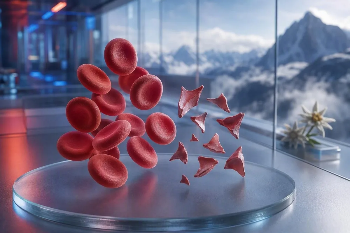
Schistotsüüdid on tükeldunud punased vererakud, mis võivad viidata ohtlikule väikeste veresoonte trombimisele, kuid üksnes määrdekommentaar ei ole diagnoos. Kiireloomulisus sõltub fragmendi protsendist, trombotsüütide arvust, hemoglobiini trendist, neerufunktsioonist, hemolüüsi markeritest ja sümptomitest.


- Schistotsüütide piirväärtus: Rohkem kui 1% schistotsüüti hästi tehtud täiskasvanu perifeersel määrdel on kahtlane trombootilise mikroangiopaatia suhtes, kui puuduvad muud punaliblede kuju muutused.
- Kiireloomuline klaster: Schistotsüüdid koos trombotsüütidega alla 150 × 10^9/L, langev hemoglobiin, tõusnud LDH ja madal haptoglobiin vajavad sama päeva jooksul arsti hinnangut.
- TTP vihje: Raske ADAMTS13 aktiivsus alla 10% toetab immuunset trombootilist trombotsütopeenilist purpurat, seisundit, mida ravitakse enne kinnitavate uuringute tulemuste saabumist, kui kliiniline kahtlus on suur.
- Mitte iga fragment pole ohtlik: Mehaanilised südameklapid, raske hüpertensioon, dialüüsiringid, põletused ja proovi kahjustus võivad tekitada tükeldunud punaseid vererakke.
- Määrd on käsitsi tehtud tõendus: Automaatne analüsaator ei suuda šistotsüüte usaldusväärselt diagnoosida; kogenud morfoloog peaks preparaati vaatama.
- Rasedus muudab panused suuremaks: Šistotsüüdid koos kõrge vererõhuga, valk uriinis või madalate trombotsüütidega pärast 20. rasedusnädalat vajavad kohest erakorralist sünnitusabi hindamist.
- Kordustestimine aitab: Värskelt kogutud EDTA proov ja korduv preparaat võivad eristada kogumisest tingitud artefakti tõelisest jätkuvast erütrotsüütide fragmenteerumisest.
- Ärge ravige ise: Raud, foolhape või toidulisandid ei ravi mikroangiopaatilist hemolüüsi ja võivad segada kiiret hindamist.
Kui tükeldunud punased vererakud vajavad kiiret arstlikku ülevaatust
Šistotsüüdid vajavad kiiret, samal päeval ülevaatust, kui need ilmuvad koos madala trombotsüütide arvu, aneemia, biokeemilise hemolüüsi, neerukahjustuse, neuroloogiliste sümptomite, palaviku või raske hüpertensiooniga. Pelgalt preparaadi märkus võib olla healoomuline või artefaktne; ohtlik leid on muster. Seisuga 7. august 2026 käsitleksin uusi šistotsüüte koos trombotsüütidega alla 100 × 10^9/L kui „helista arstile kohe” tulemust, mitte kui tavalist järelkontrolli.
Trombotsüütide langus on sageli kiireim kiireloomulisuse vihje. Hemoglobiin 92 g/L trombotsüütidega 68 × 10^9/L ja äsja teatatud šistotsüütidega on oluliselt murettekitavam kui 1% fragmendid hemoglobiiniga 145 g/L ja stabiilsed trombotsüüdid 260 × 10^9/L. Kantesti on AI-põhine vereanalüsaator, mis loeb selle klastriga seotud andmed üle täieliku verepildi (complete blood count) asemel, et tõsta morfoloogia kommentaari esile eraldi.
Minu 15-aastase kliinilise praktika jooksul on kiireloomulised otsused olnud patsientidel, kelle näitajad muutusid päevade jooksul: langev hemoglobiin, tõusev kreatiniin ja tõusev laktaatdehüdrogenaas ehk LDH. Dr. Thomas Kleini praktiline reegel on lihtne: segasus, uus nõrkus, valu rinnus, õhupuudus, väga vähe uriini või tugev peavalu koos selle mustriga tähendab erakorralist hindamist juba täna.
Šistotsüüdid ei ole põhjus oodata kordustesti, kui inimene on haige. Normaalne kaalium või normaalne leukotsüütide arv ei tühista trombootilise mikroangiopaatia riski. Patsiendid, kes püüavad mõista madalat näitajat, saavad vaadata ka meie juhendit madala punaste vereliblede sümptomite kohta, kuid ägedad sümptomid on alati tähtsamad kui veebipõhine tõlgendus.
Helistage kohe hädaabiteenistustele, kui
tekib uus ühepoolne nõrkus, minestus, krambihoog, tugev õhupuudus, rõhuv valu rinnus, mustad väljaheited või kiiresti süvenev segasus – need nõuavad hädaabiteenistusi, mitte sõnumit polikliinikusse. Need sümptomid võivad viidata organite haaratusele ning ükski šistotsüütide protsent ei välista seda ohutult.
Mida schistotsüüdid perifeersel määrdel tegelikult tähendavad
Šistotsüüdid on ebakorrapärased erütrotsüütide fragmendid, mis tekivad siis, kui vereringes olevad rakud lõigatakse füüsiliselt katki, kõige sagedamini fibriinikiudude või ebanormaalsete suure nihkejõuga pindade tõttu väikestes veresoontes. Neil on tavaliselt teravad nurgad, kiivrikujulised vormid või väikesed kolmnurksed kujundid ning puudub terve erütrotsüüdi keskne kahvatus.
Tõelised šistotsüüdid on morfoloogiline leid, mitte standardse CBC analüsaatori poolt toodetud väärtus. Rahvusvaheline hematoloogia standardiseerimise nõukogu (International Council for Standardization in Haematology) soovitab kvantifitseerimise korral loendada vähemalt 1000 punast vererakku optimaalses preparaadi tsoonis (Zini et al., 2021). See detail selgitab, miks üks labor võib raporteerida “harvad fragmendid”, samas kui teine raporteerib protsendi.
Klassikaline mure on mikroangiopaatiline hemolüütiline aneemia, kus punased vererakud puutuvad vereringes kokku trombotsüütide-rikaste mikro-trombid ja fibriiniga. Ometi võivad raske aordistenoos, mehaaniline klapp, kehavälised vereringeahelad ja ulatuslik termiline vigastus tekitada tõelisi fragmente teisel mehhanismil. Seetõttu ei ole küsimus “Kas fragmendid on reaalsed?” vaid “Mis põhjustab fragmentatsiooni?”
Kantesti on tehisintellektil põhinev vereanalüüsi tulemuste tõlgendamise platvorm, mis saab üleslaaditud täieliku vereanalüüsi (CBC) paigutada kõrvuti mida CBC sisaldab ja esile tõsta puuduvaid kaasnevaid tulemusi, nagu bilirubiin, retikulotsüüdid või haptoglobiin. Ta ei saa ise slaidi vaadata, mis jääb koolitatud laborispetsialisti tööks.
Kui palju schistotsütoosi on täiskasvanu määrdel murettekitav
Schistotsüütide arv üle 1% täiskasvanu perifeersel verepreparaadil on kahtlane trombootilise mikroangiopaatia suhtes, kui fragmentide osakaal on peamine punaliblede ebanormaalsus. Arvud alla 1% ei välista haigust, eriti kui preparaat tehti pärast vedelikke, transfusiooni või arenevat haigust.
1% piirväärtus on diagnostiline suunaja, mitte raskusastme skaala. ICSH juhised kasutavad tervete täiskasvanute ja täisaegsete vastsündinute puhul referentsvahemikku alla 1%; enneaegsetel imikutel võib olla kuni 5% fragmente ilma sama tähenduseta (Zini et al., 2021). Tulemus 2% koos raske trombotsütopeeniaga on murettekitavam kui 4% pärast mehaanilise vereringe toetusseadme paigaldamist.
Laborid võivad esitada “üksikuid”, “väheseid” või “1–2 kõrge suurendusega vaateväljas” protsendi asemel. Need väljendid ei ole omavahel asendatavad, sest vaatevälja suurus, määrde paksus ja raporteerimise poliitika võivad erineda. Küsimine, kas labor tegi ICSH-stiilis protsendiloenduse, on sageli kasulikum kui püüda ise kirjeldavat väljendit ümber arvutada.
Kõrge RDW ei mõõda schistotsüüte. RDW peegeldab rakkude suuruse varieeruvust ja võib tõusta retikulotsütoosi, rauavaeguse, transfusiooni või fragmentatsiooni korral. Meie põhjalik RDW ja erütrotsüütide indeksite juhend selgitab, miks kõrge RDW ei toeta üksi ei TTP-d ega artefakti.
Vereanalüüsi muster, mis toetab tõelist hemolüüsi
Tõeline punaliblede fragmentatsioon põhjustab tavaliselt kõrge LDH, madala haptoglobiini, suurenenud kaudse bilirubiini ja retikulotsüütide vastuse koos aneemiaga. Ükski üksik marker ei ole lõplik, sest maksahaigus, põletik, hiljutine vereülekanne ja luuüdihaigus võivad pilti moonutada.
LDH on tundlik, kuid mitte spetsiifiline. LDH üle 600 IU/L toetab sageli aktiivset hemolüüsi õiges kontekstis, kuid lihaskahjustus, maksakahjustus, infektsioon ja vähk võivad seda samuti tõsta. Haptoglobiin alla labori alumise piiri, sageli alla 0,3 g/L, lisab kaalu, sest seob vaba hemoglobiini, mis vabaneb plasmasse.
Retikulotsüütide arv peaks tavaliselt perifeerse hävimise korral tõusma, kuid see võib jääda madalaks foolhappepuuduse, rauapuuduse, neeruhaiguse või ägeda luuüdi supressiooni korral. See näiline vastuolu on lihtne märkamata jätta: patsiendil võib esineda tõeline hemolüüs ja samal ajal sobimatult madal retikulotsüütide vastus. haptoglobiini tulemuse juhend käsitleb levinud tõlgendamisalaseid lõkse.
Kantesti AI tõlgendab fragmenteerunud punaste vereliblede mustreid, võrreldes hemoglobiini, MCV-d, trombotsüüte, LDH-d, bilirubiini, kreatiniini ja retikulotsüüte samast ajapunktist. Hemoglobiini langus rohkem kui 20 g/L jooksul 48–72 tunni jooksul on kliiniliselt informatiivsem kui kergelt ebanormaalne väärtus ilma varasema võrdluseta.
Miks bilirubiin võib olla normaalne
Kaudne bilirubiin võib hemolüüsi alguses olla normaalne, pärast intravenoosse vedeliku lahjendust või siis, kui maks eemaldab bilirubiini tõhusalt. Seetõttu ei tohiks normaalne bilirubiin kõrvale jätta veenvaid fragmente, madalat haptoglobiini ja langevat hemoglobiini.
Miks TTP-d käsitletakse hematoloogia erakorralise seisundina
Trombootiline trombotsütopeeniline purpur, ehk TTP, on kahtluse all, kui schistotsüüdid esinevad koos trombotsütopeeniaga ja mikroangiopaatilise hemolüütilise aneemiaga ilma selgema alternatiivita. Ravimata immuunne TTP võib progresseeruda kiiresti, mistõttu võivad kliinikud alustada plasmafereesi ja immuunravi juba enne ADAMTS13 tulemuste saabumist.
ADAMTS13 aktiivsus alla 10% toetab TTP-d tugevalt, kuid tulemus võib võtta aega tundidest kuni päevadeni. ISTH diagnostiline juhis soovitab hinnata pretesti tõenäosust kliiniliste vahenditega, nagu PLASMIC skoor, samal ajal kui võetakse proov enne plasma ravi (Zheng et al., 2020). Skoor on sild tegevuseni, mitte asendus eriarsti otsusele.
Tuntud “pentad” palavikust, neuroloogilistest nähtudest, neerukahjustusest, trombotsütopeeniast ja hemolüütilisest aneemiast on paljudel tegelikel patsientidel puudulik. George ja Nester kirjeldasid trombootilist mikroangiopaatiat sündroomina, millel on kattuvad põhjused, mitte viie kohustusliku lahtrina (George & Nester, 2014). Praktikas on ohtlik vana harjumus oodata kõigi viie tunnuse saabumist.
Trombotsüüdid alla 30 × 10^9/L koos hemolüüsiga väärivad eriti kiiret arutelu hematoloogiaga. Trombotsüütide ülekannet tavaliselt välditakse kahtlustatud TTP korral, välja arvatud eluohtliku verejooksu korral või kui invasiivne protseduur ei saa oodata. Seotud uuringute jaoks meie hüübimistesti juhendist selgitab, miks PT ja aPTT võivad TTP korral olla normaalsed.
Muud ohtlikud põhjused, mida arstid schistotsüütide korral kaaluvad
Schistotsüüte esineb ka dissemineeritud intravaskulaarses koagulatsioonis, Shiga-toksiini hemolüütilises ureemilises sündroomis, raskes hüpertensioonis, rasedusega seotud mikroangiopaatias, teatud ravimite korral ning siirdamisega seotud trombootilises mikroangiopaatias. Seotud trombotsüütide, koagulatsiooni, neerude ja kliiniliste leidude kombinatsioon eristab neid seisundeid tavaliselt.
DIC kombineerib sageli fragmente koos pikenenud PT-ga, pikenenud aPTT-ga, madala või langeva fibrinogeeniga ning märkimisväärselt tõusnud D-dimeeriga. TTP-l on rutiinsed hüübimisajad sagedamini suhteliselt normaalsed. Fibrinogeen alla 1,5 g/L haige patsiendi korral nihutab muret tarbimiskoagulopaatia suunas, kuigi fibrinogeen võib alguses näida normaalne, sest see tõuseb ägeda põletiku ajal.
Raske hüpertensioon võib rõhu toimel lõhkuda punaseid vereliblesid, kui see kahjustab mikroveresoonkonda; näidud väärtusega 180/120 mmHg või üle selle koos peavalu, nägemise muutuse, rindkerevalu või neerufunktsiooni häirega on erakorraline olukord. Sellepärast kuulub vererõhu näit koos vereäigekese (smear) raportiga. Patsiendid võivad üle vaadata sekundaarse hüpertensiooni laboratoorsed vihjed pärast seda, kui vahetu oht on kõrvaldatud.
Kõhulahtisus, millele järgneb vähenenud uriinieritus, aneemia ja trombotsütopeenia, tekitab kahtluse hemolüütilise ureemilise sündroomi suhtes. Väljaheite testimine võib aidata tuvastada Shiga toksiini, kuid vedeliku staatus, kreatiniin, kaalium ja neuroloogiline hindamine peaksid enne kiiret abi andma juhised. Kõrge laktaat või ebastabiilsed elutähised nõuavad sepsise ja DIC-i hindamist, mitte „oota ja vaata“ lähenemist; vaata neid sepsise markerite mustrite kohta.
Tükeldunud punaste vererakkude mehaanilised põhjused
Mehaaniline destruktsioon võib tekitada tõelisi šistotsüüte ilma TTP-ta, eriti proteesiliste südameklappide, raske aordistenoosi, vatsakeste abiseadmete, dialüüsiringide või ekstrakorporaalse membraani oksügenatsiooni korral. Need põhjused väärivad endiselt meditsiinilist ülevaatust, kuid ravi sihib mehaanilist allikat, mitte immuunset TTP-d.
Mehaanilise klapi muster hõlmab sageli kroonilist kerget LDH tõusu ja madalat haptoglobiini stabiilsete trombotsüütide korral. Uus aneemia, hingeldus süvenemine, tume uriin või muutus klapi helis võivad viidata paravalvulaarsele lekkele või seadme talitlushäirele ning peaksid ajendama kardioloogia ülevaatust. Määrdumine annab vihje; ehhokardiograafia leiab põhjuse.
Sportlased küsivad mõnikord, kas maraton võib fragmente selgitada. Jalalöögist tingitud hemolüüs võib pärast pikka distantsi tekitada mööduvat vaba hemoglobiini ja madalat haptoglobiini, kuid veenva šistotsütoosi ei ole midagi, mida ma juhuslikult treeninguga seostaksin—eriti kui trombotsüüdid langevad. Võrdle seda laiemaga maratonijooksja laboratoorne juhend.
Rasked põletused ja suuremahuline veresoonte kirurgia võivad tekitada fragmenteerunud rakke füüsilise ja endoteeli kahjustuse kaudu. Nendes olukordades võib laboratoorne trend iga 6–24 tunni järel olla informatiivsem kui üksainus määrdumine. Pärast esile kutsunud sündmust stabiilne CBC on rahustav; pidevalt langev hemoglobiin ei ole.
Kuidas kogumis- ja preparaadiartefaktid matkivad schistotsüüte
Halb proovivõtt, määrdumise ettevalmistamise viibimine, alitäidetud antikoagulantide tuubid ja kahjustunud määrdumise serv võivad tekitada kujundeid, mis sarnanevad šistotsüütidega, ilma et see peegeldaks vereringe tasemel toimuvat hemolüüsi. Puhtalt kogutud ja monokihis üle vaadatud kordusproov on tavaliselt esimene kontroll, kui kliiniline pilt on rahulik.
Krenatsiooniga rakud, määrdumise serva moonutus, kuivamisartefakt ja kattuvad rakud on tavalised valenägud. Need ilmuvad sageli hajusalt või ainult sulgjast serva piirkonnas, mitte ühtlaselt ideaalses lugemistsoonis. Kogenud morfoloog eristab tõelise fragmendi teravat geomeetriat rakust, mis on slaidi ettevalmistamise käigus lihtsalt moonutatud.
Raskesti kogutud proov võib tuubi hemolüseerida ja valesti tõsta kaaliumi või LDH-d, kuid see ei tekita automaatselt tõelisi vereringes ringlevaid šistotsüüte. Kui tulemus sisaldab hemolüüsi indeksit või “proov on kahjustatud” märkust, koguvad kliinikud tulemuse tavaliselt uuesti enne haiguse diagnoosimist. Meie artikkel teemal kaaliumi võtmisega seotud vigade kohta selgitab, miks sama preanalüütiline probleem võib eksitada korraga mitut tulemust.
EDTA-st sõltuv trombotsüütide klompumine võib valesti alandada trombotsüütide arvu ja tekitada murettekitava välimusega kombinatsiooni. Tsitraattube trombotsüütide arv, mis on korrigeeritud lahjenduse suhtes, ja värske määrdumine võivad selle paljastada. Tähtis on nüanss: artefakt võib eksisteerida koos tõelise haigusega, seega ei tohiks kordustest sümptomaatilise inimese puhul kunagi hooldust edasi lükata.
Miks analüsaatori märgistused ja labori sõnastus võivad eksitada
Automaatne vereanalüsaator võib märgata ebanormaalseid erütrotsüütide populatsioone, kuid ta ei suuda iseseisvalt diagnoosida šistotsüüte piisava usaldusväärsusega, et välistada trombootiline mikroangiopaatia. Kliinilise küsimuse korral hemolüüs või TTP on vajalik jääda käsitsi perifeerse vere määrdumise ülevaatuse juurde.
Kommentaar “killustatud erütrotsüüdid esinevad” peaks tekitama küsimusi koguse, määrde kvaliteedi ja kaasneva morfoloogia kohta. Rauavaegusest, megaloblastilisest aneemiast või pärilikest erütrotsüütide häiretest tingitud väljendunud anisopoiküotsütoos muudab 1% reegli vähem spetsiifiliseks. Killud on kõige diagnostilisemalt kasulikud siis, kui need domineerivad ebanormaalses morfoloogias, mitte ei esine paljude omavahel mitteseotud kujundite hulgas.
Kantesti on tehisintellektil põhinev laboritulemuste tõlgendamise teenus, mis on loodud esile tooma vastuolulisi kombinatsioone, näiteks madal trombotsüütide arv koos kommentaariga, et trombotsüütide klombid esinevad. See ei asenda labori mikroskoopilist ülevaatust ega kliinikut, kes saab tellida kordusmäärde. Meie lähenemine kvaliteedikontrollile on kirjeldatud meditsiinilise valideerimise ülevaade.
Delta-check võrdleb praegust tulemust varasema tulemusega ja võib paljastada äkilise ebatõenäolise muutuse. Hemoglobiini langus 138 g/L-lt 87 g/L-ni 24 tunni jooksul on kliiniliselt oluline, kuid lahjendus intravenoossetest vedelikest või valesti märgistatud proov jääb siiski võimalikuks. Loe lisaks äkilistele laborimuutustele.
Mida teha pärast seda, kui vereanalüüsi tulemused mainivad schistotsüüte
Võta samal päeval ühendust arsti või teenistusega, kes uuringu tellis, kui šistotsüüte onäsäselt esile toodud, ning otsi viivitamatult erakorralist abi, kui sul on neuroloogilisi sümptomeid, õhupuudust, rindkerevalu, tume uriin, tugev peavalu, palavik või vähenenud uriinieritus. Ära tõlgenda määrde kommentaari iseenesest diagnoosina.
Hoia käepärast tegelik raport, sh hemoglobiin, trombotsüüdid, kreatiniin, bilirubiin, LDH ja kõik hemolüüsi indeksid. Arvesta ka hiljutise rasedusega, kõhulahtisusega kulgenud haigusega, väga kõrgete vererõhu näitudega, uute ravimitega, klapikirurgiaga, dialüüsiga, transfusiooniga ja autoimmuunhaigusega. Need detailid muudavad diferentsiaaldiagnoosi rohkem kui üksainus sõna morfoloogia väljal.
Ma näen patsiente kaotamas väärtuslikku aega, kui nad alustavad rauaravi, sest nad loevad “aneemia” enne seda, kui keegi on tuvastanud põhjuse. Rauavaegus võib esineda koos, kuid see ei selgita kiiret trombotsüütide langust koos fragmentidega. Dr. Thomas Klein soovitab teha foto kogu raportist, mitte ümber kirjutada ainult esile tõstetud numbreid; ümberkirjutamisel kaob sageli labori kommentaar, mis muudab mustri sidusaks.
Vältida aspiriini, ibuprofeeni ja ilma retseptita antikoagulante kuni kliiniku poolt on öeldud teisiti, kui trombotsüüdid on väga madalad või kui esineb verejooks. Ära katkesta määratud antikoagulanti ilma kiireloomulise meditsiinilise juhiseta. Väsimuse või pearinglusega inimesed saavad kasutada meie vereanalüüsid pearingluse jaoks juhend küsimuste ettevalmistamiseks pärast seda, kui kiireloomulised põhjused on välistatud.
Schistotsüüdid raseduse, vastsündinute ja laste korral
Raseduse ajal esinevad šistotsüüdid vajavad kiireloomulist hindamist, kui nendega kaasneb hüpertensioon, proteinuuria, kõrgenenud maksensüümid, madalad trombotsüüdid, peavalu, nägemissümptomid või ülakõhuvalu. HELLP-sündroom, preeklampsia, TTP, atüüpiline HUS ja DIC võivad kattuda ning ajastus sünnituse ümber aitab, kuid ei lahenda diagnoosi.
HELLP-sündroom on klassikaliselt määratletud hemolüüsi, kõrgenenud maksensüümide ja madalate trombotsüütide järgi, sageli pärast 20. rasedusnädalat või varsti pärast sünnitust. Trombotsüütide arv alla 100 × 10^9/L või AST üle 70 IU/L võib toetada muret, kuid kohalikud sünnitusabi meeskonnad tõlgendavad kogu kliinilist pilti. Ära oota selle kombinatsiooniga tavalist ambulatoorset sünnieelset vastuvõttu.
Täisaegsetel vastsündinutel võib olla vähem kui 1% šistotsüüte, samas kui enneaegsetel imikutel võib neid olla kuni 5%, sest vastsündinu erütrotsüütide morfoloogia on erinev (Zini jt, 2021). Lapsel, kellel on kõhulahtisus, kahvatus, vähenenud uriinieritus ja trombotsütopeenia, võivad fragmendid siiski viidata HUS-ile ning vajavad kohest pediaatrilist hindamist.
Trombotsüütide ja hemoglobiini referentsvahemikud muutuvad vanuse ja raseduse staadiumi järgi märkimisväärselt. Tulemuse märgistus “normaalne” üldise rakenduse poolt võib kontekstis siiski olla kliiniliselt murettekitav. Meie ressursid teemal raseduse trombotsüütide vahemikud ja samal päeval raseduse labori märguanded esitab kasulikku tausta, mitte ei asenda sünnitusabi triaaži.
Uuringud, mida kliinikud tellivad tõeliste schistotsüütide selgitamiseks
Esimene uuringute komplekt, kui kahtlustatakse mikroangiopaatilist hemolüüsi, hõlmab tavaliselt korduvat CBC-d ja verepreparaati, retikulotsüütide arvu, LDH-d, haptoglobiini, bilirubiini fraktsioone, kreatiniini, uriinianalüüsi, PT-d, aPTT-d, fibrinogeeni ja D-dimeeri. ADAMTS13 aktiivsuse ja inhibiitori uuringud tuleks võtta enne plasmafereesi, kui kahtlustatakse TTP-d, kuid ravi ei tohi oodata.
Otsene antikehade test, mida nimetatakse ka DAT-iks või Coombsi testiks, aitab eristada immuunset hemolüüsi mitteimmuunsest fragmenteerumisest. Positiivne DAT ei välista teist protsessi, kuid klassikaline TTP on tavaliselt DAT-negatiivne. Uriini mikroskoopia võib näidata valgu- või rakulisi silindreid, kui neeru mikroveresoonte kahjustus on aktiivne; vt meie selgitust granulaarsed uriinisilindrid.
Väljaheite Shiga-toksiini testimist peetakse asjakohaseks pärast sobivat seedetrakti haigust, samal ajal kui komplementtestid, ravimi ülevaade ja geneetilised või funktsionaalsed uuringud võivad olla kasutusel atüüpilise HUS-i korral. Järjekord varieerub riigiti ja haiglati. Kreatiniini tõus 26,5 µmol/L 48 tunni jooksul vastab ühele ägeda neerukahjustuse kriteeriumile ja väärib tähelepanu isegi enne, kui see muutub dramaatiliseks numbriks.
Retikulotsüütide produktsiooni indeks alla 2 olulise aneemia korral viitab ebapiisavale luuüdi vastusele, isegi kui toore retikulotsüütide protsent näib kõrge. See arvutus korrigeerib aneemia korral madalamat erütrotsüütide massi. The retikulotsüütide ja hematoloogia markerite juhend aitab selgitada, miks protsendid võivad olla eksitavad.
Millal on mõistlik määrret korrata ja millal mitte
Korduv verepreparaat 24 tunni jooksul on mõistlik, kui fragmente on vähe, patsient tunneb end hästi, trombotsüüdid ja hemoglobiin on stabiilsed ning proovi kvaliteedis on kahtlus. Kordus ei ole ohutu viivitus, kui esineb trombotsütopeenia, aktiivne hemolüüs, organikahjustus või murettekitavad sümptomid.
Hea kordusproov kogutakse värskelt õigesti täidetud EDTA-torusse ja tehakse kiiresti preparaat. Taotlus peaks konkreetselt paluma hinnata kahtlustatavaid šistotsüüte ja trombotsüütide klompumist, kui kumbki on võimalik. Labor võib võrrelda mõlemat preparaati kõrvuti, mis on palju kasulikum kui lihtsalt automatiseeritud CBC uuesti tegemine.
Oluline on trendi suund. Trombotsüütide arv, mis liigub 210-lt 165-le ja seejärel 98 × 10^9/L-le kolme päeva jooksul, on murettekitav, isegi kui kaks esimest näitu võivad jääda kohaliku referentsvahemiku piiridesse. Kantesti AI kasutab nende trajektooride esiletoomiseks pikisuunalist konteksti, põhimõte, mida käsitletakse meie vereanalüüsi trendianalüüsi juhendit.
Ülekanne võib ajutiselt lahjendada või varjata algset morfoloogiat. Kui saite teise proovi eel punaseid vererakke, öelge meeskonnale kuupäev ja ülekantud ühikute arv. Meie juhend väärtuste muutumisest pärast haiglast väljakirjutamist selgitab, miks postravi trendid vajavad teistsugust lähtepunkti.
Seisundid, mis võivad sarnaneda, olemata schistotsüütide haigus
Rauavaegus, vitamiin B12 vaegus, maksahaigus, pärilikud erütrotsüütide häired ja raske infektsioon võivad tekitada ebanormaalseid erütrotsüütide kujusid ilma trombootilise mikroangiopaatia spetsiifilise fragmenteerumismustriga. Verepreparaadi täielik morfoloogia ja kliiniline ajatelg väldivad fragmentide ülehindamist.
Raske vitamiin B12 vaegus võib põhjustada pseudo-trombootilist mikroangiopaatiat koos aneemia, kõrge LDH, madalate trombotsüütide ja aeg-ajalt esinevate fragmentidega. See põhjustab sageli väljendunud makrotsütoosi, madala retikulotsüütide vastuse ja LDH väärtused, mis võivad mõnikord olla üle 2 500 IU/L—need tunnused peaksid käivitama B12 ja metüülmaloonhappe testimise, mitte automaatse TTP ravi. Our B12 vs folaadi juhendis selgitab seda olulist jäljendajat.
Rauapuudus põhjustab tavaliselt mikrotsütoosi, hüpokroomiat, pliiatsilaadseid rakke ja kõrget RDW-d; fragmente võib rasketel juhtudel raporteerida, kuid need on harva domineerivaks tunnuseks. Ferritiin alla 15 µg/L toetab tugevalt rauavarude vähenemist muidu tüsistusteta täiskasvanul, kuigi põletik võib puudulikkust varjata, tõstes ferritiini.
Minu kogemuse põhjal on kõige rahustavam tulemus mitte ainult “puuduvad skistotsüüdid”, vaid järjepidev alternatiivne selgitus koos stabiilsete trombotsüütidega, stabiilse neerufunktsiooniga ja puuduvate biokeemiliste hemolüüsi tunnustega. Kui leiud jäävad lahknevaks, võib hematoloog vaadata tegelikku preparaati, mitte tugineda lõpparuande sõnastusele.
AI tõlgenduse kasutamine nii, et ei jääks vahele määrde erakorralist olukorda
AI saab aidata korraldada vereanalüüsi tulemusi ja tuvastada ohtlikke kombinatsioone, kuid ta ei saa kinnitada skistotsüüdi diagnoosi CBC PDF-ist ega asendada kiiret kliinilist hindamist. Perifeerne verepreparaat on visuaalne mikroskoopia ning algne laboratoorne klaasplaat jääb tõe allikaks.
Kantesti on tehisintellektil põhinev vereanalüüsi tööriist, mis kaardistab omavahel seotud tulemused üle aruannete, sh trombotsüütide dünaamika, hemoglobiini muutused, kreatiniin, bilirubiin ja LDH. See võib vähendada levinud viga, kui loetakse iga ebanormaalset markerit eraldi. Ta ei suuda näha, kas skistotsüüdiks nimetatud kuju on tõeline fragment, preparaadi serva artefakt või raporteerimise konventsioon.
Üleslaadimise kvaliteet on oluline, sest puudu jäänud ühik või komakoht võib tulemuse tähenduse ümber pöörata. Enne aruande jagamist kontrollige patsiendi identifikaatoreid, kogumise kuupäeva, ühikuid, võrdlusvahemikke ja seda, kas tulemus ütleb “hinnanguline”, “ootel” või “proov hemolüüsitud”. Meie AI aruande täpsuse kontrollnimekirja on selleks kasulik lõplikuks inimkontrolliks.
Kantesti kliinilisi meetodeid on üle vaadatud koos sisendiga meie Meditsiininõukogu, ning meie tehnoloogia on kirjeldatud AI tõlgendamise tehnoloogia juhend. Dr. Thomas Kleinina sooviksin ma siiski, et arst võtaks patsiendi puhul, kellel on uued fragmendid, trombotsüüdid alla 100 × 10^9/L ja sümptomid, võtaks laboriga otse ühendust—sest just siis on kiirus ja kliiniline läbivaatus kõige olulisemad.
Korduma kippuvad küsimused
Milline tase schistotsüütide puhul on ohtlik?
Rohkem kui 1% skistosüüte hoolikalt üle vaadatud täiskasvanu perifeersel verepreparaadil on kahtlane trombootilise mikroangiopaatia suhtes, kui fragmenteerunud rakud on peamine erütrotsüütide kõrvalekalle. Iga skistosüütide protsent muutub kiireloomuliseks, kui see esineb koos trombotsüütide arvuga alla 150 × 10^9/L, langeva hemoglobiinisisaldusega, kõrge LDH-ga, madala haptoglobiiniga, neerukahjustusega või neuroloogiliste sümptomitega. Tulemused alla 1% ei välista usaldusväärselt arenevat seisundit. Kliiniline muster määrab ohu rohkem kui üksnes protsent.
Kas šistotsüüte võib põhjustada halb vereproovide võtmine?
Raskesti kogutud proov, hilinenud ettevalmistus, ebapiisavalt täidetud EDTA-toru või halvasti valmistatud preparaat võib tekitada rakukujusid, mis matkivad skistotsüüte, kuigi see ei tekita tõelist ringlevat fragmenteerumist. Laborid saavad seda selgitada värskelt kogutud prooviga ja optimaalse määrdeala käsitsi ülevaatusega. Hemolüüsitud proov võib samuti valelikult suurendada kaaliumi ja LDH taset, mis võib muuta raporti näiliselt murettekitavamaks. Kordustestimine ei tohiks edasi lükata erakorralist hindamist, kui esinevad sümptomid, trombotsütopeenia või aneemia.
Kas šistotsüüdid tähendavad alati TTP-d?
Schistotsüüdid ei tähenda alati trombootilist trombotsütopeenilist purpurat ehk TTP-d. Neid võib esineda DIC-i (dissemineeritud intravaskulaarse koagulatsiooni), raske hüpertensiooni, Shiga-toksiini HUS-i, rasedusega seotud häirete, mehaaniliste südameklappide, dialüüsiringide ja ulatusliku koetrauma korral. TTP muutub suure prioriteediga mureks, kui schistotsüüdid esinevad koos trombotsütopeeniaga ja mitteimmuunse hemolüütilise aneemiaga, eriti kui ADAMTS13 aktiivsus on alla 10%. Kuna ADAMTS13 uuring võib võtta aega, nõuab kahtlustatav TTP kiiret erialaspetsialisti hindamist enne tulemuse saabumist.
Millised vereanalüüsid on kõrged šistotsüütse hemolüüsi korral?
Tõeline fragmentatsiooniline hemolüüs tõstab tavaliselt LDH-d ja kaudset bilirubiini, langetab haptoglobiini ning kutsub esile suurenenud retikulotsüütide arvu, samal ajal kui hemoglobiin langeb. LDH võib aktiivse hemolüüsi korral ületada 600 RÜ/L, kuid see ei ole spetsiifiline, sest lihaste, maksa ja muude kudede kahjustus tõstab seda samuti. Haptoglobiin alla ligikaudu 0,3 g/L toetab intravaskulaarset hemolüüsi, kui maksa süntees on piisav. Kreatiniin, trombotsüütide arv, PT, aPTT, fibrinogeen ja D-dimeer aitavad tuvastada aluseks oleva põhjuse.
Kas skistosüüte on raseduse ajal normaalne?
Schistotsüüte ei peeta raseduse tavapäraseks normaalseks leiuks, kui need esinevad koos hüpertensiooniga, proteinuuriaga, maksaensüümide tõusuga, langevate trombotsüütidega, peavaluga, nägemishäiretega või ülakõhuvaluga. See kombinatsioon võib viidata HELLP-sündroomile, preeklampsiale, TTP-le, atüüpilisele HUS-ile või DIC-ile ning nõuab kohest sünnitusabi hindamist. Trombotsüütide arv alla 100 × 10^9/L selles olukorras on eriti murettekitav. Rasedusele spetsiifilised võrdlusvahemikud ei muuda hemolüüsi mustrit ohutuks, mida võiks eirata.
Kas mehaaniline südameklapp võib põhjustada šistotsüüte?
Mehaanilised südameklapid ja raske aordistenoos võivad põhjustada tõelisi šistotsüüte, sest suure nihkejõuga verevool lõhub punaseid vereliblesid füüsiliselt. See põhjustab sageli kerget kroonilist hemolüüsi, mille korral on suurenenud LDH ja vähenenud haptoglobiin, samal ajal kui trombotsüütide arv püsib stabiilsena. Uus aneemia, tume uriin, õhupuudus või muutus klapi töös nõuab südame ülevaatust, sest lekkimine või seadme probleem võib olla põhjuseks. Leidu ei tohiks automaatselt märgistada TTP-ks, ilma et arvestataks südame ajalugu.
Hangi AI-toega vereanalüüsi analüüs juba täna
Liitu enam kui 2 miljoni kasutajaga üle maailma, kes usaldavad Kantesti-d kohese ja täpse laborianalüüsi jaoks. Laadi üles oma vereanalüüsi tulemused ja saad põhjaliku tõlgenduse 15,000+ biomarkerite kohta sekunditega.
📚 Viidatud teaduspublikatsioonid
Klein, T., Mitchell, S., & Weber, H. (2026). Kantesti vereanalüüsi tõlgendamise mootori automatiseeritud tehniline võrdlusuuring, mis põhineb eelregistreeritud rubriigil, 100 000 sünteetilise testjuhtumi peal. Kantesti AI Medical Research.
Klein, T., Mitchell, S., & Weber, H. (2026). Kliinilise valideerimise raamistik v2.0 (meditsiinilise valideerimise leht). Kantesti AI Medical Research.
📖 Välised meditsiinilised viited
Zheng XL jt. (2020). ISTH juhised trombootilise trombotsütopeenilise purpura diagnoosimiseks. Tromboosi ja hemostaasi ajakiri.
⚕️ Meditsiiniline lahtiütlus
Käesolev artikkel on mõeldud üksnes hariduslikel eesmärkidel ega kujuta endast meditsiinilist nõuannet. Diagnoosi ja ravivalikute otsuste tegemiseks konsulteeri alati kvalifitseeritud tervishoiutöötajaga.
E-E-A-T usaldussignaalid
Kogemus
Arsti juhitud kliiniline ülevaade labori tõlgendamise töövoogudest.
Ekspertiis
Laborimeditsiin keskendub sellele, kuidas biomarkerid käituvad kliinilises kontekstis.
Autoriteetsus
Kirjutanud dr Thomas Klein, ülevaade: dr Sarah Mitchell ja prof dr Hans Weber.
Usaldusväärsus
Tõenduspõhine tõlgendus selgete edasiste sammudega, et vähendada ärevust.