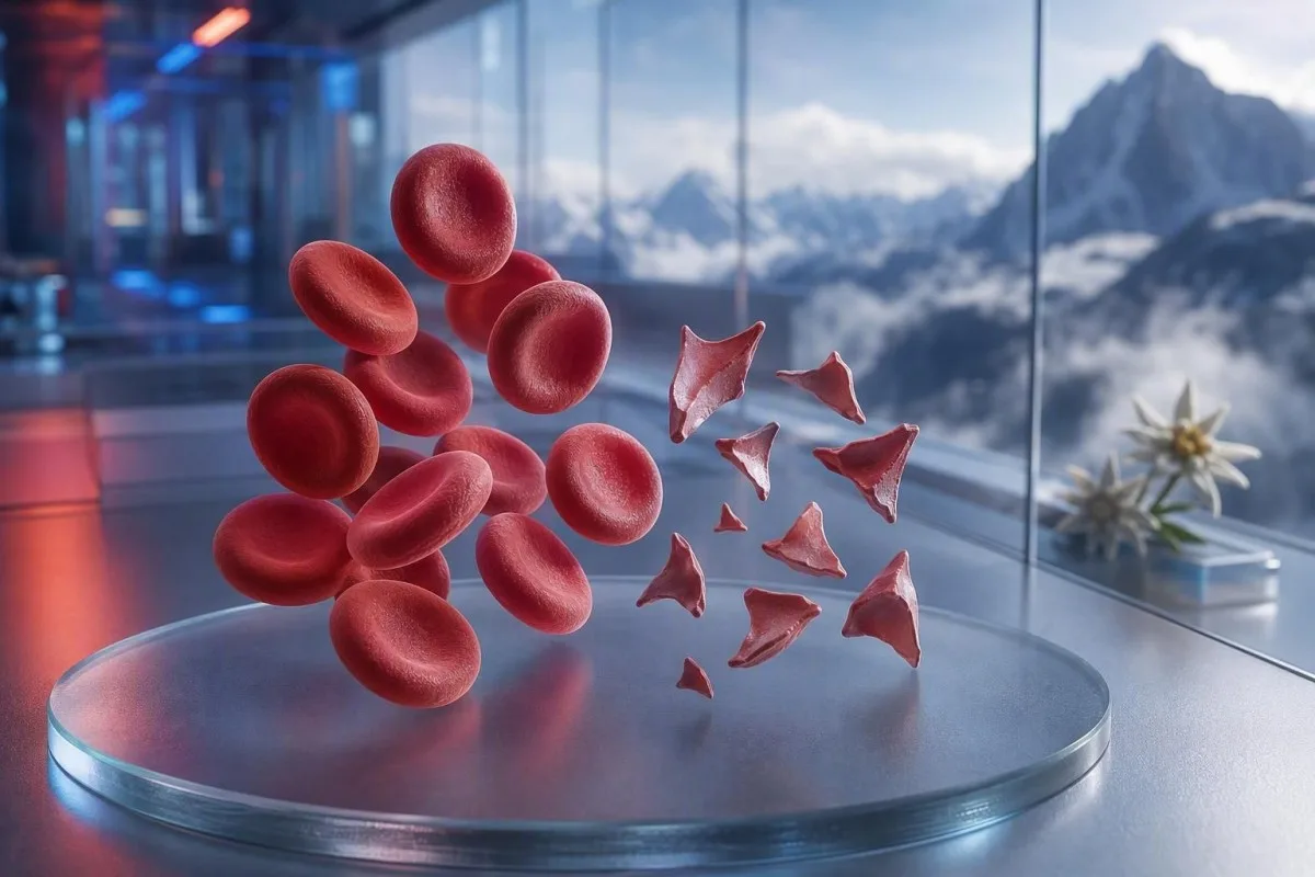
Skistocitoj estas fragmentitaj ruĝaj ĉeloj, kiuj povas signali danĝeran koaguliĝon en malgrandaj sangaj glasoj, sed ununura komento pri ŝmiraĵo ne estas diagnozo. Urĝeco dependas de la procento de fragmentoj, trombocitkalkulo, tendenco de hemoglobino, rena funkcio, hemolizaj signoj kaj simptomoj.


- Sojlo por skistocitoj: Pli ol 1% skistocitoj sur bone farita plenkreska periferia ŝmiraĵo estas suspektaj pri tromba mikroangiopatio kiam aliaj ŝanĝoj de formo de ruĝaj ĉeloj forestas.
- Urĝa areto: Skistocitoj kune kun trombocitoj sub 150 × 10^9/L, falanta hemoglobino, levita LDH, kaj malalta haptoglobino postulas takson de klinikisto en la sama tago.
- Indico por TTP: Severa ADAMTS13-agado sub 10% subtenas imunan trombotan trombocitopenian purpuron, kondiĉon traktatan antaŭ ol konfirmtestoj revenas kiam klinika suspekto estas alta.
- Ne ĉiu fragmento estas danĝera: Mekanikaj korvalvoj, severa hipertensio, dializaj cirkvitoj, brulvundoj, kaj damaĝo de la specimeno povas produkti fragmentitajn ruĝajn sangajn ĉelojn.
- Ŝmiraĵo estas manfarita pruvo: Automatiski analizatoroj flagas ne povas fidinde diagnozi skistocitojn; sperta morfologo devus inspekti la preparon.
- Gravedeco ŝanĝas la riskon: Skistocitoj kun alta sangopremo, proteino en urino, aŭ malaltaj trombocitoj post 20 semajnoj postulas tujan obstetrikan taksadon.
- Ripeta testado helpas: Freŝe kolektita EDTA-provaĵo kaj ripeta ŝmiraĵo povas apartigi kolektan artefakton de vera daŭra fragmentiĝo de ruĝaj sangĉeloj.
- Ne memtraktu: Fero, folato aŭ suplementoj ne traktas mikroangiopatian hemolizon kaj povas distri de urĝa taksado.
Kiam fragmentitaj ruĝaj ĉeloj bezonas urĝan medicinan revizion
Skistocitoj bezonas urĝan revizion en la sama tago kiam ili aperas kune kun malalta trombocitokalkulo, anemio, biokemia hemolizo, rena damaĝo, neŭrologiaj simptomoj, febro, aŭ severa hipertensio. Nur komento pri ŝmiraĵo povas esti bonkora aŭ artefakta; la danĝera trovo estas la ŝablono. Ĝis la 7-a de aŭgusto 2026, mi traktus novajn skistocitojn kune kun trombocitoj sub 100 × 10^9/L kiel rezulton “voku la klinikiston nun”, ne kiel rutinan sekvan kontrolon.
Falo de trombocitoj ofte estas la plej rapida indiko de urĝeco. Hemoglobino de 92 g/L kun trombocitoj de 68 × 10^9/L kaj ĵus raportitaj skistocitoj estas signife pli maltrankviliga ol 1% fragmentoj kun hemoglobino 145 g/L kaj stabilaj trombocitoj de 260 × 10^9/L. Kantesti estas AI-sanga testanalizilo, kiu legas ĉi tiun aron tra la kompleta sangokalkulado anstataŭ levi morfologian komenton izolite.
En miaj 15 jaroj da klinika praktiko, la alvokoj kiujn mi faras rapide estas por pacientoj kies rezultoj ŝanĝiĝis dum tagoj: falanta hemoglobino, kreskanta kreatinino, kaj kreskanta laktatdehidrogenazo, aŭ LDH. La praktika regulo de d-ro Thomas Klein estas simpla: konfuzo, nova malforteco, brusta doloro, manko de spiro, tre malmulta urino, aŭ severa kapdoloro kune kun ĉi tiu ŝablono signifas kriz-taksadon hodiaŭ.
Skistocitoj ne estas kialo atendi ripetan teston se la persono estas malsana. Normala kalio aŭ normala leŭkocitokalkulo ne nuligas la riskon de trombota mikroangiopatio. Pacientoj kiuj provas kompreni malaltan kalkulon ankaŭ povas revizii nian gvidilon al simptomoj de malaltaj ruĝaj sangĉeloj, sed akutaj simptomoj ĉiam superas interpreton rete.
Voku krizajn servojn nun por
Nova unuflanka malforteco, svenado, epilepsiatako, severa manko de spiro, premanta brusta malkomforto, nigraj fekoj, aŭ rapide plimalboniĝanta konfuzo postulas krizajn servojn prefere ol mesaĝon al kliniko. Tiuj simptomoj povas reflekti implikiĝon de organoj, kaj neniu skistocita procento sekure ekskludas tion.
Kion skistocitoj sur periferia ŝmiraĵo vere signifas
Skistocitoj estas neregulaj fragmentoj de ruĝaj sangĉeloj kreitaj kiam cirkulantaj ĉeloj estas fizike tonditaj, plej ofte de fibrinaj fadenoj aŭ nenormale alt-ŝeraĵaj surfacoj en malgrandaj vaskuloj. Ili kutime havas akrajn angulojn, formojn kiel kaskoj, aŭ malgrandajn triangulajn formojn kaj mankas la centra paleco de sendifekta ruĝa sangĉelo.
Veraj skistocitoj estas morfologia trovo, ne valoro produktita de la norma analizilo de CBC. La Internacia Konsilio por Normigado en Hematologio rekomendas kalkuli almenaŭ 1,000 ruĝajn ĉelojn en la optimuma zono de la ŝmiraĵo kiam kvantigo gravas (Zini et al., 2021). Tiu detalo klarigas kial unu laboratorio povas raporti “maloftajn fragmentojn” dum alia raportas procenton.
La klasika zorgo estas mikroangiopatia hemolitika anemio, kie ruĝaj ĉeloj renkontas trombocit-riĉajn mikro-trombojn kaj fibrinon en la cirkulado. Tamen severa aorta stenozо, mekanika valvo, eksterkoraj cirkvitoj, kaj grava termika vundo povas krei verajn fragmentojn per malsama mekanismo. La demando do ne estas “Ĉu fragmentoj estas realaj?” sed “Kio kaŭzas la fragmentiĝon?”
Kantesti estas AI-sanganaliza interpretada platformo, kiu povas meti alŝutitan kompletan sangokalkulon apud kion inkluzivas CBC kaj reliefigi mankantajn kunajn rezultojn kiel bilirubino, retikulocitoj, aŭ haptoglobino. Ĝi ne povas inspekti la preparon mem, kio restas la tasko de trejnita laboratoria profesiulo.
Kiom da skistocitozo estas koncerna ĉe plenkreskulo
Kalkulo de skistocitoj super 1% sur plenkreska periferia ŝmiraĵo estas suspektinda pri trombota mikroangiopatio kiam fragmentoj estas la domina ruĝĉela anomalio. Kalkuloj sub 1% ne ekskludas malsanon, precipe se la filmo estis farita post fluidoj, transfuzo, aŭ evoluanta malsano.
La 1%-limo estas diagnoza atentigo, ne severeca skalo. Gvidlinioj de ICSH uzas referencintervalon sub 1% por sanaj plenkreskuloj kaj plentempaj novnaskitoj; antaŭtempaj beboj povas havi ĝis 5% fragmentojn sen la sama implico (Zini et al., 2021). Rezulto de 2% kun severa trombocitopenio estas pli alarmiga ol 4% post enmeto de mekanika cirkula subtena aparato.
Laboratorioj povas raporti “okazajn,” “malmultajn,” aŭ “1–2 per alta-potenca kampo” anstataŭ procenton. Tiuj frazoj ne estas interŝanĝeblaj, ĉar kampograndeco, ŝmiraĵa dikeco, kaj raportada politiko varias. Demandi ĉu la laboratorio faris ICSH-stilan procentkalkulon ofte estas pli utila ol provi mem konverti priskriban frazon.
Alta RDW ne mezuras skistocitojn. RDW reflektas variecon en ĉela grandeco kaj povas pliiĝi kun retikulocitozo, fera manko, transfuzo, aŭ fragmentiĝo. Nia detala RDW kaj ruĝĉela indeksa gvidilo klarigas kial alta RDW subtenas nek TTP nek artefakton per si mem.
La ŝablono de sangotestoj kiu subtenas veran hemolizon
Vera ruĝĉela fragmentiĝo kutime produktas altan LDH, malaltan haptoglobino, pliigitan nerektan bilirubinon, kaj retikulocitan respondon kune kun anemio. Neniu ununura markilo estas definitiva, ĉar hepata malsano, inflamo, lastatempa transfuzo kaj malsano de la osta medolo povas distordi la bildon.
LDH estas sentema, sed ne specifa. LDH super 600 IU/L ofte subtenas aktivan hemolizon en la ĝusta kunteksto, tamen muskola vundo, hepata vundo, infekto kaj kancero ankaŭ povas ĝin altigi. Haptoglobino sub la malsupra limo de la laboratorio, ofte sub 0,3 g/L, pezigas la interpreton, ĉar ĝi ligas liberan hemoglobinon liberigitan en la plasmon.
Retikulocita kalkulo kutime devus pliiĝi dum detruo en la periferio, sed ĝi povas resti malalta ĉe manko de folato, manko de fero, rena malsano aŭ akuta subpremado de la osta medolo. Tiu ŝajna kontraŭdiro estas facile preteratentebla: paciento povas havi veran hemolizon kaj samtempe neadekvatan, malaltan retikulocitan respondon. La haptoglobina rezulto gvidas tra la oftaj interpretaj kaptiloj.
Kantesti AI interprets fragmentitajn ŝablonojn de ruĝaj sangaj ĉeloj komparante hemoglobinon, MCV, trombocitojn, LDH, bilirubinon, kreatininon kaj retikulocitojn de la sama tempopunkto. Faliĝanta hemoglobino je pli ol 20 g/L en 48 ĝis 72 horoj estas klinike pli informa ol iomete nenormala valoro sen antaŭa komparo.
Kial bilirubino povas esti normala
Indirekta bilirubino povas esti normala frue en hemolizo, post diluo per intravejna fluido, aŭ kiam la hepato forigas bilirubinon efike. Tial normala bilirubino ne devus superregi konvinkajn fragmentojn, malaltan haptoglobinon kaj falantan hemoglobinon.
Kial TTP estas traktata kiel hematologia kriz-situacio
Trombota trombocitopenia purpuro, aŭ TTP, estas suspektata kiam skistocitoj akompanas trombocitopenion kaj mikroangiopatian hemolizan anemion sen pli klara alternativo. Netrajtita imuna TTP povas rapide progresi, do klinikistoj povas komenci plasmoŝanĝon kaj imunoterapion antaŭ ol la rezultoj de ADAMTS13 revenas.
ADAMTS13-agado sub 10% forte subtenas TTP, sed la rezulto povas daŭri horojn aŭ tagojn. La diagnoza gvidlinio de ISTH konsilas taksi antaŭtestan verŝajnecon per klinikaj iloj kiel la PLASMIC-poentaro, dum oni prenas specimenon antaŭ plasmo-traktado (Zheng et al., 2020). Poentaro estas ponto al ago, ne anstataŭaĵo por specialista juĝo.
La konata “pentado” de febro, neŭrologiaj signoj, rena difekto, trombocitopenio kaj hemoliza anemio estas nekompleta ĉe multaj realaj pacientoj. George kaj Nester priskribis trombotan mikroangiopation kiel sindromon kun interkovrantaj kaŭzoj prefere ol kvin devigaj skatoloj (George & Nester, 2014). En praktiko, atendi ĉiujn kvin trajtojn estas danĝera malnova kutimo.
Trombocitoj sub 30 × 10^9/L kun hemolizo meritas precipe urĝan diskuton kun hematologio. Trombocita transfuzo estas kutime evitata en suspektata TTP, krom se estas vivdanĝera sangado aŭ enpenetra proceduro ne povas atendi. Por rilata testado, nia gvidilo pri koaguliĝaj testoj klarigas kial PT kaj aPTT povas esti normalaj en TTP.
Aliaj danĝeraj kaŭzoj de skistocitoj kiujn kuracistoj konsideras
Skistocitoj ankaŭ okazas en disvastigita intravaskula koaguliĝo, Shiga-toksina hemoliza uremia sindromo, severa hipertensio, graved-rilata mikroangiopatio, iuj medikamentoj, kaj transplant-rilata trombota mikroangiopatio. La asociitaj trombocitaj, koagulaj, renaj kaj klinikaj trovoj kutime distingas tiujn kondiĉojn.
DIC ofte kombinas fragmentojn kun plilongigita PT, plilongigita aPTT, malalta aŭ falanta fibrinogeno, kaj markite altigita D-dimero. TTP pli ofte havas relative normalajn rutinajn koagulajn tempojn. Fibrinogeno sub 1,5 g/L ĉe malsana paciento ŝanĝas la zorgon al konsuma koagulopatio, kvankam fibrinogeno komence povas aspekti normala ĉar ĝi pliiĝas dum akuta inflamo.
Severa hipertensio povas disŝiri ruĝajn sangajn ĉelojn kiam premo damaĝas la mikroangiojn; mezuroj je aŭ super 180/120 mmHg kun kapdoloro, ŝanĝo de vido, brusta doloro aŭ rena misfunkcio estas krizo. Tial sangoprema mezuro apartenas apud la raporto de la ŝmiraĵo. Pacientoj povas revizii sekundara hipertensio laboratoriaj indicoj post kiam tuja danĝero estis traktita.
Diareo sekvata de malpliigita urina eligo, anemio, kaj trombocitopenio levas suspekton pri hemolitika uremia sindromo. Feka testado povas helpi identigi Shiga-toksinon, sed la stato de hidratigo, kreatinino, kalio, kaj neŭrologia takso unue gvidas urĝan prizorgon. Alta laktato aŭ malstabilaj vivsignoj postulas taksadon por sepsa kaj DIC prefere ol atendema aliro; vidu ĉi tiujn sepsiso-signaj ŝablonoj.
Mekanikaj kaŭzoj de fragmentitaj ruĝaj sangaj ĉeloj
Mekanika detruo povas krei verajn ŝistocitojn sen TTP, precipe kun prostetaj korvalvoj, severa aorta stenozо, ventriklaj asistaj aparatoj, dializaj cirkvitoj, aŭ eksterkorpa membranoksigenado. Tiuj kaŭzoj ankoraŭ meritas medicinan revizion, sed la traktado celas la mekanikan fonton prefere ol imunecan TTP.
Mekanvalva ŝablono ofte inkluzivas kronikan mildan LDH-altigon kaj malaltan haptoglobinon kun stabilaj trombocitoj. Nova anemio, plimalboniĝanta manko de spiro, malhela urino, aŭ ŝanĝo en la valvsono povas sugesti paravalvulan likon aŭ misfunkcion de la aparato kaj devus instigi kardiologian revizion. La sangomakulo donas indicon; ekokardiografio trovas la fonton.
Atletoj foje demandas ĉu maratono povas klarigi fragmentojn. Piedfrapa hemolizo povas kaŭzi paseman liberan hemoglobinon kaj malaltan haptoglobinon post longdistanca kurado, sed konvinka ŝistocitozo ne estas io, kion mi facile atribuus al ekzercado—precipe se trombocitoj falas. Komparu ĝin kun la pli vasta maratonkuristo laboratorigvidilo.
Severaj brulvundoj kaj grava vaskula kirurgio povas produkti fragmentitajn ĉelojn per fizika kaj endotelia vundo. En tiuj situacioj, laboratorie sekvata tendenco ĉiun 6 ĝis 24 horojn povas esti pli informa ol unu sola sangomakulo. Stabila CBC post la komenca evento estas trankviliga; kontinue falanta hemoglobino ne estas.
Kiel kolektaj kaj vitraj (slide) artefaktoj imitas skistocitojn
Malbona specimen-kolektado, prokrastita preparado de la sangomakulo, neplene plenigitaj antikoagulantaj tuboj, kaj difektita rando de la makulo povas krei formojn kiuj similas ŝistocitojn sen reflekti hemolizon je cirkuladnivelo. Ripeta specimeno kolektita pure kaj reviziita en la monotavolo estas la kutima unua kontrolo kiam la klinika bildo estas trankvila.
Krenitaj ĉeloj, distordo de la rando de la sangomakulo, sekiĝa artefakto, kaj interkovrantaj ĉeloj estas oftaj falsaj imitaĵoj. Ili ofte aperas difuze aŭ nur ĉe la plumeca rando prefere ol egale en la ideala legadozono. Spertulo pri morfologio distingas la akran geometrion de vera fragmento de ĉelo kiu simple estis distordita dum preparado de la lumbildo.
Malfacila kolektado povas hemolizi la tubon kaj false altigi kalion aŭ LDH, sed ĝi ne aŭtomate produktas verajn cirkulantajn ŝistocitojn. Se rezulto inkluzivas hemoliz-indekson aŭ komenton “specimeno kompromitita”, klinikistoj kutime re-kolektas antaŭ diagnozi malsanon. Nia artikolo pri eraroj dum kalio-eltirado klarigas kial la sama antaŭanaliza problemo povas misgvidi plurajn rezultojn samtempe.
EDTA-dependa trombocita kungluiĝo povas false malaltigi la trombocitnombron kaj fabriki alarmaspektan kombinaĵon. Trombocita nombro en citrata tubo, korektita por diluo, kaj freŝa filmo povas tion malkovri. La nuanco gravas: artefakto povas kunekzisti kun vera malsano, do ripeta testo neniam devus prokrasti prizorgon por iu kiu estas simptoma.
Kial avertoj de analizilo kaj laboratoriaj vortigoj povas misgvidi
Aŭtomataj sangaj analiziloj povas marki nenormalajn populaciojn de eritrocitoj, sed ili ne povas sendepende diagnozi ŝistocitojn kun sufiĉa fidindeco por ekskludi tromban mikroangiopation. Mana revizio de periferia sangomakulo restas necesa kiam la klinika demando estas hemolizo aŭ TTP.
Komento “fragmentitaj eritrocitoj ĉeestantaj” devus instigi demandojn pri kvanto, ŝmirmemora kvalito kaj samtempa morfologio. Markita anizopoikilocitozo pro feromanko, megaloblasta anemio aŭ heredaj eritrocitaj malordoj igas la regulon 1% malpli specifa. Fragmentoj estas plej diagnoze utilaj kiam ili regas la nenormalan morfologion, prefere ol aperi inter multaj senrilataj formoj.
Kantesti estas AI-servo por interpreto de laboratoriaj testrezultoj, desegnita por marki malkongruajn kombinojn, kiel malalta trombocitokalkulo kune kun komento kiu diras trombocitajn klumpaĵojn. Ĝi ne anstataŭas la mikroskopan revizion de la laboratorio nek la klinikiston, kiu povas peti ripetan ŝmirmemoraĵon. Nia aliro al kontrolado de kvalito estas priskribita en la superrigardo pri medicina validigo.
Delta-kontrolo komparas la nunan rezulton kun antaŭa rezulto kaj povas elmontri subitan neplausiblan ŝanĝon. Falo de hemoglobino de 138 g/L al 87 g/L ene de 24 horoj estas klinike signifa, sed diluo pro intravejnaj fluidoj aŭ misetikeditaj specimenoj restas ebla. Legu pli pri subitaj laboratoriaj ŝanĝoj.
Kion fari post sangotestrezultoj kiuj mencias skistocitojn
Kontaktu la klinikiston aŭ servon kiu ordonis la teston la saman tagon se skistocitoj estas nove raportitaj, kaj serĉu urĝan medicinan prizorgon tuj se vi havas neŭrologiajn simptomojn, mankon de spiro, brustan doloron, malhelan urinon, severan kapdoloron, febron aŭ reduktitan urinproduktadon. Ne interpretu la ŝmirmemoran komenton kiel diagnozon per si mem.
Havu la realan raporton disponebla, inkluzive de hemoglobino, trombocitoj, kreatinino, bilirubino, LDH, kaj ajnan hemolizan indicon. Ankaŭ notu lastatempan gravedecon, diarean malsanon, severajn sangopremajn valorojn, novajn medikamentojn, valvan kirurgion, dializon, transfuzon, kaj aŭtoimunan malsanon. Tiuj detaloj ŝanĝas la diferencialon pli ol unu sola vorto en la morfologia kampo.
Mi vidas pacientojn perdi valoran tempon komencante feron ĉar ili legas “anemion” antaŭ ol iu identigas la kaŭzon. Feromanko povas kunesti, sed ĝi ne klarigas rapidan falon de trombocitoj kun fragmentoj. D-ro Thomas Klein rekomendas preni foton de la tuta raporto anstataŭ transskribi nur la elstarigitajn nombrojn; transskribo ofte preterlasas la laboratorian komenton kiu faras la ŝablonon kohera.
Evitu aspirinon, ibuprofenon, kaj nekonforme preskribajn antikoagulantojn ĝis klinikisto konsilas alie se trombocitoj estas tre malaltaj aŭ se sangado ĉeestas. Ne ĉesu preskribitan antikoagulanton sen urĝa medicina gvido, ankaŭ ne. Homoj kun laceco aŭ kapturniĝemo povas uzi nian sangotestoj por kapturniĝo gvidas por prepari demandojn post kiam urĝaj kaŭzoj estas ekskluditaj.
Skistocitoj dum gravedeco, ĉe novnaskitoj kaj ĉe infanoj
Skistocitoj dum gravedecon postulas urĝan taksadon kiam ili okazas kune kun hipertensio, proteinurio, altigitaj hepataj enzimoj, malaltaj trombocitoj, kapdoloro, vidaj simptomoj, aŭ doloro en la supra abdomeno. HELLP-sindromo, antaŭ-eklampsio, TTP, atipika HUS, kaj DIC povas interkovri, kaj la tempigo ĉirkaŭ akuŝo helpas sed ne solvas la diagnozon.
HELLP-sindromo estas klasike difinita per hemolizo, altigitaj hepataj enzimoj, kaj malaltaj trombocitoj, ofte post 20 semajnoj da gravedecon aŭ baldaŭ post akuŝo. Trombocitokalkulo sub 100 × 10^9/L aŭ AST super 70 IU/L povas subteni zorgon, sed lokaj obstetrikaj teamoj interpretas la tutan klinikan bildon. Ne atendu rutinan antaŭnaskan rendevuon kun ĉi tiu kombino.
Term-naskitaj novnaskitoj povas havi malpli ol 1% skistocitojn, dum antaŭtempaj beboj povas havi ĝis 5% ĉar la morfologio de novnaskitaj eritrocitoj estas malsama (Zini et al., 2021). Tamen, en infano kun diareo, paleco, malpliigita urino, kaj trombocitopenio, fragmentoj povas indiki HUS kaj postulas tujan pediatrian taksadon.
Referencaj intervaloj por trombocitoj kaj hemoglobino ŝanĝiĝas signife laŭ aĝo kaj gravedeja stadio. Rezulto markita “normala” de ĝenerala aplikaĵo ankoraŭ povas esti klinike maltrankviliga en kunteksto. Niaj rimedoj pri trombocitaj intervaloj dum gravedecon kaj samtagaj laboratoriaj flagoj dum gravedecon provizi utilan fonon, ne anstataŭaĵon por akuŝa triado.
Testoj kiujn klinikistoj ordonas por esplori verajn skistocitojn
La unua esploraro por suspektata mikroangiopata hemolizo kutime inkluzivas ripetan CBC kaj ŝmirmakulo, retikulocitan nombron, LDH, haptoglobinon, bilirubinajn frakciojn, kreatininon, urinanalizon, PT, aPTT, fibrinogenon kaj D-dimeron. ADAMTS13-agado kaj inhibitor-testado devas esti prenitaj antaŭ plasma interŝanĝo se oni suspektas TTP, sed kuracado ne devas atendi.
Rekta kontraŭglobulina testado, ankaŭ nomata DAT aŭ Coombs-testado, helpas distingi imun-hemolizon de ne-imuna fragmentiĝo. Pozitiva DAT ne ekskludas duan procezon, sed klasika TTP kutime estas DAT-negativa. Urina mikroskopio povas montri proteinajn aŭ ĉelajn rolantarojn kiam rena mikroangia vundo estas aktiva; vidu nian klarigon de grajnecaj urinaj cilindroj.
Testado pri feka Shiga-toksino estas konsiderata post kongrua gastrointestina malsano, dum komplementa testado, revizio de medikamentoj, kaj genetikaj aŭ funkciaj studoj povas esti uzataj por atipika HUS. La ordo varias laŭ lando kaj hospitalo. Pliiĝo de kreatinino je 26,5 µmol/L ene de 48 horoj plenumas unu kriterio de akuta rena vundo kaj meritas atenton eĉ antaŭ ol ĝi fariĝas drama nombro.
Retikulocita produktada indico sub 2 ĉe signifa anemio sugestas neadekvatan respondon de la osta medolo, eĉ se la kruda procento de retikulocitoj ŝajnas alta. Tiu kalkulo korektas por la pli malalta maso de ruĝaj globuloj en anemio. La gvidilo pri retikulocitoj kaj hematologiaj signoj helpas klarigi kial procentoj povas esti trompaj.
Kiam ripeti la ŝmiraĵon estas saĝe kaj kiam ne
Ripeti ŝmirmakulon ene de horoj ĝis 24 horoj estas saĝa kiam fragmentoj estas malabundaj, la paciento fartas bone, trombocitoj kaj hemoglobino estas stabilaj, kaj oni pridubas la kvaliton de la specimeno. Ripeto ne estas sekura prokrasto kiam trombocitopenio, aktiva hemolizo, organa vundo, aŭ koncernaj simptomoj ĉeestas.
Bona ripeta specimeno estas freŝe kolektita en ĝuste plenigitan EDTA-tubon kaj rapide farita en ŝmirmakulon. La peto devus specife demandi pri revizio de suspektataj ŝistocitoj kaj trombocita kungluiĝo se unu el ili estas plausible. Laboratorio povas kompari ambaŭ ŝmirmakulojn flank-al-flanke, kio estas multe pli utila ol simple reruli la aŭtomatigitan CBC.
Gravas la direkto de la tendenco. Trombocitokalkulo kiu moviĝas de 210 al 165 al 98 × 10^9/L dum tri tagoj estas koncerna eĉ se la unuaj du mezuroj povas fali ene de loka referenca intervalo. Kantesti AI uzas longitudan kuntekston por elmontri tiujn trajektoriojn, principo diskutita en nia průvodce analýzou trendů krevních testů.
Transfuzo povas provizore dilui aŭ obskuri la originan morfologion. Se vi ricevis eritrocitojn antaŭ la dua specimeno, diru al la teamo la daton kaj nombron de unuoj. Nia gvidilo al ŝanĝantaj valoroj post hospitala eliro klarigas kial posttraktadaj tendencoj postulas malsaman bazlinion.
Kondiĉoj kiuj povas aspekti simile sen esti skistocita malsano
Fera manko, manko de vitamino B12, hepata malsano, heredaj malordoj de ruĝaj globuloj, kaj severa infekto povas krei nenormalajn formojn de ruĝaj globuloj sen la specifa fragmentiĝa ŝablono de trombota mikroangiopatio. La plena morfologio de la ŝmirmakulo kaj la klinika tempolinio malhelpas troan nomadon de fragmentoj.
Severa manko de vitamino B12 povas kaŭzi pseŭdotrombatan mikroangiopation kun anemio, alta LDH, malaltaj trombocitoj, kaj foje fragmentoj. Ĝi ofte produktas markitan makrocitozon, malaltan retikulocitan respondon, kaj LDH-valorojn foje super 2,500 IU/L—trajtoj kiuj devus ekigi testadon de B12 kaj metilmalona acido anstataŭ aŭtomatan TTP-kuracadon. Nia Gvidilo pri B12 kontraŭ folato klarigas ĉi tiun gravan imitaĵon.
Fera manko kutime produktas mikrocitozon, hipokromion, plumformajn ĉelojn, kaj altan RDW; fragmentoj povas esti raportitaj en severaj kazoj, sed ili malofte estas la domina trajto. Ferritino sub 15 µg/L forte subtenas malplenigitajn ferajn rezervojn ĉe alie nekomplika plenkreskulo, kvankam inflamo povas maski mankon per altigo de ferritino.
Laŭ mia sperto, la plej trankviliga rezulto ne estas nur “neniuj skistocitoj”, sed kohera alternativa klarigo kun stabilaj trombocitoj, stabila rena funkcio, kaj neniu biokemia hemolizo. Se la trovoj restas malkongruaj, hematologo povas revizii la realan preparon prefere ol fidi je la vortumo de la fina raporto.
Uzi interpreton per AI sen preteratenti ŝmiraĵan kriz-situacion
AI povas helpi organizi rezultojn de sangoanalizo kaj identigi danĝerajn kombinaĵojn, sed ĝi ne povas konfirmi skistocitan diagnozon el CBC PDF aŭ anstataŭi urĝan klinikan taksadon. Periferia sangomakulo estas vida mikroskopio, kaj la originala laboratoriaj preparo restas la fonto de vero.
Kantesti estas AI-movita ilo por analizo de sango-testoj, kiu mapigas rilatajn rezultojn tra raportoj, inkluzive de trombocitaj trajektorioj, ŝanĝoj en hemoglobino, kreatinino, bilirubino, kaj LDH. Tio povas redukti la oftan eraron legi ĉiun anomalion aparte. Ĝi ne povas vidi ĉu formo nomata skistocito estas vera fragmento, prepar- randa artefakto, aŭ raportada konvencio.
Alŝuta kvalito gravas, ĉar preterlasita unuo aŭ decimala signo povas renversi la signifon de rezulto. Antaŭ ol kunhavigi raporton, kontrolu identigilojn de la paciento, daton de kolekto, unuojn, referencajn intervalojn, kaj ĉu rezulto diras “taksite”, “atendata”, aŭ “specimeno hemolizita”. Nia kontrolilon pri precizeco de AI-raporto utilas por tiu fina homa kontrolo.
La klinikaj metodoj de Kantesti estas reviziitaj kun enigo de nia Medicina Konsila Komisiono, kaj nia teknologio estas priskribita en la gvidilo pri AI-interpretada teknologio. Kiel d-ro Thomas Klein, mi tamen volus, ke klinikisto voku la laboratorion rekte por paciento kun novaj fragmentoj, trombocitoj sub 100 × 10^9/L, kaj simptomoj—ĉar ĝuste tiam plej gravas rapideco kaj klinika ekzameno.
Oftaj Demandoj
Kio estas danĝera nivelo de skistocitoj?
Pli ol 1% skistocitoj sur zorge reviziita plenkreska periferia sangomakulo estas suspektaj pri trombota mikroangiopatio kiam fragmentitaj ĉeloj estas la ĉefa ruĝĉela anomalio. Ĉiu procento de skistocitoj iĝas urĝa kiam ĝi okazas kun trombocitokalkulo sub 150 × 10^9/L, malpliiĝanta hemoglobino, alta LDH, malalta haptoglobino, rena vundo aŭ neŭrologiaj simptomoj. Rezulto sub 1% ne fidinde ekskludas evoluantan kondiĉon. La klinika ŝablono determinas danĝeron pli ol la procento sole.
Ĉu skistocitoj povas esti kaŭzitaj de malbona sangopreno?
Malfacila kolekto, prokrastita preparado, neplene plenigita EDTA-tubo, aŭ malbone farita preparpreparo povas krei ĉelajn formojn kiuj imitas skistocitojn, kvankam ĝi ne kreas veran cirkulantan fragmentiĝon. Laboratorioj povas klarigi tion per ĵus kolektita specimeno kaj mana revizio de la optimuma areo de la ŝmiraĵo. Hemolizita specimeno ankaŭ povas malvere pliigi kalion kaj LDH, kio povas igi la raporton aspekti pli alarmiga. Ripeta testado ne devas prokrasti urĝan taksadon se ĉeestas simptomoj, malaltaj trombocitoj, aŭ anemio.
Ĉu skistocitoj ĉiam signifas TTP?
Šistocitoj ne ĉiam signifas trombotan trombocitopenian purpuron, aŭ TTP. Ili povas okazi kun DIC, severa hipertensio, Shiga-toksina HUS, malsanoj rilataj al gravedeco, mekanikaj korvalvoj, dializaj cirkvitoj, kaj grava hista vundo. TTP iĝas altpriorita zorgo kiam ŝistocitoj kunekzistas kun trombocitopenio kaj ne-imuna hemoliza anemio, precipe kiam la aktiveco de ADAMTS13 estas sub 10%. Ĉar testado de ADAMTS13 povas daŭri, suspektata TTP postulas urĝan specialistan taksadon antaŭ ol la rezulto alvenas.
Kiaj sangokontroloj estas altaj ĉe ŝistocita hemolizo?
Vera fragmenta hemoliza ofte altigas LDH kaj nerektan bilirubinon, malaltigas haptoglobinon, kaj ekigas pliigitan retikulocitan nombron dum hemoglobino falas. LDH povas superi 600 IU/L en aktiva hemolizo, sed ĝi ne estas specifa ĉar muskola, hepata kaj alia hista vundo ankaŭ altigas ĝin. Haptoglobino sub proksimume 0,3 g/L subtenas intravaskulan hemolizon kiam hepata produktado estas adekvata. Kreatinino, trombocitnombro, PT, aPTT, fibrinogeno kaj D-dimero helpas identigi la suban kaŭzon.
Ĉu skistocitoj estas normalaj dum gravedeco?
Skistocitoj ne estas konsiderataj rutina normala trovo en gravedeco kiam ili okazas kune kun hipertensio, proteinurio, altigitaj hepataj enzimoj, falantaj trombocitoj, kapdoloro, vidŝanĝoj aŭ supra abdomena doloro. Ĉi tiu kombinaĵo povas signali HELLP-sindromon, antaŭeklampsion, TTP, atipan HUS aŭ DIC kaj postulas tujan obstetrikan taksadon. Trombocitkalkulo sub 100 × 10^9/L en ĉi tiu situacio estas precipe maltrankviliga. Gravedec-specifaj referencaj intervaloj ne igas hemolizan ŝablonon sekura por ignori.
Ĉu mekanika korvalvo povas kaŭzi ŝistocitojn?
Meĥanikaj korvalvoj kaj severa aorta stenozeco povas kaŭzi verajn ŝistocitojn ĉar alt-ŝera sangofluo fizike fragmentigas ruĝajn globulojn. Tio ofte kaŭzas mildan kronikan hemolizon kun levita LDH kaj malaltigita haptoglobino, dum trombocitkalkuloj restas stabilaj. Nova anemio, malhela urino, manko de spiro aŭ ŝanĝo en valva funkcio postulas kardiologian kontrolon ĉar liko aŭ problemo de la aparato povas esti respondeca. La trovo ne aŭtomate estu etikedita kiel TTP sen konsideri la kardiologian historion.
Akiru hodiaŭ AI-movitan analizon de sangoanalizo
Aliĝu al pli ol 2 milionoj da uzantoj tutmonde, kiuj fidas je Kantesti por tuja, preciza analizo de laboratoriaj testoj. Alŝutu viajn rezultojn de sangoanalizo kaj ricevu ampleksan interpretadon de 15,000+-biomarkiloj en sekundoj.
📚 Referencitaj esplorpublikaĵoj
Klein, T., Mitchell, S., & Weber, H. (2026). Antaŭregistrita, rubrik-bazita aŭtomatigita teknika komparnormo de la Kantesti-sanga-testa interpretmotoro sur 100,000 sintezaj testkazoj. Kantesti AI Medicina Esploro.
Klein, T., Mitchell, S., & Weber, H. (2026). Klinika Valida Kadro v2.0 (Medicina Valida Paĝo). Kantesti AI Medicina Esploro.
📖 Eksteraj medicinaj referencoj
Zheng XL et al. (2020). ISTH-gvidlinioj por la diagnozo de trombota trombocitopenia purpuro. Journal of Thrombosis and Haemostasis.
⚕️ Medicina Deklaro
Ĉi tiu artikolo estas nur por edukaj celoj kaj ne konsistigas medicinan konsilon. Ĉiam konsultu kvalifikitan sanprovizanton por diagnozaj kaj traktadaj decidoj.
Signaloj de fido E-E-A-T
Sperto
Kuracista gvidata klinika revizio de laboratoriaj interpretaj laborfluoj.
Kompetenteco
Laboratoria medicino fokusiĝas pri kiel biomarkiloj kondutas en klinika kunteksto.
Aŭtoritateco
Skribita de d-ro Thomas Klein kun revizio de d-ro Sarah Mitchell kaj prof. d-ro Hans Weber.
Fidindeco
Evident-bazita interpretado kun klaraj sekvaj vojoj por redukti alarmon.