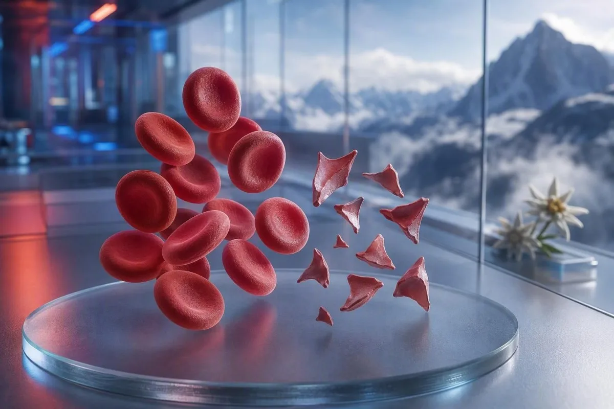
Şistositlər təhlükəli xırda damar laxtalanmasını göstərə bilən parçalanmış eritrositlərdir, lakin tək bir yaxma şərhi diaqnoz deyil. Təcili vəziyyət fraqment faizindən, trombosit sayından, hemoglobin dinamikasından, böyrək funksiyasından, hemoliz markerlərindən və simptomlardan asılıdır.


- Şistosit həddi: Yaxşı hazırlanmış yetkin periferik yaxmada 1%-dən çox şistosit digər eritrosit forma dəyişiklikləri yoxdursa, trombotik mikroangiopatiya üçün şübhəlidir.
- Təcili qrup: Şistositlər plus trombositlər 150 × 10^9/L-dən aşağı, düşən hemoglobin, yüksəlmiş LDH və aşağı haptoglobin eyni gün klinisyen tərəfindən qiymətləndirilməlidir.
- TTP ipucu: ADAMTS13 aktivliyinin 10%-dən aşağı olması immun trombotik trombositopenik purpuranı dəstəkləyir; klinik şübhə yüksək olduqda təsdiqləyici testlərin nəticələri gələnə qədər müalicə edilir.
- Hər fraqment təhlükəli deyil: Mexaniki ürək qapaqları, ağır hipertenziya, dializ dövrələri, yanıq (burns) və nümunə zədələnməsi parçalanmış eritrositlər yarada bilər.
- Yaxma əl ilə sübutdur: Avtomatlaşdırılmış analizator şistositləri etibarlı şəkildə diaqnoz edə bilməz; təcrübəli morfoloq preparatı mütləq gözlə yoxlamalıdır.
- Hamiləlik riskləri artırır: 20 həftədən sonra yüksək qan təzyiqi, sidikdə protein və ya aşağı trombositlərlə müşayiət olunan şistositlər təcili obstetrik qiymətləndirmə tələb edir.
- Təkrar test kömək edir: Təzə götürülmüş EDTA nümunəsi və təkrar yaxma (film) toplanma artefaktını davam edən həqiqi eritrosit parçalanmasından ayıra bilər.
- Özbaşına müalicə etməyin: Dəmir, folat və ya əlavələr mikrovaskulyar angiopatik hemolizi müalicə etmir və təcili qiymətləndirmədən yayındıra bilər.
Parçalanmış eritrositlər təcili tibbi qiymətləndirmə tələb edəndə
Şistositlər aşağı trombosit sayı, anemiya, biokimyəvi hemoliz, böyrək zədələnməsi, nevroloji simptomlar, qızdırma və ya ağır hipertenziya ilə birlikdə göründükdə eyni gün təcili baxış tələb edir. Yalnız yaxma şərhi həm zərərsiz, həm də artefakt ola bilər; təhlükəli tapıntı nümunədir. 7 avqust 2026-cı il tarixinə kimi, yeni şistositlər və trombositlər 100 × 10^9/L-dən aşağı olduqda bunu rutin təqib yox, “indi həkimə zəng et” nəticəsi kimi qiymətləndirərdim.
Trombositlərin düşməsi çox vaxt ən sürətli təcili xəbərdarlıq işarəsidir. Hemoglobin 92 q/L, trombosit 68 × 10^9/L və yeni bildirilmış şistositlər, hemoglobin 145 q/L olan 1% fraqmentləri və trombositləri 260 × 10^9/L səviyyəsində sabit olan vəziyyətdən xeyli daha çox narahatedicidir. Kantesti bu klasteri tam qan sayımı (complete blood count) üzrə oxuyan və təkbaşına morfologiya şərhini yüksəltməyən AI qan test analizatorudur.
Klinik praktikada 15 illik təcrübəmdə tez verdiyim qərarlar nəticələri günlər ərzində dəyişən xəstələr üçündür: hemoglobinin düşməsi, kreatininin yüksəlməsi və laktat dehidrogenazanın (LDH) yüksəlməsi. Dr. Thomas Klein-in praktik qaydası sadədir: bu nümunə ilə yanaşı çaşqınlıq, yeni zəiflik, sinə ağrısı, nəfəs darlığı, çox az sidik və ya ağır baş ağrısı varsa, bu gün təcili qiymətləndirmə deməkdir.
Şistositlər insanın vəziyyəti pisdirsə, təkrar test gözləmək üçün səbəb deyil. Normal kalium və ya normal leykosit sayı trombotik mikrovaskulyar angiopatiya riskini ləğv etmir. Aşağı göstəricini anlamağa çalışan xəstələr də bizim bələdçimizə baxa bilərlər: aşağı qırmızı qan hüceyrəsi simptomları, amma kəskin simptomlar onlayn şərhdən həmişə üstündür.
Təcili yardım xidmətlərinə indi zəng edin:
Yeni birtərəfli zəiflik, huşun itməsi, qıcolma, ağır nəfəs darlığı, döş qəfəsində sıxıcı narahatlıq, qara nəcis və ya sürətlə pisləşən çaşqınlıq klinikaya mesajdan çox təcili yardım tələb edir. Bu simptomlar orqan tutulmasını göstərə bilər və şistosit faizinin “heç bir təhlükəsiz istisnası” yoxdur.
Periferik yaxmada şistositlər əslində nə deməkdir
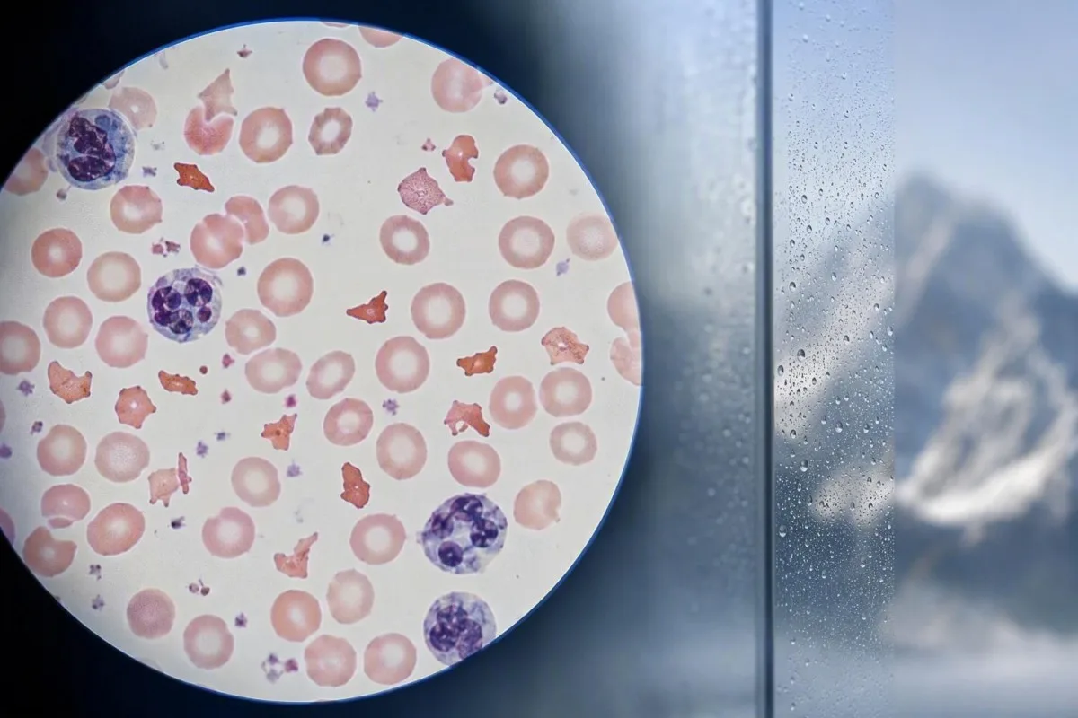
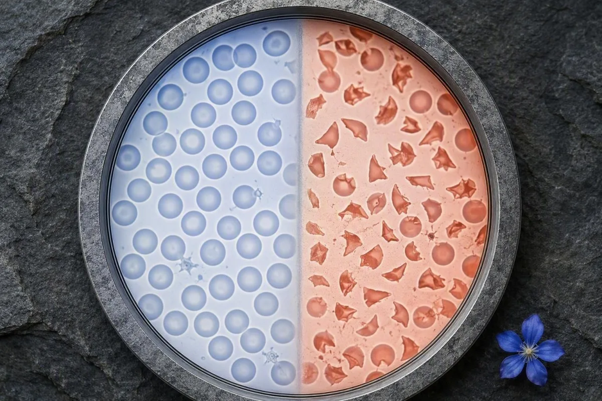
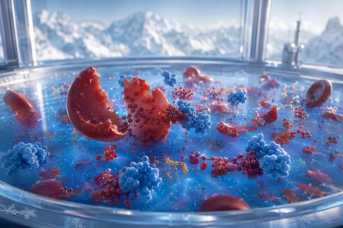
Şistositlər dövran edən hüceyrələr fiziki olaraq kəsildikdə yaranan qeyri-düzgün eritrosit fraqmentləridir; ən çox fibrin telləri və ya kiçik damarların anormal yüksək-şırma (shear) səthləri tərəfindən törədilir. Adətən iti bucaqlar, dəbilqə kimi formalar və ya kiçik üçbucaqşəkilli görünüş olur və bütöv eritrositdəki mərkəzi solğunluğu olmur.
Həqiqi şistositlər morfoloji tapıntıdır; standart CBC analizatorunun verdiyi bir dəyər deyil. Hematologiyada Standartlaşdırma üzrə Beynəlxalq Şura (International Council for Standardization in Haematology) kəmiyyətləndirmə vacib olduqda optimal yaxma zonasında ən azı 1,000 eritrositin sayılmasını tövsiyə edir (Zini et al., 2021). Bu detal izah edir ki, bir laboratoriya “nadir fraqmentlər” barədə, digəri isə faiz barədə hesabat verə bilər.
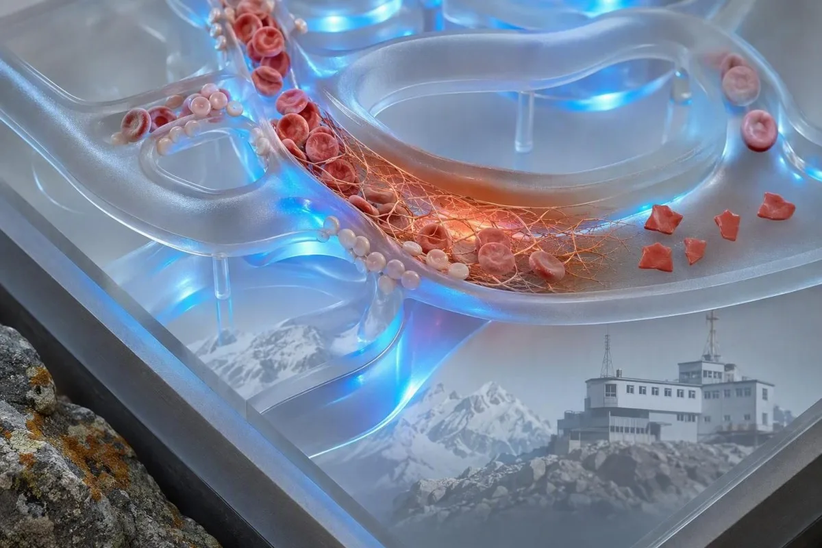
Klassik narahatlıqdır: mikrovaskulyar angiopatik hemolitik anemiya, burada qırmızı qan hüceyrələri dövran zamanı trombositlə zəngin mikrotromblar və fibrinlə təmas edir. Lakin ağır aortal stenoz, mexaniki qapaq, ekstrakorporal dövran sistemləri və ciddi termal zədələnmə fərqli mexanizm vasitəsilə həqiqi fraqmentlər yarada bilər. Buna görə sual “Fraqmentlər doğrudanmı var?” deyil, “Fraqmentləri nə yaradır?”
Kantesti tam qan sayımı (CBC) nəticələrini yüklənmiş halda yan-yana yerləşdirə bilən və CBC-nin nələri əhatə etdiyini bilirubin, retikulositlər və ya haptoglobin kimi çatışmayan yoldaş nəticələri vurğulaya bilən AI qan analizi nəticələri platformasıdır. O, slaydı özü yoxlaya bilmir; bu, təlim keçmiş laboratoriya mütəxəssisinin işidir.
Yetkin yaxmada şistositoz nə qədər narahatedicidir
Yetkin şəxsin periferik yaxmasında 1%-dən yuxarı şistosit sayı, fraqmentlər qırmızı qan hüceyrələri üzrə əsas anormallıq olduqda, trombotik mikroangiopatiya üçün şübhəlidir. 1%-dən aşağı göstəricilər xəstəliyi istisna etmir, xüsusən də film mayelər verildikdən, köçürmə (transfuziya) edildikdən və ya gedən xəstəlik fonunda hazırlanıbsa.
1% kəsim göstəricisi diaqnostik siqnaldır, ağırlıq (severity) şkalası deyil. ICSH rəhbərliyi sağlam yetkinlər və müddətli doğulmuş yeni doğulmuşlar üçün 1%-dən aşağı referens intervaldan istifadə edir; vaxtından əvvəl doğulmuş körpələr eyni məna daşımadan 5%-ə qədər fraqmentə malik ola bilər (Zini et al., 2021). Ağır trombositopeniya ilə 2% nəticəsi, mexaniki dövran dəstəyi cihazı yerləşdirildikdən sonra 4% nəticəsindən daha çox narahatedicidir.
Laboratoriyalar faiz əvəzinə “bəzən”, “az miqdarda” və ya “yüksək böyütmə sahəsində 1–2” kimi hesabat verə bilər. Bu ifadələr bir-birini əvəz etmir, çünki sahənin ölçüsü, yaxmanın qalınlığı və hesabatvermə siyasəti fərqlənir. Laboratoriyanın ICSH tipli faiz hesablaması edib-etmədiyini soruşmaq, təsviri ifadəni özünüz çevirməyə çalışmaqdan çox vaxt daha faydalıdır.
Yüksək RDW şistositləri ölçmür. RDW hüceyrə ölçüsündə dəyişkənliyi əks etdirir və retikulositoz, dəmir çatışmazlığı, transfuziya və ya fraqmentləşmə ilə arta bilər. Bizim ətraflı RDW və eritrosit indeksi bələdçisi yüksək RDW-nin təkbaşına nə TTP-ni, nə də artefaktı dəstəkləmədiyini izah edir.
Həqiqi hemolizi dəstəkləyən qan analizi nümunəsi
Həqiqi qırmızı qan hüceyrəsi fraqmentləşməsi adətən yüksək LDH, aşağı haptoglobin, artmış indirekt bilirubin və anemiya ilə yanaşı retikulosit cavabı yaradır. Tək bir markerin qəti diaqnostik dəyəri yoxdur, çünki qaraciyər xəstəliyi, iltihab, yaxın zamanda köçürmə və sümük iliyi xəstəlikləri mənzərəni təhrif edə bilər.
LDH həssasdır, amma spesifik deyil. LDH-nin 600 IU/L-dən yuxarı olması uyğun klinik şəraitdə çox vaxt aktiv hemolizi dəstəkləyir, lakin əzələ zədələnməsi, qaraciyər zədələnməsi, infeksiya və xərçəng də onu yüksəldə bilər. Laboratoriyanın aşağı həddindən aşağı, adətən 0,3 g/L-dən aşağı olan haptoglobin əlavə çəki verir, çünki plazmaya buraxılan sərbəst hemoglobini bağlayır.
Retikulositlərin sayı adətən periferik destruksiya zamanı yüksəlməlidir, amma folat çatışmazlığında, dəmir çatışmazlığında, böyrək xəstəliyində və ya kəskin sümük iliyi supressiyasında aşağı qala bilər. Bu görünən ziddiyyət asanlıqla qaçırılır: xəstədə həqiqi hemoliz ola bilər və retikulosit cavabı qeyri-münasib şəkildə aşağı ola bilər. haptoglobin nəticəsi bələdçisi ümumi interpretasiya tələlərini əhatə edir.
Kantesti AI eyni zaman nöqtəsindən hemoglobin, MCV, trombositlər, LDH, bilirubin, kreatinin və retikulositləri müqayisə edərək fraqmentləşmiş eritrosit nümunələrini şərh edir. 48–72 saat ərzində 20 q/L-dən çox düşən hemoglobin, əvvəlcədən müqayisə olmadan yüngül dərəcədə anormal dəyərdən klinik olaraq daha informativdir.
Bilirubinin niyə normal ola bilməsi
Hemolizin erkən mərhələsində, venadaxili maye ilə durulaşmadan sonra və ya qaraciyər bilirubini effektiv şəkildə təmizlədikdə indirekt bilirubin normal ola bilər. Buna görə də normal bilirubin inandırıcı fraqmentləri, aşağı haptoglobini və düşən hemoglobini üstələməməlidir.
Niyə TTP hematologiya təcili kimi müalicə olunur
Şistositlər trombositopeniya ilə birlikdə və daha aydın alternativ olmadan mikrovaskulyar patoloji ilə gedən hemolitik anemiya müşahidə olunarsa, trombotik trombositopenik purpura (TTP) şübhə altına alınır. Müalicə olunmamış immun TTP sürətlə proqress edə bilər, buna görə klinisyenlər ADAMTS13 nəticələri geri qayıtmadan əvvəl plazma mübadiləsi və immun müalicəyə başlaya bilərlər.
ADAMTS13 aktivliyi 10%-dən aşağı TTP-ni güclü şəkildə dəstəkləyir, amma nəticə saatlar və ya günlər çəkə bilər. ISTH diaqnostik təlimatı, plazma müalicəsindən əvvəl nümunə götürərkən (Zheng et al., 2020) PLASMIC skoru kimi klinik alətlərlə pretest ehtimalının qiymətləndirilməsini tövsiyə edir. Skor ixtisaslı qərara körpüdür, mütəxəssis mühakiməsinin əvəzi deyil.
Qızdırma, nevroloji əlamətlər, böyrək çatışmazlığı, trombositopeniya və hemolitik anemiyadan ibarət tanış “pentad” bir çox real xəstədə natamam olur. George və Nester trombotik mikroangiopatiyanı beş məcburi qutudan daha çox, üst-üstə düşən səbəbləri olan bir sindrom kimi təsvir etmişdir (George & Nester, 2014). Praktikada beş xüsusiyyətin hamısını gözləmək təhlükəli köhnə vərdişdir.
Hemoliz fonunda trombositlərin 30 × 10^9/L-dən aşağı olması hematologiya ilə xüsusilə təcili müzakirə tələb edir. TTP şübhəsi olduqda trombosit köçürülməsi adətən həyat üçün təhlükəli qanaxma olmadıqca və ya invaziv prosedur gözləyə bilmədikcə aparılmır. Əlaqəli testlər üçün bizim koaqulyasiya test bələdçimizdən PT və aPTT-nin TTP-də niyə normal ola biləcəyini izah edir.
Həkimlərin nəzərdən keçirdiyi şistositlərin digər təhlükəli səbəbləri
Şistositlər həmçinin disseminə olunmuş damardaxili laxtalanmada (DIC), Şiga-toksin hemolitik uremik sindromda, ağır hipertenziyada, hamiləliklə əlaqəli mikrovaskulyar patalogiyada, bəzi dərmanlarda və transplantasiya ilə əlaqəli trombotik mikroangiopatiyada da rast gəlinir. Əlaqəli trombosit, koaqulyasiya, böyrək və klinik tapıntılar adətən bu vəziyyətləri bir-birindən ayırır.
DIC çox vaxt fraqmentləri uzanmış PT, uzanmış aPTT, aşağı və ya düşən fibrinogen və kəskin yüksəlmiş D-dimer ilə birləşdirir. TTP-də daha çox nisbətən normal rutin koaqulyasiya göstəriciləri olur. Xəstə vəziyyətdə fibrinogenin 1,5 g/L-dən aşağı olması istehlak koaqulopatiyası ilə bağlı narahatlığı artırır, baxmayaraq ki, fibrinogen əvvəlcə normal görünə bilər, çünki kəskin iltihab zamanı yüksəlir.
Ağır hipertenziya təzyiq mikrovaskulyar zədələnmə törətdikdə eritrositləri parçalaya bilər; baş ağrısı, görmə dəyişikliyi, sinə ağrısı və ya böyrək disfunksiyası ilə birlikdə 180/120 mmHg və ya daha yüksək göstəricilər təcili vəziyyətdir. Buna görə də qan təzyiqi göstəricisi yaxma hesabatının yanında verilməlidir. Xəstələr nəzərdən keçirə bilər ikincili hipertenziya üçün laborator göstərici ipucları dərhal təhlükə aradan qaldırıldıqdan sonra.
Diareya, ardınca sidik ifrazının azalması, anemiya və trombositopeniya hemolitik uremik sindromdan şübhələnməyə səbəb olur. Nəcisdə Shiga toksininin aşkarlanması kömək edə bilər, lakin təcili yardımı əvvəlcə hidratasiya vəziyyəti, kreatinin, kalium və nevroloji qiymətləndirmə istiqamətləndirir. Yüksək laktat və ya qeyri-sabit həyati göstəricilər gözləmə yanaşması əvəzinə sepsis və DİK üçün qiymətləndirmə tələb edir; bunlara baxın sepsis marker nümunələri üzrə ayrıca bələdçimiz.
Parçalanmış eritrositlərin mexaniki səbəbləri
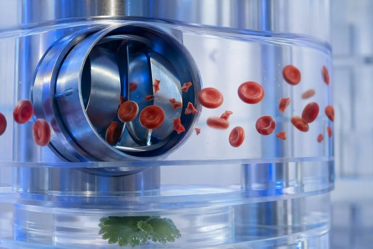
Mexaniki destruksiya TTP olmadan da real şistositlər yarada bilər; xüsusilə protez ürək qapaqları, ağır aortal stenoz, ventrikulyar assist cihazları, dializ dövrələri və ya ekstrakorporal membran oksigenasiyası zamanı. Bu səbəblər yenə də tibbi qiymətləndirmə tələb edir, lakin müalicə immun TTP-yə deyil, mexaniki mənbəyə hədəflənir.
Mexaniki qapaq nümunəsi çox vaxt xronik yüngül LDH yüksəlməsi və stabil trombositlərlə birlikdə aşağı haptoglobin ilə xarakterizə olunur. Yeni anemiya, nəfəs darlığının pisləşməsi, tünd rəngli sidik və ya qapaq səsinin dəyişməsi paravalvulyar sızma və ya cihaz disfunksiyasını göstərə bilər və kardiologiya baxışı tələb edir. Smear ipucu verir; exokardioqrafiya mənbəni tapır.
İdmançılar bəzən marafonun fraqmentləri izah edib-etməyəcəyini soruşurlar. Ayaq zərbəsi ilə bağlı hemoliz uzunmüddətli qaçışdan sonra keçici sərbəst hemoglobin və aşağı haptoglobin yarada bilər, amma inandırıcı şistositozu məşqə sadəcə “aid etmək” istəməzdim—xüsusən də trombositlər düşürsə. Daha geniş marafon qaçışçısı laborator bələdçisi.
Ağır yanıq xəsarətləri və böyük damar cərrahiyyəsi fiziki və endotel zədələnməsi vasitəsilə fraqmentləşmiş hüceyrələr yarada bilər. Bu hallarda hər 6–24 saatdan bir laborator trendə baxmaq tək bir smear-dən daha məlumatverici ola bilər. Tetikləyici hadisədən sonra CBC-nin stabil qalması ürəkaçandır; hemoglobinin davamlı düşməsi isə deyil.
Toplanma və şüşə (slayd) artefaktlarının şistositləri necə təqlid etməsi
Nümunənin zəif toplanması, smearın hazırlanmasının gecikdirilməsi, az doldurulmuş antikoaqulyant borular və zədələnmiş smear kənarı dövran səviyyəli hemolizi əks etdirmədən şistositlərə bənzəyən formalar yarada bilər. Təmiz toplanmış və monolayerdə nəzərdən keçirilmiş təkrar nümunə klinik mənzərə sakit olduqda adətən ilk yoxlamadır.
Krenat hüceyrələr, smear-kənar təhrif, quruma artefaktı və üst-üstə düşən hüceyrələr tez-tez rast gəlinən yalan oxşarlardır. Onlar çox vaxt diffuz şəkildə və ya ideal oxu zonasında bərabər paylanmaqdan çox “tüklü” (feathered) kənarda görünür. Təcrübəli morfoloq həqiqi fraqmentin iti həndəsəsini slayd hazırlanması zamanı sadəcə təhrif olunmuş hüceyrədən ayırd edir.
Çətin toplanma borunu hemoliz edə və kaliumu və ya LDH-ni yalançı olaraq yüksəldə bilər, amma bu, avtomatik olaraq həqiqi dövran edən şistositlər yaratmır. Nəticəyə hemoliz indeksi və ya “nümunə zədələnib” kimi qeyd daxil edilirsə, klinisyenlər adətən xəstəliyi diaqnoz etməzdən əvvəl nümunəni yenidən götürürlər. Məqaləmizdə kalium götürmə (qan alma) səhvləri eyni pre-analitik problemin niyə bir neçə nəticəni eyni anda çaşdıra biləcəyini izah edirik.
EDTA-dan asılı trombosit yığılması trombosit sayını yalançı olaraq azalda və həyəcanverici görünən bir kombinasiyaya “hazırlaya” bilər. Diluziya nəzərə alınaraq düzəldilmiş sitrat borusunda trombosit sayı və təzə film bunu üzə çıxara bilər. İncə məqam önəmlidir: artefakt həqiqi xəstəliklə yanaşı mövcud ola bilər, buna görə təkrar test simptomlu bir şəxs üçün qayğını heç vaxt gecikdirməməlidir.
Niyə analizatorun işarələməsi və laboratoriya ifadəsi yanıltıcı ola bilər
Avtomatlaşdırılmış qan analizatorları anormal eritrosit populyasiyalarını işarələyə bilər, lakin trombotik mikroangiopatiyanı istisna etmək üçün şistositləri müstəqil şəkildə kifayət qədər etibarlılıqla diaqnoz edə bilməz. Klinik sual hemoliz və ya TTP olduqda əl ilə periferik smearin yenidən yoxlanması yenə də zəruridir.
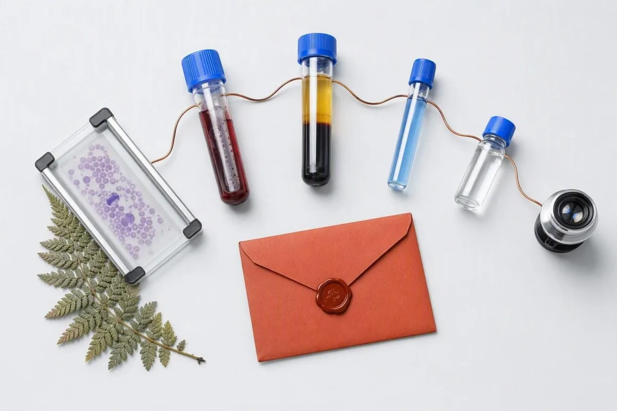
“Parçalanmış qırmızı hüceyrələr mövcuddur” şərhi miqdar, yaxmanın keyfiyyəti və yanaşı mövcud olan morfologiya barədə suallar doğurmalıdır. Dəmir çatışmazlığı, meqaloblast anemiya və ya irsi eritrosit pozğunluqları ilə əlaqəli ifadəli anizopoikilositoz 1% qaydasını daha az spesifik edir. Parçalar anormal morfologiyanın içində üstünlük təşkil etdikdə ən çox diaqnostik əhəmiyyət daşıyır; bir çox müxtəlif, əlaqəsiz formalardan yalnız biri kimi görünəndə isə daha az faydalıdır.
Kantesti uyğunsuz kombinasiyaları, məsələn, “trombosit yığılmaları var” şərhi ilə yanaşı trombosit sayının aşağı olması kimi halları işarələmək üçün nəzərdə tutulmuş AI laboratoriya testinin şərh xidmətidir. Bu, laboratoriyanın mikroskopik baxışını və ya təkrar preparat istəyə bilən klinisistin yerini tutmur. Keyfiyyət yoxlamalarına yanaşmamızda təsvir olunur: tibbi validasiya icmalı.
Delta yoxlaması cari nəticəni əvvəlki nəticə ilə müqayisə edir və qəfil, inandırıcı olmayan dəyişiklikləri üzə çıxara bilər. Hemoglobinin 138 q/L-dən 87 q/L-ə 24 saat ərzində düşməsi klinik baxımdan əhəmiyyətlidir, lakin venadaxili mayelərdən durulaşma və ya səhv etiketlənmiş nümunə də mümkündür. Daha ətraflı: qəfil laborator dəyişikliklər.
Qan analizi nəticələrində şistositlər qeyd olunandan sonra nə etmək lazımdır
Şistositlər ilk dəfə bildirilibsə həmin gün testi sifariş edən klinisistə və ya xidmətə müraciət edin; nevroloji simptomlar, nəfəs darlığı, sinə ağrısı, tünd rəngli sidik, güclü baş ağrısı, qızdırma və ya sidik ifrazının azalması varsa dərhal təcili tibbi yardım axtarın. Yaxma şərhini təkbaşına diaqnoz kimi şərh etməyin.
Hemoglobin, trombositlər, kreatinin, bilirubin, LDH və hər hansı hemoliz indeksi daxil olmaqla, faktiki hesabatı hazır saxlayın. Həmçinin yaxınlarda hamiləlik, diareya xəstəliyi, ağır qan təzyiqi göstəriciləri, yeni dərmanlar, qapaq cərrahiyyəsi, dializ, transfuziya və autoimmun xəstəliyi də qeyd edin. Bu detallar morfologiya sahəsindəki tək bir sözə nisbətən differensial diaqnozu daha çox dəyişir.
Mən görürəm ki, səbəb müəyyən edilməmişdən əvvəl “anemiya” sözünü oxuyub dəmirə başlamaq xəstələrə dəyərli vaxt itirdir. Dəmir çatışmazlığı yanaşı mövcud ola bilər, amma parçalarla birlikdə sürətli trombosit azalmasını izah etmir. Dr. Thomas Klein yalnız vurğulanmış rəqəmləri köçürməkdənsə, tam hesabatın şəklini çəkməyi tövsiyə edir; köçürmə çox vaxt nümunəni bir-biri ilə uyğunlaşdıran laborator şərhini itirir.
Trombositlər çox aşağıdırsa və ya qanaxma varsa, klinisist başqa cür məsləhət verməyənədək aspirin, ibuprofen və reseptsiz antikoaqulyantlardan çəkinin. Təyin olunmuş antikoaqulyantı da təcili tibbi rəhbərlik olmadan dayandırmayın. Yorğunluğu və ya başgicəllənməni olan insanlar bizim başgicəllənmə üçün qan testləri təcili səbəblər istisna edildikdən sonra suallar hazırlamaq üçün istifadə edə bilərlər.
Hamiləlikdə, yenidoğulmuşlarda və uşaqlarda şistositlər
Hamiləlik zamanı şistositlər, hipertoniya, proteinuriya, yüksəlmiş qaraciyər fermentləri, aşağı trombositlər, baş ağrısı, görmə simptomları və ya yuxarı qarın ağrısı ilə birlikdə olduqda təcili qiymətləndirmə tələb edir. HELLP sindromu, preeklampsiya, TTP, atipik HUS və DİK (DIC) üst-üstə düşə bilər; doğuş vaxtı ətrafında qiymətləndirmə kömək edir, amma diaqnozu qəti şəkildə aydınlaşdırmır.
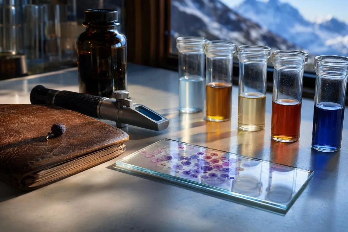
HELLP sindromu klassik olaraq hemoliz, yüksəlmiş qaraciyər fermentləri və aşağı trombositlərlə müəyyən edilir; çox vaxt hamiləliyin 20-ci həftəsindən sonra və ya doğuşdan qısa müddət sonra baş verir. Trombosit sayı 100 × 10^9/L-dən aşağı və ya AST 70 IU/L-dən yuxarı olduqda narahatlıq üçün əsas ola bilər, lakin yerli obstetrik qruplar bütün klinik mənzərəni şərh edir. Bu kombinasiya ilə rutin antenatal qəbulunuzu gözləməyin.
Term (vaxtında doğulmuş) yeni doğulmuşlarda 1% şistosit daha az ola bilər, halbuki vaxtından əvvəl doğulmuş körpələrdə neonatal eritrosit morfologiyası fərqli olduğuna görə (Zini et al., 2021) 5%-ə qədər ola bilər. Lakin uşaqda diareya, solğunluq, sidiyin azalması və trombositopeniya varsa, parçalar HUS-a işarə edə bilər və dərhal pediatrik qiymətləndirmə tələb edir.
Trombositlər və hemoglobin üçün istinad intervalları yaşa və hamiləlik mərhələsinə görə əhəmiyyətli dərəcədə dəyişir. Ümumi tipli tətbiqdə “normal” kimi işarələnmiş nəticə kontekstdə yenə də klinik baxımdan narahatedici ola bilər. Hamiləliklə bağlı resurslarımız: hamiləlikdə trombosit diapazonları və eyni gün hamiləlik laboratoriya işarələri faydalı məlumat fonu təqdim edin, mama triage-nin əvəzi deyil.
Həkimlərin həqiqi şistositləri araşdırmaq üçün istədiyi testlər
Şübhəli mikroangiopatik hemoliz üçün ilk müayinə dəsti adətən təkrar CBC və yaxma, retikulosit sayı, LDH, haptoglobin, bilirubin fraksiyaları, kreatinin, sidik analizi, PT, aPTT, fibrinogen və D-dimeri əhatə edir. TTP şübhəsi olduqda plazma mübadiləsindən əvvəl ADAMTS13 aktivliyi və inhibitor testləri götürülməlidir, lakin müalicə gözləməməlidir.

Birbaşa antiglobulin testi, DAT və ya Kumbs testi də adlanır, immun hemolizi qeyri-immun fraqmentləşmədən ayırmağa kömək edir. Müsbət DAT ikinci bir prosesi istisna etmir, lakin klassik TTP adətən DAT-mənfidir. Sidik mikroskopiyası böyrək mikrovaskulyar zədələnmə aktiv olduqda zülal və ya hüceyrə silindrlərini göstərə bilər; izahımıza baxın dənəvari sidik silindrləri.
Nəcisdə Şiga-toksin testi uyğun qastrointestinal xəstəlikdən sonra nəzərdən keçirilir, atipik HUS üçün isə komplement testləri, dərmanların nəzərdən keçirilməsi və genetik və ya funksional tədqiqatlar istifadə oluna bilər. Sıra ölkə və xəstəxanadan asılı olaraq dəyişir. 48 saat ərzində 26,5 µmol/L kreatinin artımı kəskin böyrək zədələnməsi meyarlarından birini ödəyir və dramatik rəqəmə çevrilmədən belə diqqətə layiqdir.
Əhəmiyyətli anemiyada retikulosit istehsal indeksi 2-dən aşağı olması, xam retikulosit faizinin yüksək görünməsinə baxmayaraq, qeyri-kafi sümük iliyi cavabını göstərir. Bu hesablamalar anemiyada daha aşağı eritrosit kütləsini düzəldir. retikulosit və hematologiya marker bələdçimiz faizlərin niyə yanıltıcı ola biləcəyini izah etməyə kömək edir.
Yaxmanı təkrar etməyin məntiqli olduğu və olmadığı hallar
Fraqmentlər az olduqda, xəstə özünü yaxşı hiss etdikdə, trombositlər və hemoglobin sabit olduqda və nümunə keyfiyyəti şübhə doğurduqda, bir neçə saatdan 24 saata qədər müddətdə yaxmanı təkrar etmək məntiqlidir. Trombositopeniya, aktiv hemoliz, orqan zədələnməsi və ya narahatedici simptomlar varsa təkrarın təhlükəsiz gecikmə olmur.
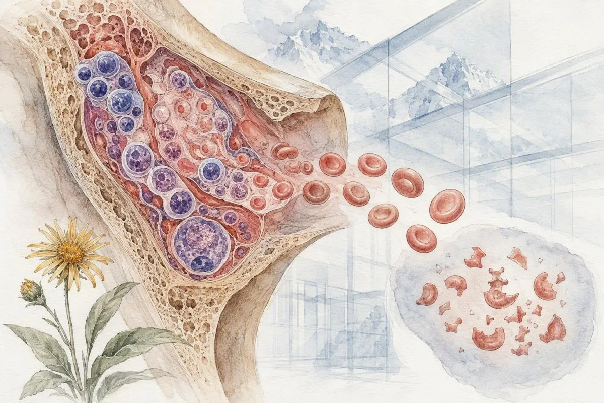
Yaxşı təkrar nümunə düzgün doldurulmuş EDTA borusuna təzə götürülməli və dərhal filmə çevrilməlidir. Sorğu xüsusi olaraq şübhəli şistositlərin və trombosit yığılmasının (clumping) nəzərdən keçirilməsini istəməlidir, əgər bunlardan biri mümkündürsə. Laboratoriya hər iki filmi yan-yana müqayisə edə bilər ki, bu da sadəcə avtomatlaşdırılmış CBC-ni yenidən işlətməkdən qat-qat daha faydalıdır.
Trendlərin istiqaməti önəmlidir. Üç gün ərzində trombosit sayının 210-dan 165-ə, sonra 98 × 10^9/L-ə düşməsi narahatedicidir; baxmayaraq ki, ilk iki göstərici yerli referens intervalına daxil ola bilər. Kantesti AI bu trayektoriyaları üzə çıxarmaq üçün uzununa kontekstdən istifadə edir; bu prinsip bizim qan testi trendlərinin analizi bələdçimizdən istifadə edərək müqayisə edin.
Transfuziya ilkin morfologiyanı müvəqqəti olaraq durulaşdıra və ya gizlədə bilər. İkinci nümunədən əvvəl eritrosit almısınızsa, komandaya tarixi və neçə vahid (unit) olduğunu bildirin. Bələdçimiz xəstəxanadan çıxışdan sonra dəyərlərin dəyişməsi müalicədən sonrakı trendlərin niyə fərqli baza (baseline) tələb etdiyini izah edir.
Şistosit xəstəliyi olmadan oxşar görünə bilən vəziyyətlər
Dəmir çatışmazlığı, vitamin B12 çatışmazlığı, qaraciyər xəstəliyi, irsi eritrosit pozğunluqları və ağır infeksiya trombotik mikroangiopatiyanın spesifik fraqmentləşmə nümunəsi olmadan belə anormal eritrosit formaları yarada bilər. Yaxmanın tam morfologiyası və klinik zaman xətti fraqmentlərin həddən artıq qiymətləndirilməsinin qarşısını alır.
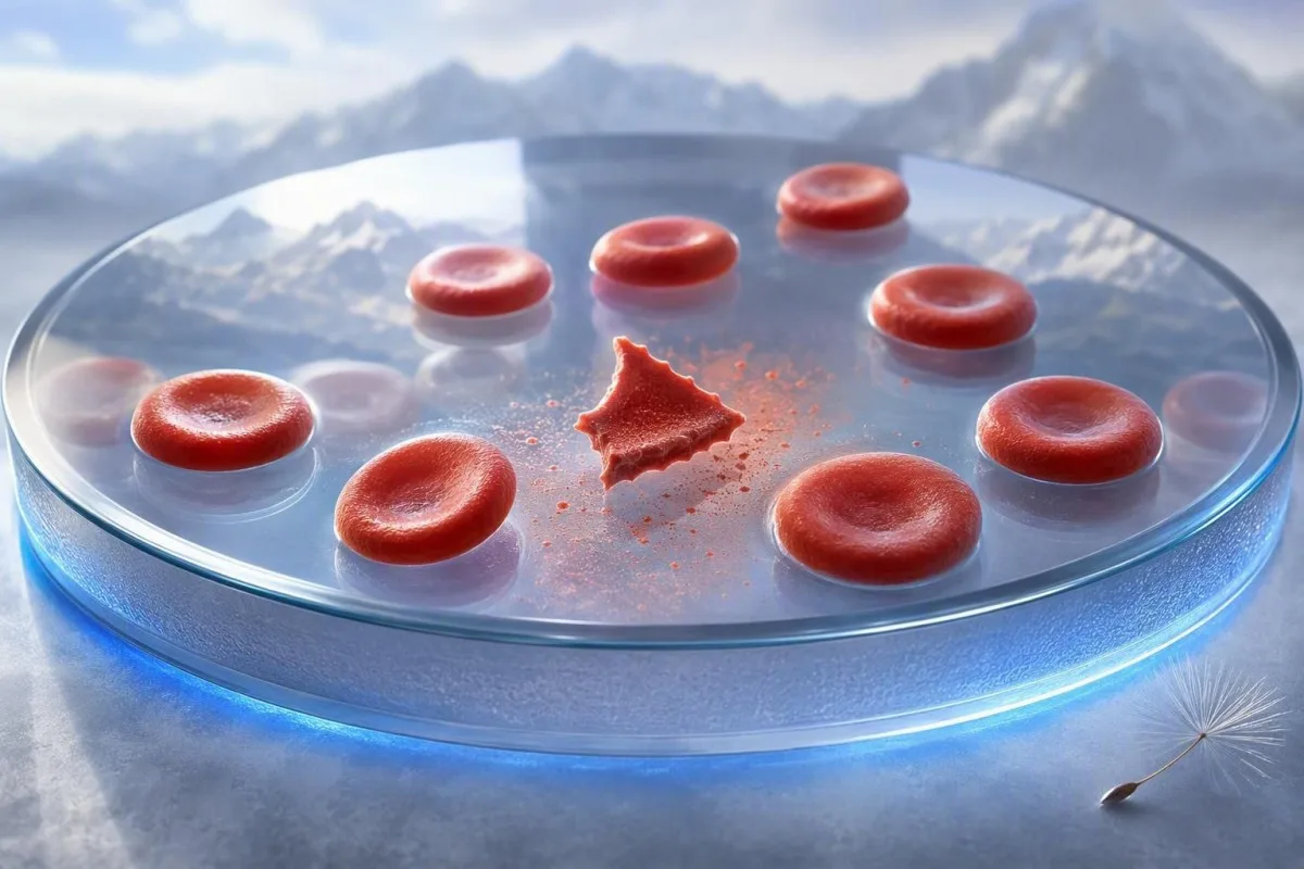
Ağır vitamin B12 çatışmazlığı anemiya, yüksək LDH, aşağı trombositlər və bəzən fraqmentlərlə psevdotrombotik mikroangiopatiya yarada bilər. Çox vaxt ifadəli makrositoz, aşağı retikulosit cavabı və bəzən 2,500 IU/L-dən yuxarı LDH göstəriciləri əmələ gətirir—bu xüsusiyyətlər avtomatik TTP müalicəsindən daha çox B12 və metilmalonik turşu testlərini tələb etməlidir. Bizim B12 ilə folat bələdçisində izah olunur bu mühüm imitatoru izah edir.
Dəmir çatışmazlığı adətən mikrositoz, hipoxromiya, qələmvari hüceyrələr və yüksək RDW yaradır; ağır hallarda fraqmentlər qeyd oluna bilər, lakin onlar nadir hallarda əsas xüsusiyyət olur. Ferritin 15 µg/L-dən aşağı olması, başqa cür ağırlaşmamış yetkin insanda dəmir ehtiyatlarının tükənməsini güclü şəkildə dəstəkləyir, baxmayaraq ki, iltihab ferritini yüksəltməklə çatışmazlığı “örtə” bilər.
Mənim təcrübəmə görə, ən rahatlaşdırıcı nəticə təkcə “şistositlərin olmaması” deyil, sabit trombositlərlə, sabit böyrək funksiyası ilə və biokimyəvi hemoliz olmadan uyğun alternativ izahın olmasıdır. Əgər tapıntılar uyğunsuz qalarsa, hematoloq yekun hesabatın ifadəsinə güvənməkdənsə, faktiki şüşə preparatı (slaydı) yenidən nəzərdən keçirə bilər.
Şəkil (yaxma) təcili vəziyyətini qaçırmadan AI şərhindən istifadə
AI qan analizinin nəticələrini təşkil etməyə və təhlükəli kombinasiyaları müəyyən etməyə kömək edə bilər, amma CBC PDF-dən şistosit diaqnozunu təsdiqləyə və ya təcili klinik qiymətləndirməni əvəz edə bilməz. Periferik yaxma vizual mikroskopiyadır və ilkin laboratoriya slaydı həqiqətin mənbəyidir.
Kantesti hesabatlar üzrə əlaqəli nəticələri xəritələşdirən, AI ilə işləyən qan analizi alətidir; o, trombosit trayektoriyalarını, hemoglobin dəyişikliklərini, kreatinini, bilirubini və LDH-ni də daxil edir. Bu, hər bir anormal markerin ayrı-ayrılıqda oxunması ilə bağlı yayğın səhvi azalda bilər. O, şistosit adlanan formanın həqiqi fraqment olub-olmadığını, yaxma kənarı artefaktını və ya hesabat vermə konvensiyasını görə bilmir.
Yükləmə keyfiyyəti önəmlidir, çünki buraxılmış vahid və ya onluq rəqəm nəticənin mənasını tərsinə çevirə bilər. Hesabatı paylaşmazdan əvvəl pasiyent identifikatorlarını, götürülmə tarixini, vahidləri, istinad diapazonlarını və nəticənin “estimated” (təxmini), “pending” (gözlənilir) və ya “sample haemolysed” (nümunə hemolizə uğramış) yazıb-yazmadığını yoxlayın. Bizim AI hesabatının dəqiqlik yoxlama siyahısına baxın. həmin yekun insan yoxlaması üçün faydalıdır.
Kantesti-nin klinik metodları bizim Tibbi Məsləhət Şurası, tərəfindən verilən töhfələrlə nəzərdən keçirilir və texnologiyamızda AI şərh texnologiyası bələdçisi. təsvir olunur. Dr. Thomas Klein kimi, mən yenə də klinisistin yeni fraqmentləri olan, trombositləri 100 × 10^9/L-dən aşağı olan və simptomları olan bir pasiyent üçün laboratoriyaya birbaşa zəng etməsini istərdim—çünki məhz bu zaman sürət və klinik müayinə ən çox önəm daşıyır.
Tez-tez verilən suallar
Şistositlərin hansı səviyyəsi təhlükəlidir?
Diqqətlə yenidən nəzərdən keçirilmiş yetkin periferik yaxmada 1%-dən çox şistositin olması, parçalanmış hüceyrələr əsas qırmızı qan hüceyrəsi anomaliyası olduqda, trombotik mikroangiopatiya üçün şübhəlidir. Şistosit faizinin istənilən səviyyəsi, trombosit sayı 150 × 10^9/L-dən aşağı olduqda, hemoglobinin azalması, yüksək LDH, aşağı haptoglobin, böyrək zədələnməsi və ya nevroloji simptomlarla birlikdə baş verdikdə təcili qiymətləndirmə tələb edir. 1%-dən aşağı nəticə inkişaf edən vəziyyəti etibarlı şəkildə istisna etmir. Klinik mənzərə yalnız faiz göstəricisindən daha çox təhlükəni müəyyən edir.
Şiztositlər pis qan götürülməsi ilə yarana bilərmi?
Çətin toplanmış nümunə, gecikdirilmiş hazırlıq, az doldurulmuş EDTA borusu və ya zəif hazırlanmış şüşə slayd hüceyrə formalarını şistositlərə bənzədə bilər, baxmayaraq ki, bu, həqiqi dövran edən fraqmentasiya yaratmır. Laboratoriyalar bunu təzə toplanmış nümunə və optimal yaxma sahəsinin əl ilə nəzərdən keçirilməsi ilə dəqiqləşdirə bilərlər. Hemoliz olunmuş nümunə həmçinin kalium və LDH göstəricilərini yalançı olaraq artıra bilər ki, bu da hesabatın daha həyəcanverici görünməsinə səbəb ola bilər. Təkrar test təcili qiymətləndirməni gecikdirməməlidir; əgər simptomlar, trombositlərin aşağı olması və ya anemiya mövcuddursa.
Şistositlər həmişə TTP-ni ifadə edirmi?
Şistositlər hər zaman trombotik trombositopenik purpuraya və ya TTP-yə işarə etmir. Onlar DİK, ağır hipertenziya, Şiga-toksin HUS, hamiləliklə bağlı pozğunluqlar, mexaniki ürək qapaqları, dializ dövrələri və böyük toxuma zədələnməsi zamanı da baş verə bilər. Şistositlər trombositopeniya və qeyri-immun hemolitik anemiya ilə birlikdə olduqda, xüsusilə ADAMTS13 aktivliyi 10%-dən aşağı olduqda TTP yüksək prioritetli narahatlıq kimi qiymətləndirilməlidir. ADAMTS13 testinin nəticəsi vaxt tələb edə bildiyindən, şübhəli TTP nəticə çıxana qədər təcili ixtisaslı qiymətləndirmə tələb edir.
Şistosit hemolizi ilə hansı qan analizləri yüksək olur?
Doğru parçalanma (fragmentasiya) hemolizi adətən LDH və indirekt bilirubini yüksəldir, haptoglobini azaldır və hemoglobin düşərkən retikulositlərin sayının artmasına səbəb olur. Aktiv hemoliz zamanı LDH 600 IU/L-dən çox ola bilər, lakin o, spesifik deyil, çünki əzələ, qaraciyər və digər toxumaların zədələnməsi də onu yüksəldə bilər. Qaraciyər tərəfindən istehsal adekvat olduqda, təxminən 0,3 q/L-dən aşağı haptoglobin intravaskulyar hemolizi dəstəkləyir. Kreatinin, trombositlərin sayı, PT, aPTT, fibrinogen və D-dimer əsas səbəbi müəyyən etməyə kömək edir.
Hamiləlikdə şistositlər normaldırmı?
Şistositlər hipertoniya, proteinuriya, yüksəlmiş qaraciyər fermentləri, düşən trombositlər, baş ağrısı, görmə dəyişiklikləri və ya yuxarı qarın ağrısı ilə birlikdə olduqda, rutin normal hamiləlik tapıntısı hesab edilmir. Bu kombinasiyа HELLP sindromu, preeklampsiya, TTP, atipik HUS və ya DİK-i göstərə bilər və dərhal mamalıq qiymətləndirilməsi tələb edir. Bu vəziyyətdə trombosit sayının 100 × 10^9/L-dən aşağı olması xüsusilə narahatedicidir. Hamiləliyə xas referens aralıqlar hemoliz nümunəsini nəzərdən qaçırmağı təhlükəsiz etmir.
Mexaniki ürək qapağı şistositlərə səbəb ola bilərmi?
Mexaniki ürək qapaqları və ağır aortal stenoz yüksək sürətli qan axını nəticəsində eritrositlərin fiziki parçalanmasına görə həqiqi şistositlərə səbəb ola bilər. Bu, çox vaxt LDH-nin yüksəlməsi və haptoglobinin azalması ilə yüngül xroniki hemoliz yaradır, trombositlərin sayı isə sabit qalır. Yeni yaranan anemiya, tünd rəngli sidik, nəfəs darlığı və ya qapağın funksiyasında dəyişiklik ürək baxışını tələb edir, çünki sızma və ya cihaz problemi səbəb ola bilər. Bu tapıntı, ürək tarixçəsi nəzərə alınmadan avtomatik olaraq TTP kimi etiketlənməməlidir.
Əsas səbəb müalicə edildikdən sonra şistositlər nə qədər tez yox olmalıdır?
Əsas proses nəzarətə alındıqdan sonra şistositlər günlər ərzində azala bilər, lakin vaxtlama səbəbdən və hemolizin davam edib-etməməsindən asılı olaraq dəyişir. Həkimlər yalnız fragmenlərin yox olmasını gözləmək əvəzinə trombosit sayını, hemoglobin, LDH, böyrək funksiyasını və təkrar yaxma nəticələrini izləyirlər.
Bu gün AI ilə gücləndirilmiş qan analizi nəticələrinin şərhini əldə edin
Ani və dəqiq laborator analiz şərhinə güvənən dünya üzrə 2 milyondan çox istifadəçiyə qoşulun. Qan analizinin nəticələrini yükləyin və bir neçə saniyəyə 15,000+ biomarkerlərinin hərtərəfli şərhini alın.
📚 İstinad edilən tədqiqat nəşrləri
Klein, T., Mitchell, S., & Weber, H. (2026). Kantesti Qan Testi Şərh Mühərrikinin 100.000 sintetik sınaq halında əvvəlcədən qeydiyyatdan keçmiş, rubrikaya əsaslanan avtomatlaşdırılmış texniki benchmarkı. Kantesti AI Tibbi Tədqiqatlar.
Klein, T., Mitchell, S., & Weber, H. (2026). Klinik Validasiya Çərçivəsi v2.0 (Tibbi Validasiya Səhifəsi). Kantesti AI Tibbi Tədqiqatlar.
📖 Xarici Tibbi İstinadlar
Zheng XL və b. (2020). Trombotik trombositopenik purpura diaqnozu üçün ISTH təlimatları. Trombosis və Hemostaz Jurnalı.
⚕️ Tibbi Məsuliyyətdən İmtina
Bu məqalə yalnız təhsil məqsədlidir və tibbi məsləhət təşkil etmir. Diaqnoz və müalicə qərarları üçün həmişə ixtisaslı səhiyyə işçisi ilə məsləhətləşin.
E-E-A-T Etibar Siqnalları
Təcrübə
Həkim rəhbərliyi ilə laborator şərh iş axınlarının klinik icmalı.
Ekspertiza
Biomarkerlərin klinik kontekstdə necə davrandığına dair laboratoriya təbabəti diqqəti.
Səlahiyyətlilik
Dr. Thomas Klein tərəfindən yazılıb; Dr. Sarah Mitchell və Prof. Dr. Hans Weber tərəfindən nəzərdən keçirilib.
Etibarlılıq
Aydın izləmə yolları ilə həyəcanı azaltmağa yönəlmiş sübuta əsaslanan şərh.