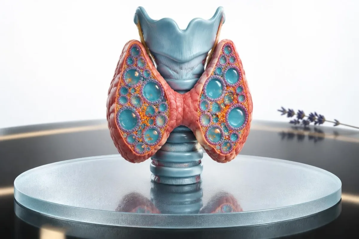
A low TSH result does not automatically mean permanent thyroid disease. The next decision depends on repeat results, thyroid hormone levels, age, symptoms, heart rhythm and bone health.
Key takeaways
- Definition: Subclinical hyperthyroidism means TSH is below the laboratory range while free T4 and free T3 remain within range.
- Severity: TSH of 0.10–0.39 mIU/L is usually called mild or grade 1 suppression; TSH below 0.10 mIU/L is grade 2 suppression.
- Repeat testing: A persistent result should usually be confirmed with TSH, free T4 and T3 after about 2–3 months, or sooner when symptoms are marked.
- Transient causes: Acute illness, thyroiditis, pregnancy-related hCG, glucocorticoids, dopamine, excess thyroid medication and biotin interference can temporarily lower TSH.
- Heart risk: Persistent TSH below 0.10 mIU/L is associated with a higher risk of atrial fibrillation, especially in adults aged 65 years or older.
- Bone risk: Postmenopausal adults with sustained TSH below 0.10 mIU/L deserve particular attention to fracture risk and bone-density assessment.
- Treatment: Guidelines generally favour treatment or specialist review for persistent TSH below 0.10 mIU/L in older adults or people with heart disease, osteoporosis or significant symptoms.
- Urgency: Chest pain, fainting, severe breathlessness or a sustained very rapid or irregular heartbeat needs urgent medical assessment, regardless of the thyroid result.
What does low TSH with normal T4 and T3 mean?
Low TSH with normal free T4 and free T3 fits the biochemical pattern called subclinical hyperthyroidism, but one abnormal sample does not prove persistent thyroid overactivity. Kantesti is an AI blood test analyzer that can place a thyroid result beside its laboratory range, related hormones and previous measurements; it cannot replace clinical assessment or confirm a diagnosis.
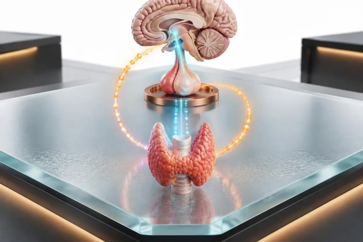
TSH is a pituitary signal that tells the thyroid how strongly to work. A TSH below about 0.4 mIU/L with free T4 and free T3 inside their laboratory ranges is the usual definition of subclinical hyperthyroidism, although laboratories use different assays and cutoffs.
For example, a hypothetical 52-year-old with TSH 0.18 mIU/L, free T4 15 pmol/L and free T3 4.8 pmol/L may have mild biochemical suppression rather than overt hyperthyroidism. The result becomes more meaningful if the same pattern remains present after 2–3 months.
The term subclinical describes the blood-test pattern, not necessarily the person’s experience. Some people have palpitations or heat intolerance, while others feel entirely well; symptoms alone cannot distinguish transient suppression from Graves disease.
| Level | Range | What it means |
|---|---|---|
| Typical TSH range | About 0.4–4.0 mIU/L | TSH is within a common adult laboratory interval; free hormones still require context. |
| Mild suppression | 0.10–0.39 mIU/L | Often called grade 1 subclinical hyperthyroidism; repeat testing commonly clarifies persistence. |
| Marked suppression | 0.01–0.09 mIU/L | Often called grade 2; persistent results carry greater heart and bone concern. |
| Undetectable TSH | <0.01 mIU/L | Needs prompt clinical review, particularly if symptoms or raised free hormones develop. |
Why the free hormones matter
Free T4 and free T3 represent the unbound fractions available to tissues, and both can remain normal while the pituitary detects a small excess of thyroid hormone. A TSH of 0.06 mIU/L with normal free hormones is therefore different from TSH 0.06 mIU/L with free T4 above 22 pmol/L.
Reference intervals vary by assay, age and pregnancy status; a result near the edge of one laboratory’s range may be inside another’s. Always read the units and ranges printed beside the result rather than importing a range from a different website.
Our guide to T4 and T3 explains why total and free hormone measurements are not interchangeable.
When can a low TSH be temporary?
A low TSH can be temporary after acute illness, during thyroiditis, with pregnancy-related hormonal changes, or because of medicines and supplements. The practical response is usually to identify the possible trigger and repeat a complete thyroid panel rather than label the result permanent disease immediately.
Non-thyroidal illness can briefly disrupt pituitary signalling, especially during hospital-level illness, calorie restriction or recovery from infection. Glucocorticoids and dopamine can also reduce TSH secretion; the effect may occur without true excess thyroid hormone.
Subacute thyroiditis can release preformed hormone for several weeks, followed by a low-TSH phase and sometimes temporary hypothyroidism. TSH may stay suppressed after free T4 has normalised, so a single recovery-phase result can look like ongoing subclinical hyperthyroidism.
A hypothetical 39-year-old tested two weeks after a viral illness has TSH 0.09 mIU/L and normal free T4, but no palpitations or weight loss. Rechecking after recovery may show TSH returning to 1.4 mIU/L, which supports a transient process rather than Graves disease.
Timing the repeat after illness
For a stable person without major symptoms, repeating TSH, free T4 and free or total T3 after approximately 2–3 months is often more informative than testing again after a few days. A repeat at 2–6 weeks is reasonable when TSH is nearly undetectable, symptoms are significant or pregnancy changes the clinical context.
The European Thyroid Association guideline describes persistence as a central part of diagnosis because transient low TSH is common in several settings. Our article on subacute thyroiditis results covers the expected sequence in more detail.
Do not stop prescribed thyroid medicine solely because of a low TSH result. The prescriber must compare the dose, indication and timing with the laboratory pattern.
What causes subclinical hyperthyroidism?
The main causes are early Graves disease, an autonomous thyroid nodule or multinodular goitre, thyroiditis, excess thyroid replacement and medication or supplement effects. The cause matters because a temporary thyroiditis needs a different plan from persistent hormone production by nodules.
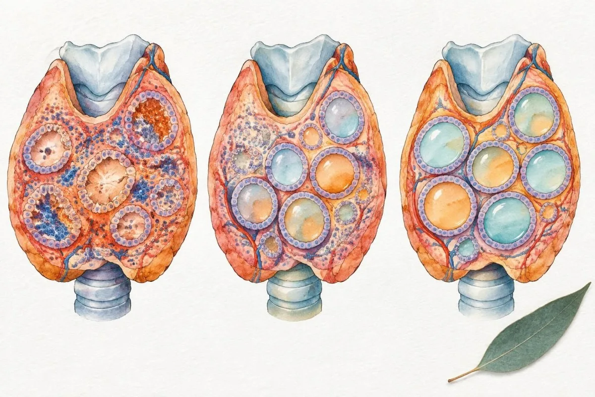
Graves disease is an autoimmune cause in which thyroid-stimulating antibodies activate the TSH receptor. A toxic nodule or multinodular thyroid can produce hormone independently of pituitary control; this becomes more common with increasing age and iodine exposure.
Excess levothyroxine or liothyronine is a frequent explanation in people being treated for hypothyroidism, particularly after a dose change or weight loss. Amiodarone, iodine-containing products, glucocorticoids, dopamine agonists and some cancer therapies can complicate interpretation.
Lithium usually causes hypothyroidism but can also contribute to thyroiditis or hyperthyroidism in selected people. Medication history should include over-the-counter products, thyroid supplements, weight-loss preparations and the exact time each dose was taken; our lithium monitoring guide explains why thyroid results need medication context.
Biotin and assay interference
High-dose biotin can interfere with some immunoassays, producing a falsely low TSH and falsely high free T4 or free T3 pattern. Common hair-and-nail products may contain 5–10 mg, far above the nutritional requirement, so record the dose before testing.
A clinician may advise stopping non-essential biotin for at least 48 hours before repeat testing, but the correct interval depends on dose, kidney function and the assay used. Do not interrupt essential treatment without professional advice.
If the numbers conflict with symptoms—for example, a low TSH despite no clinical clues and an unexpected hormone pattern—laboratory consultation or testing on an alternative assay can prevent an unnecessary diagnosis.
What symptoms can subclinical hyperthyroidism cause?
Subclinical hyperthyroidism symptoms are often absent or mild, but palpitations, tremor, heat intolerance, sweating, anxiety, weight loss, frequent stools and sleep disturbance can occur. The normal free-hormone levels explain why symptoms are usually less pronounced than in overt hyperthyroidism.
A resting pulse above 100 beats per minute, new exercise intolerance or an irregular heartbeat deserves attention even if free T4 is normal. Conversely, fatigue and anxiety are non-specific; thyroid symptoms should not be attributed to a low TSH without checking sleep, anaemia, medicines and cardiovascular factors.
Older adults may show fewer classic symptoms and instead develop worsening breathlessness, reduced stamina, confusion or a new arrhythmia. A normal pulse in the clinic does not exclude intermittent atrial fibrillation, which may require an electrocardiogram or ambulatory monitoring.
A hypothetical 68-year-old with TSH 0.07 mIU/L reports only poor sleep, but a home monitor repeatedly shows pulse irregularity. The clinically useful next step is rhythm assessment, not simply reassurance based on normal free T4.
Symptoms versus the laboratory pattern
Symptoms do not reliably predict the degree of biochemical suppression. People with TSH below 0.10 mIU/L can feel normal, while people with TSH 0.25 mIU/L may notice tremor because of caffeine, medication or anxiety.
A low free T3 result points away from classic thyroid overproduction and may reflect illness, medication or impaired conversion; our low free T3 explanation discusses that different pattern.
Write down the timing, duration and trigger of symptoms for 7 days before the appointment. That record can reveal intermittent palpitations or a relationship to caffeine, decongestants and thyroid medication.
How should low TSH be retested?
Repeat testing should usually include TSH, free T4 and T3, with timing adjusted for symptom severity, pregnancy, recent illness and medication changes. Repeating only TSH can miss a transition to overt hyperthyroidism or a recovery phase after thyroiditis.

For stable adults with mildly low TSH, a repeat panel after roughly 2–3 months is commonly reasonable. Marked suppression, new palpitations, pregnancy, suspected medication excess or a free hormone near the upper limit warrants earlier clinical review.
Use the same laboratory when possible because assay methods and reference intervals differ. Record whether the sample was taken before or after levothyroxine, liothyronine, biotin or other relevant medicines; liothyronine can create a short-lived post-dose T3 rise.
The 2016 American Thyroid Association guideline recommends confirming the biochemical pattern and investigating the cause rather than treating every isolated low TSH result. Our schedule for checking thyroid levels helps place repeat testing alongside medication and symptom changes.
What to bring to the repeat test
Bring the original report, the laboratory’s ranges, a complete medication and supplement list, and the date of any dose change. A repeat TSH of 0.31 mIU/L after an earlier value of 0.08 mIU/L is a trend toward recovery, even if it remains slightly below range.
Pregnancy and recent delivery change TSH interpretation because hCG can stimulate the thyroid and postpartum thyroiditis can create shifting phases. Pregnancy-specific ranges should be used rather than automatically applying a non-pregnant adult interval.
Do not deliberately fast unless the laboratory or clinician requests it; fasting is not generally required for TSH, free T4 or free T3. Hydration and consistent medication timing are usually more useful than fasting.
Why does the TSH level and degree of suppression matter?
TSH of 0.10–0.39 mIU/L usually represents grade 1 subclinical hyperthyroidism, whereas TSH below 0.10 mIU/L represents grade 2 suppression. Grade 2 results are more consistently associated with atrial fibrillation and bone loss, particularly when persistent.

The 0.10 mIU/L threshold is not a magical biological switch; risk tends to rise as TSH becomes lower and remains suppressed. Still, clinicians use it because studies repeatedly find stronger associations below this level.
A TSH of 0.22 mIU/L in a healthy 42-year-old is not equivalent to a TSH of 0.02 mIU/L in a 78-year-old with atrial fibrillation. Age, duration, free hormones, symptoms and comorbidities alter the likely benefit of treatment.
Total T3 can help when free T3 is unavailable or the result is borderline, but binding proteins can affect total measurements. See our total T3 test guide before comparing a total result with a free-hormone reference range.
Why one decimal place can mislead
TSH values of 0.09 and 0.10 mIU/L may be analytically close even though they fall on different sides of a guideline category. Laboratory imprecision, biological variation and the patient’s trend should prevent overconfident decisions based on a single hundredth.
A sustained TSH of 0.05 mIU/L over 6 months carries a different implication from a single 0.05 mIU/L result during an acute illness. Duration is therefore a clinical variable, not just a date in the record.
Ask whether the report says less than a measurable value, such as <0.01 mIU/L, because an undetectable result may need faster assessment than a result of 0.08 mIU/L.
How does subclinical hyperthyroidism affect the heart?
Persistent subclinical hyperthyroidism can increase the risk of atrial fibrillation and other cardiovascular strain, with the strongest concern when TSH is below 0.10 mIU/L or the person is older. This risk is why an apparently small laboratory abnormality may matter even when symptoms are minimal.

A pooled analysis by Collet and colleagues in the Thyroid Studies Collaboration found an approximately 1.68-fold higher risk of atrial fibrillation in people with subclinical hyperthyroidism compared with euthyroid participants. The association was strongest with more suppressed TSH and should not be interpreted as proof that every low result causes arrhythmia.
Thyroid hormone can increase heart-rate responsiveness and shorten atrial electrical recovery, creating a setting in which pre-existing hypertension, valve disease or sleep apnoea becomes more consequential. The combination of low TSH and an irregular pulse therefore carries more meaning than either finding alone.
Kantesti’s medical validation information describes how our outputs are intended to support interpretation rather than diagnose arrhythmia; our methodology and clinical standards explain why a flagged result still needs clinician review.
When rhythm testing is sensible
An electrocardiogram is appropriate when palpitations, an irregular pulse, fainting or unexplained breathlessness accompanies low TSH. A 24-hour or longer monitor may be needed because atrial fibrillation can be intermittent and absent during a short appointment.
A normal ECG does not erase future risk if TSH remains below 0.10 mIU/L for months. Repeat thyroid testing and cardiovascular follow-up may be more useful than a single reassuring snapshot.
Chest pain, fainting, severe breathlessness or a sustained very rapid heartbeat requires urgent care rather than routine thyroid follow-up. These symptoms can have causes unrelated to the thyroid, but delay is unsafe.
What is the bone risk from low TSH?
Persistent subclinical hyperthyroidism can accelerate bone turnover, with the clearest fracture concern in postmenopausal adults and people whose TSH remains below 0.10 mIU/L. Bone risk depends on duration, age, prior fractures, calcium and vitamin D status, menopause, steroid use and baseline bone density.
Thyroid hormone stimulates bone remodelling; when breakdown outpaces formation, cortical bone at the hip and forearm may become more vulnerable. The association is less clear in younger men and premenopausal adults, where baseline fracture risk is usually lower.
Blum and colleagues’ individual-participant meta-analysis in JAMA found higher fracture risk with subclinical hyperthyroidism, particularly when TSH was below 0.10 mIU/L. The study supports risk assessment but does not prove that treating every mildly low TSH prevents fractures.
A hypothetical 70-year-old postmenopausal woman with TSH 0.06 mIU/L, a previous wrist fracture and long-term prednisolone needs a different discussion from a 35-year-old with one transient TSH of 0.28 mIU/L. Our osteoporosis blood-test guide covers related secondary causes.
When a DEXA scan may help
Bone-density scanning is more persuasive when persistent suppression coexists with age over 65, postmenopausal status, a previous low-trauma fracture, glucocorticoid exposure or other osteoporosis risks. A DEXA result is interpreted with the clinical fracture profile, not as a thyroid test.
Calcium and vitamin D measurements can identify correctable contributors, but a normal vitamin D level does not neutralise thyroid-related bone turnover. Weight-bearing exercise and adequate protein support bone health but do not substitute for assessing a persistent TSH below 0.10 mIU/L.
Treatment decisions should balance the uncertain benefit of correcting mild biochemical disease against the known risks of anti-thyroid medicines, radioiodine or surgery.
Why do age, menopause and pregnancy change decisions?
Age, menopause and pregnancy alter both the meaning of low TSH and the balance between treatment benefits and harms. Adults aged 65 years or older, postmenopausal people without bone-protective therapy and pregnant patients generally need more individualised, timely review than younger asymptomatic adults.

Older adults have a higher baseline risk of atrial fibrillation, stroke and fractures, so persistent TSH below 0.10 mIU/L is treated more seriously. Some guidelines recommend treatment in this group even when free T4 and free T3 remain normal.
Postmenopausal bone loss can amplify the skeletal effect of thyroid overactivity, especially without oestrogen or bisphosphonate therapy. A low TSH should therefore prompt a fracture-risk review rather than an assumption that normal free T4 means bones are unaffected.
Pregnancy requires specialist interpretation because hCG can lower TSH, particularly in the first trimester, while overt maternal hyperthyroidism has separate fetal and maternal risks. Pregnancy-specific ranges and thyroid-antibody results are more useful than a generic adult cutoff.
Population-specific context
A 29-year-old at 10 weeks of pregnancy with TSH 0.18 mIU/L and normal free T4 may have a normal hCG-related shift, whereas a non-pregnant adult with the same result may need repeat testing for thyroid disease. The sample’s gestational timing is therefore clinically necessary.
Adults over 60 may have fewer symptoms and more medication interactions, including anticoagulants, anti-arrhythmics and osteoporosis medicines. Our blood-test priorities for women over 60 explains why age changes the wider laboratory picture.
For menopause-specific questions, thyroid results should be separated from vasomotor symptoms because hot flushes, insomnia and anxiety can overlap with mild thyroid excess. A pattern over time is more reliable than symptom labels.
When is treatment recommended?
Treatment is generally considered for persistent TSH below 0.10 mIU/L in adults aged 65 or older, people with heart disease or osteoporosis, and people with significant hyperthyroid symptoms. For younger asymptomatic adults with TSH 0.10–0.39 mIU/L, observation and repeat testing are often reasonable.
The ATA guideline supports treating older adults with persistent TSH below 0.10 mIU/L and considering treatment in younger adults with symptoms, cardiac disease, osteoporosis or other risk factors. For TSH between 0.10 and 0.39 mIU/L, recommendations are more cautious because the evidence for benefit is weaker.
Treatment choice depends on cause: antithyroid medication may be used for Graves disease, radioiodine or surgery may suit autonomous nodules, and thyroiditis often requires observation and symptom control. Reducing an excessive prescribed thyroid dose is different from treating new endogenous hyperthyroidism.
Subclinical hypothyroidism is the opposite laboratory pattern—high TSH with low or normal free hormone—and should not be treated using the same logic. Our comparison of subclinical hypothyroidism decisions helps prevent that common mix-up.
Why clinicians may reasonably disagree
Evidence for preventing heart failure, fractures or mortality by treating mild grade 1 suppression remains limited, partly because randomised trials are small and treatment harms are not negligible. The argument for treatment becomes stronger as TSH falls, risk factors accumulate and persistence is documented.
A 48-year-old with TSH 0.12 mIU/L, normal hormones and no risk factors may reasonably receive surveillance, while a 76-year-old with TSH 0.07 mIU/L and recurrent atrial fibrillation may be offered treatment or specialist management. Both decisions can follow guideline-based reasoning.
Ask what outcome treatment is intended to prevent, how long observation is safe, and which adverse effects would trigger a change in plan.
Which tests identify the underlying cause?
The cause is investigated with medication review, repeat thyroid hormones, thyroid antibodies and selected imaging rather than with one universal test. TSH-receptor antibodies can support Graves disease, while ultrasound or a radionuclide scan may help identify nodules or uneven thyroid activity.

TSH-receptor antibodies, often reported as TRAb, are useful when Graves disease is suspected, particularly if there are eye symptoms or a diffuse thyroid enlargement. Thyroid peroxidase antibodies are more associated with autoimmune thyroid disease generally and do not by themselves prove the cause of low TSH.
Ultrasound describes thyroid structure and blood flow but cannot always prove functional autonomy. A radionuclide uptake scan can show whether nodules or the whole gland are actively producing hormone, but it is avoided or modified in pregnancy.
Eye pain, double vision, colour-vision change or difficulty closing the eyelids needs prompt review because Graves eye disease can occur alongside biochemical thyroid changes. Our Graves eye-symptom guide explains warning signs.
What the clinical examination adds
A clinician may check pulse rhythm, blood pressure, tremor, reflexes, thyroid size and eye findings; these observations can redirect testing even when TSH is only mildly low. A new irregular pulse is a stronger reason for ECG assessment than a small isolated TSH change.
Recent iodine exposure from contrast imaging, supplements or antiseptics can precipitate thyroid dysfunction in susceptible people. Tell the clinician about contrast within the previous several weeks and about products labelled thyroid support.
Do not request an ultrasound solely because TSH is low if there is no palpable thyroid abnormality; imaging should answer a clinical question rather than create incidental findings.
What follow-up schedule is usually used?
Follow-up usually means repeating thyroid tests, tracking symptoms and reviewing heart and bone risk at intervals based on the degree of TSH suppression and its cause. A persistent grade 2 result needs closer review than a recovering grade 1 result.
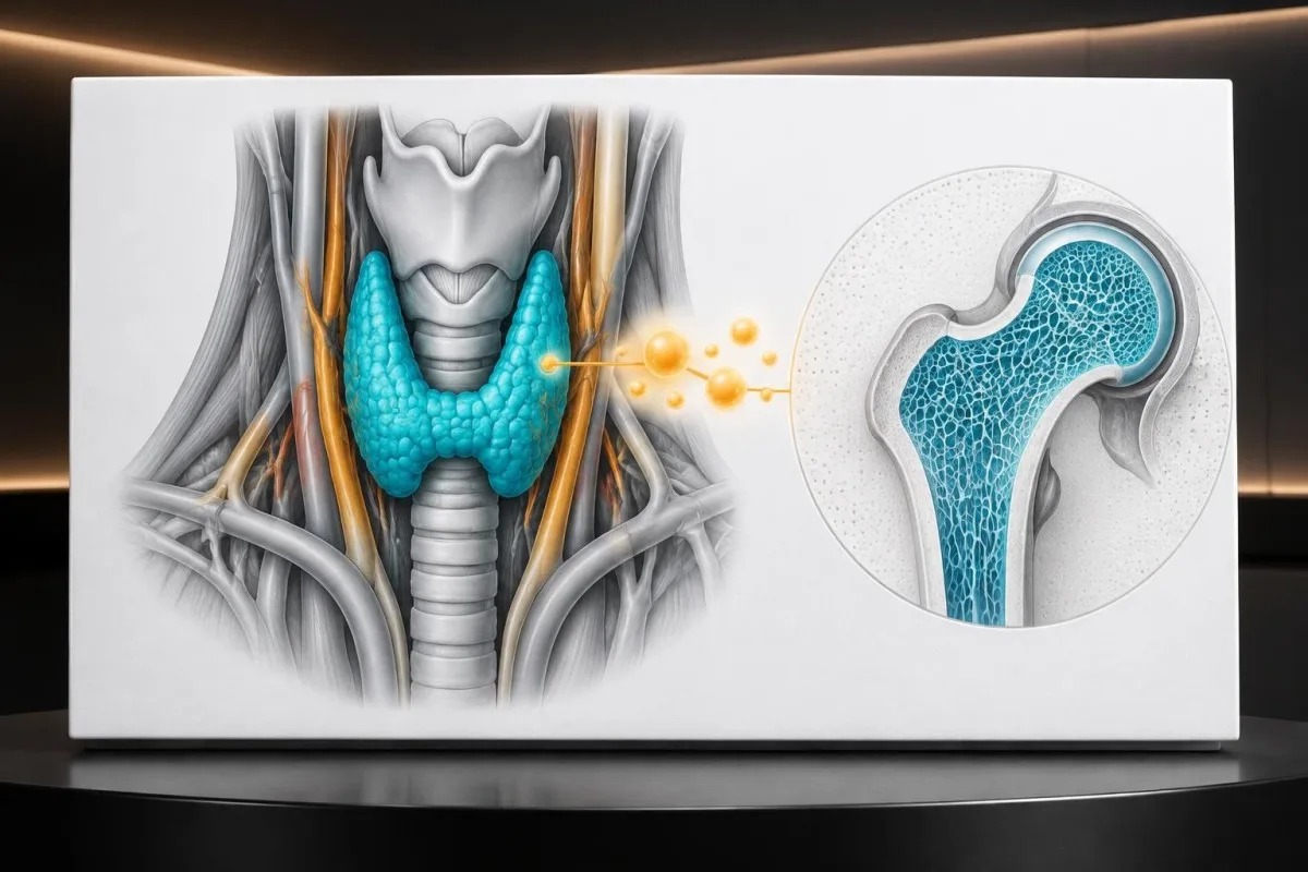
After a mildly low TSH with normal hormones, repeat testing at approximately 2–3 months often establishes whether the abnormality persists. If the result is stable and low-risk, monitoring every 6–12 months may be considered, although the interval should shorten after medication changes or new symptoms.
A trend of TSH 0.31, then 0.18, then 0.07 mIU/L suggests increasing suppression even if free T4 remains normal. Conversely, 0.06, then 0.19, then 0.42 mIU/L suggests recovery and may support a transient cause.
Kantesti can compare uploaded reports over time, but an automated trend cannot know whether a person was ill, pregnant, taking biotin or changing levothyroxine. Our AI trend analysis workflow is best used to organise questions for a clinician.
What should be tracked between tests?
Record resting pulse, palpitations, tremor, weight change, sleep, heat intolerance and medication changes for 2–4 weeks. A symptom diary is especially useful when clinic observations are normal but episodes occur at home.
For people with cardiovascular risk, an ECG or ambulatory monitor may be repeated according to symptoms rather than thyroid numbers alone. For people with skeletal risk, bone-density follow-up follows osteoporosis guidance and fracture history.
A return to TSH 1.0 mIU/L does not necessarily prove that the original result was meaningless; recovery after thyroiditis can be genuine evidence of a temporary trigger.
How should you read your thyroid report?
Read TSH, free T4 and T3 together, then compare each value with the laboratory’s own range, units, collection date and medication context. A red flag beside TSH is not the same as a diagnosis, and a result marked normal can still be clinically relevant when the trend is changing.
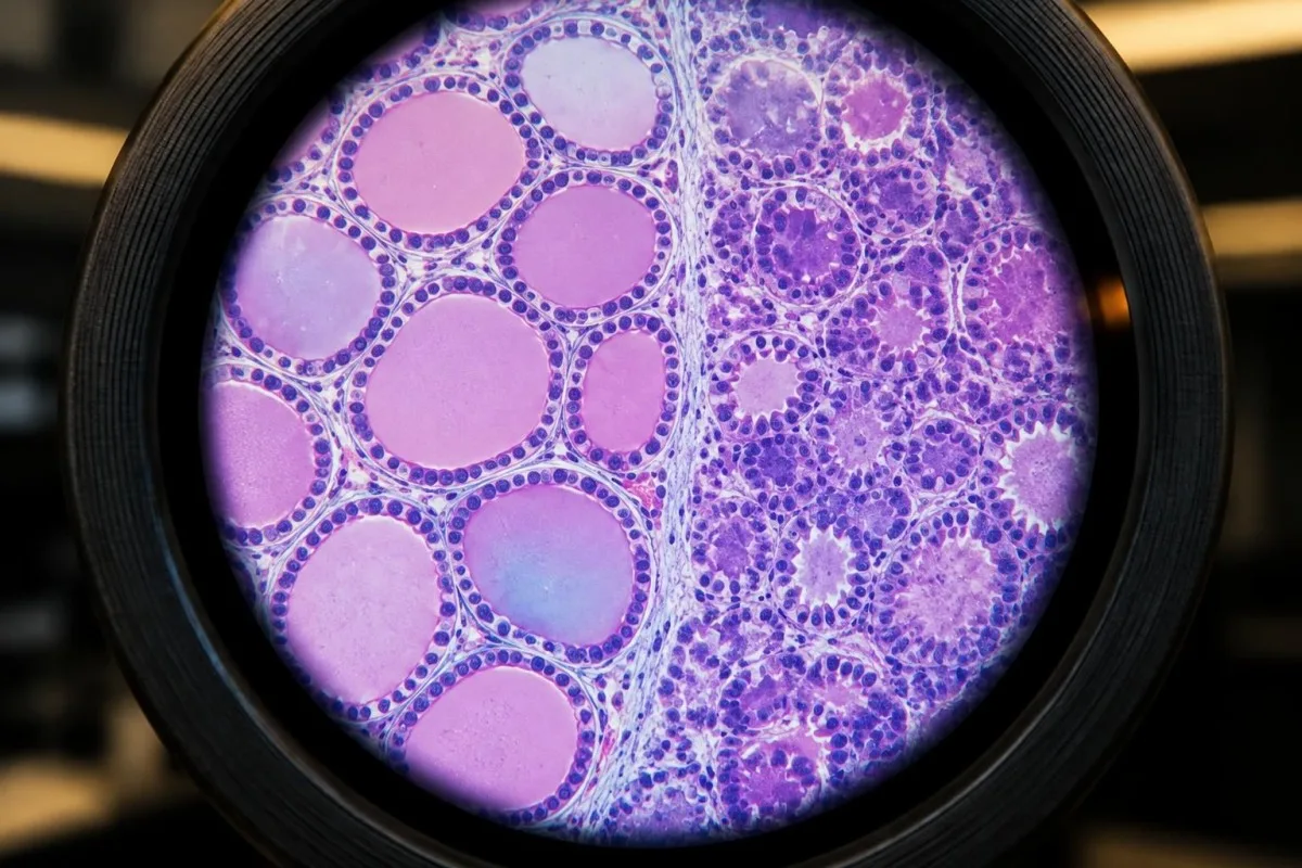
Start with the exact TSH value and whether the report uses mIU/L or another unit; then check whether free T4 and free T3 are within the printed range. A TSH of 0.28 mIU/L is not interpreted as 0.28 ng/dL, because the units describe different measurements.
Next, identify whether the person takes levothyroxine, liothyronine, amiodarone, steroids, dopamine-related medicines or high-dose biotin. The same TSH can mean dose excess in one person, early Graves disease in another and recovery from thyroiditis in a third.
Our blood-test biomarker guide explains how Kantesti’s AI blood test interpretation platform groups related results rather than presenting a single flagged value in isolation.
What automated interpretation can and cannot do
Kantesti can help translate units, identify missing companion tests and organise a chronological pattern, but it cannot palpate a thyroid, assess an irregular pulse or decide whether radioiodine is appropriate. Interpretation remains a support step, not a prescription.
A useful report summary includes the result, reference range, date, medication timing, recent illness and symptoms. That information often changes the next step more than a small difference of 0.05 mIU/L.
If the report is incomplete—for example, TSH is present but free T4 is missing—ask whether the panel should be completed before drawing conclusions.
What is a sensible next-step care plan?
A sensible plan is to confirm persistence, identify the cause, assess heart and bone risk, and agree on a monitoring or treatment interval with a clinician. Seek urgent care for severe cardiovascular symptoms rather than waiting for a routine repeat thyroid test.

For a stable adult with TSH 0.20 mIU/L, normal free hormones and no risk factors, the next step may be repeat testing in 2–3 months with medication and illness review. For persistent TSH 0.05 mIU/L in an older adult, earlier endocrinology or primary-care review is more appropriate.
Urgent symptoms include chest pain, fainting, severe breathlessness, new neurological symptoms or a sustained very rapid or irregular heartbeat. These signs may represent arrhythmia or another emergency and should not be explained away as mild thyroid disease.
Kantesti’s Medical Advisory Board information describes the role of clinical oversight in health content; our platform is an aid for understanding laboratory data, not a substitute for urgent or personalised medical care.
Questions to ask at the appointment
Ask whether the pattern is grade 1 or grade 2, whether the result has persisted for at least 2–3 months, and which cause is most likely. Ask specifically whether an ECG, thyroid antibodies, ultrasound, uptake scan or bone-density assessment is justified.
Clarify the follow-up date and the result that would change the plan—for example, TSH below 0.10 mIU/L, raised free T4, new atrial fibrillation or a new fracture. A defined trigger is safer than an open-ended instruction to monitor.
Bring every supplement and medicine, including products described as natural thyroid support. Natural does not mean free of thyroid hormone or iodine, and undisclosed ingredients can make repeat results difficult to interpret.
Frequently asked questions
What does low TSH with normal T4 and T3 mean?
Low TSH with normal free T4 and free T3 is the laboratory pattern called subclinical hyperthyroidism. TSH between 0.10 and 0.39 mIU/L is commonly considered mild suppression, while TSH below 0.10 mIU/L is more marked. One result is not enough to prove persistent disease because illness, thyroiditis, medicines, pregnancy-related hCG and biotin interference can temporarily lower TSH.
Can subclinical hyperthyroidism go away on its own?
Subclinical hyperthyroidism can resolve when the cause is temporary, such as thyroiditis, acute illness, medication excess or assay interference. A repeat TSH, free T4 and T3 after about 2–3 months often shows whether the pattern persists. Persistent TSH below 0.10 mIU/L deserves more active evaluation, especially in adults aged 65 years or older or people with heart or bone risk.
What symptoms can subclinical hyperthyroidism cause?
Subclinical hyperthyroidism symptoms may include palpitations, tremor, heat intolerance, sweating, anxiety, weight loss, frequent stools and poor sleep, but many people have no symptoms. Symptoms can occur even when free T4 and free T3 are normal, while fatigue and anxiety may have unrelated causes. A new irregular pulse or resting heart rate above 100 beats per minute warrants medical review.
When should subclinical hyperthyroidism be treated?
Treatment is generally considered when TSH remains below 0.10 mIU/L in adults aged 65 years or older, or in people with atrial fibrillation, other heart disease, osteoporosis or significant symptoms. Younger adults with TSH between 0.10 and 0.39 mIU/L and no risk factors are often monitored rather than treated immediately. The correct treatment depends on whether the cause is Graves disease, an autonomous nodule, thyroiditis or excess prescribed thyroid hormone.
Does subclinical hyperthyroidism affect the heart or bones?
Persistent subclinical hyperthyroidism is associated with a higher risk of atrial fibrillation and fractures, particularly when TSH is below 0.10 mIU/L. A pooled analysis reported an approximately 1.68-fold higher atrial-fibrillation risk, while fracture risk is most concerning in postmenopausal adults and people with other osteoporosis risks. Risk assessment should include age, prior fracture, pulse rhythm, medication use and the duration of TSH suppression.
How often should thyroid tests be repeated after a low TSH?
Stable adults with mildly low TSH and normal free hormones are often retested with TSH, free T4 and T3 after approximately 2–3 months. Testing may need to occur sooner when TSH is nearly undetectable, symptoms are significant, pregnancy is involved or thyroid medication has changed. Once a low-risk pattern is confirmed as stable, follow-up intervals may extend to 6–12 months under clinical supervision.
Get your blood test results explained
Upload your lab report and Kantesti's AI explains each biomarker in plain language within seconds. Used by more than 2 million people worldwide.
References
- Klein, T., Mitchell, S., & Weber, H. (2026). aPTT Normal Range: D-Dimer, Protein C Blood Clotting Guide. Kantesti AI Medical Research. DOI
- Klein, T., Mitchell, S., & Weber, H. (2026). Serum Proteins Guide: Globulins, Albumin & A/G Ratio Blood Test. Kantesti AI Medical Research. DOI
- Ross DS et al. (2016). 2016 American Thyroid Association Guidelines for Diagnosis and Management of Hyperthyroidism and Other Causes of Thyrotoxicosis. Thyroid. DOI PubMed
- Collet TH et al. (2012). Thyroid Function and the Risk of Coronary Heart Disease and Mortality. Archives of Internal Medicine. DOI PubMed
- Blum MR et al. (2015). Subclinical Thyroid Dysfunction and Fracture Risk: A Meta-analysis. JAMA. DOI PubMed
- 2M+Tests analysed
- 127+Countries
- 100+Languages
About the medical team


E-E-A-T Trust Signals
Experience
Physician-led clinical review of lab interpretation workflows.
Expertise
Laboratory medicine focus on how biomarkers behave in clinical context.
Authoritativeness
Medically reviewed by Dr. Thomas Klein, with contributions from Dr. Sarah Mitchell and Prof. Dr. Hans Weber.
Trustworthiness
Evidence-based interpretation with clear follow-up pathways to reduce alarm.



Medical disclaimer
This article is for educational purposes only and does not constitute medical advice. Always consult a qualified healthcare provider for diagnosis and treatment decisions.
Published · Contact the editorial team
Published by Kantesti Ltd, registered in England & Wales, company no. 17090423, London, United Kingdom.