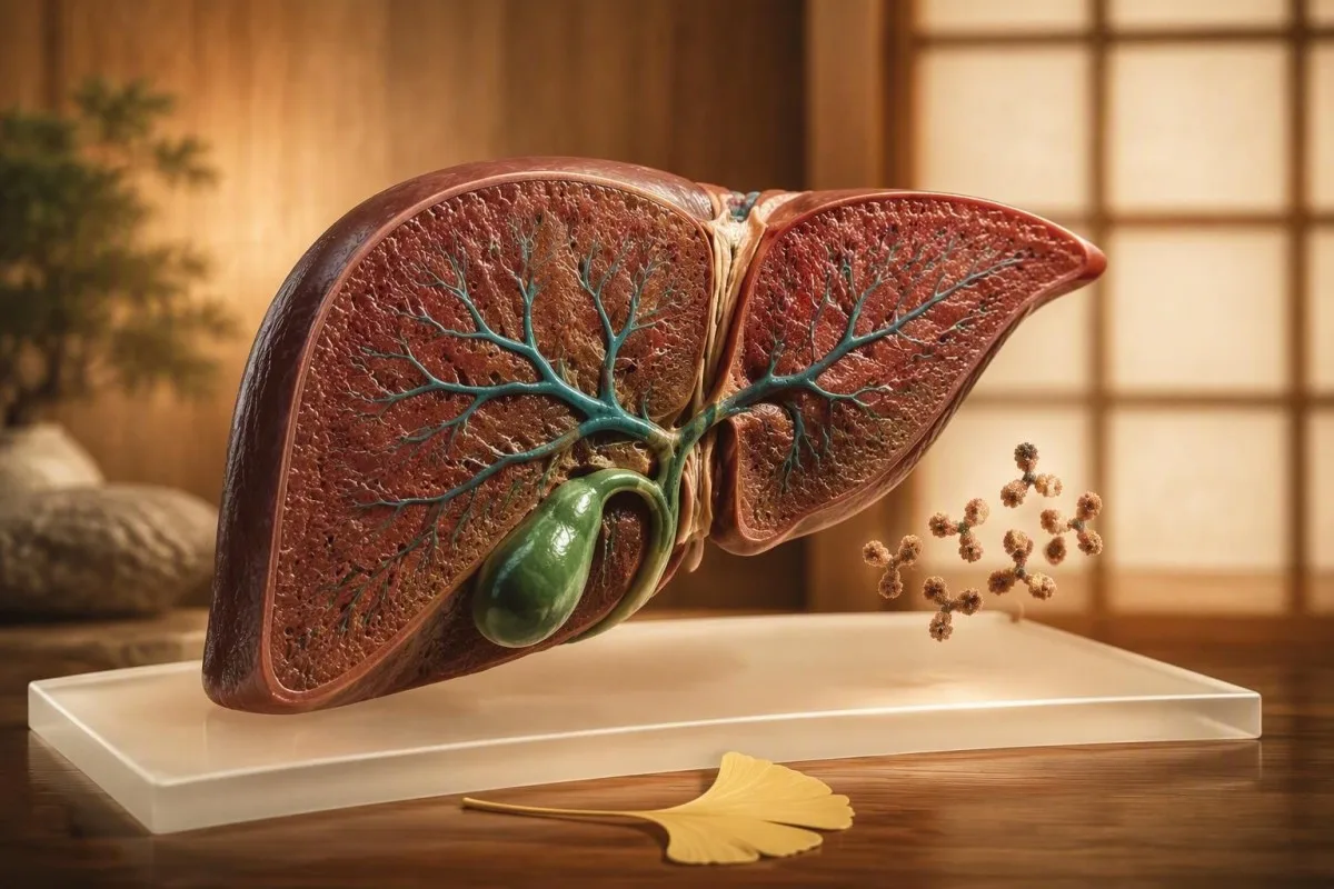
A positive AMA can be a powerful clue to primary biliary cholangitis, but it does not diagnose liver disease by itself. The alkaline phosphatase pattern, symptoms, imaging, and trend over time determine what happens next.


- Positive AMA is found in about 90-95% of people with primary biliary cholangitis (PBC), but an antibody alone does not prove active disease.
- AMA-M2 is the PBC-associated antibody subtype most often measured by ELISA or immunoblot testing.
- Alkaline phosphatase (ALP) above the laboratory upper limit, especially persistently for 6 months or more, is the central blood-test clue to active PBC.
- GGT elevation supports a cholestatic liver pattern when ALP is raised, but GGT alone is not specific for PBC.
- Normal ALP and bilirubin with a positive AMA usually calls for planned monitoring rather than immediate treatment in an otherwise well person.
- Bilirubin above the reference range, low albumin, a raised INR, or falling platelets can signal more advanced liver involvement and need prompt specialist review.
- Itching, fatigue, dry eyes, dry mouth, jaundice, dark urine, or pale stools should be discussed with a clinician even if symptoms feel mild.
- PBC treatment most commonly starts with ursodeoxycholic acid at 13-15 mg/kg/day after a hepatology assessment confirms the diagnosis.
What a positive anti-mitochondrial antibody result actually means
A positive anti-mitochondrial antibody (AMA) means your immune system has made antibodies that recognize mitochondrial proteins, most often the E2 component of pyruvate dehydrogenase. It strongly raises suspicion for primary biliary cholangitis (PBC) when alkaline phosphatase is persistently high, but it is not a diagnosis in isolation.
PBC is a chronic autoimmune condition in which small intrahepatic bile ducts are gradually damaged, causing bile-related enzymes to rise. Around 90-95% of patients with established PBC have AMA, whereas only a small minority of people with a positive test have no biochemical or clinical evidence of PBC. The positive anti-mitochondrial antibody meaning therefore depends on the entire liver panel.
When I review an AMA result, I first ask whether the report says AMA, AMA-M2, an indirect immunofluorescence titre, or a numerical ELISA index. A titre of 1:40 or greater is a commonly used positive threshold for indirect immunofluorescence, although assay cutoffs vary between laboratories and countries. A result cannot be compared safely across methods without the lab's own reference interval.
Kantesti is an AI blood test analyzer that reads an AMA result alongside ALP, GGT, bilirubin, albumin, platelets, and prior values rather than treating one antibody flag as a verdict. For a broader primer on lab flags, see our guide to positive antibody results.
Why mitochondria are involved
AMA does not mean that mitochondria throughout the body are being destroyed. In PBC, the most relevant target is the PDC-E2 antigen expressed in the small bile ducts, and the immune response appears unusually concentrated there. That tissue selectivity remains an active research question rather than a fully settled fact.
How AMA patterns relate to primary biliary cholangitis
AMA is highly specific for PBC in the right clinical setting, particularly when ALP is elevated and imaging excludes a blocked large bile duct. Current diagnostic frameworks generally require two of three features: cholestatic liver tests, PBC-specific antibodies, or compatible liver histology.
The 2017 EASL guideline identifies persistent cholestatic liver biochemistry plus AMA as sufficient for diagnosis in many patients, without routine liver biopsy (European Association for the Study of the Liver, 2017). ALP is the anchor because it reflects impaired bile flow or bile-duct injury more directly than ALT or AST. A normal abdominal ultrasound is still useful because ultrasound checks for obstruction, stones, or structural alternatives.
AMA-M2 is the subtype most closely linked with PBC, yet a strongly positive AMA-M2 result does not tell us whether fibrosis is absent, mild, or advanced. In my practice, a person with ALP 240 IU/L and an upper limit of 120 IU/L concerns me far more than someone with the same antibody result and ALP 86 IU/L. The first pattern is approximately 2 times the upper limit of normal and needs hepatology assessment.
A small number of people with PBC are AMA-negative but have PBC-specific antinuclear antibodies such as anti-gp210 or anti-sp100. This is why a negative AMA does not fully exclude PBC when ALP remains elevated. Our explanation of cholestasis blood-test patterns shows why the enzyme pattern matters.
Why an AMA positive result alone is not diagnostic
An AMA positive result with normal ALP and normal bilirubin does not automatically mean you have PBC. It may represent preclinical autoimmunity, a transient or low-level finding, assay variation, or—less often—another autoimmune or liver condition.
The risk of later PBC is real but variable in AMA-positive people whose liver tests are normal. Published cohorts differ because they use different assays, follow-up periods, and referral populations; that uncertainty is clinically meaningful. I would not tell a symptom-free patient with ALP 92 IU/L and an upper limit of 120 IU/L that they have cirrhosis because of one antibody result.
False-positive or nonspecific AMA findings can occur with other autoimmune illnesses, chronic infections, medication-related immune activation, and occasionally in people without identifiable disease. Low-level reactivity is more vulnerable to method-related disagreement than a clearly positive, repeatedly confirmed result. Repeating the identical test at the same laboratory is often more informative than chasing a different assay immediately.
The 2018 AASLD practice guidance advises periodic biochemical follow-up for AMA-positive patients with normal liver tests rather than assuming a PBC diagnosis on serology alone (Lindor et al., 2019). A practical first step is to understand what an out-of-range flag means before making lifestyle or treatment decisions.
Which liver results make a positive AMA more concerning
A positive AMA deserves more urgent follow-up when ALP and GGT are high, bilirubin rises, or liver synthetic markers such as albumin and INR become abnormal. These combinations suggest ongoing cholestasis or impaired liver function rather than an isolated immune marker.
Typical adult ALP reference ranges are roughly 30-120 IU/L, but the upper limit is assay-specific and can be higher in some laboratories. A persistent ALP above the upper limit plus AMA should trigger assessment for PBC after bone sources of ALP and extrahepatic obstruction are considered. GGT helps confirm liver origin when ALP is raised, especially in adults with no recent fracture.
Bilirubin is often normal early in PBC. A bilirubin above the laboratory limit—commonly above 17-21 micromol/L or 1.0-1.2 mg/dL—needs timely clinical review because it may reflect more significant cholestasis, obstruction, or advancing liver dysfunction. Read our focused guide to high bilirubin patterns for the practical distinctions.
Albumin below about 35 g/L, INR above 1.2 in someone not taking warfarin, or platelets below 150 x 10^9/L can indicate reduced hepatic reserve or portal hypertension, although each has non-liver causes. These are not PBC-specific tests; together, however, they change the urgency of referral.
How to read ALP, GGT, ALT and AST together
PBC usually produces a cholestatic pattern: ALP and often GGT rise more than ALT and AST. ALT or AST can be mildly high, but a predominantly hepatocellular pattern should widen the differential diagnosis.
A useful clinical calculation is the R ratio: divide ALT by its upper limit of normal, then divide ALP by its upper limit of normal. An R ratio below 2 supports cholestatic injury, while above 5 supports hepatocellular injury; results between 2 and 5 are mixed. This is a clinician's pattern-recognition tool, not a home diagnosis.
For example, ALP 280 IU/L with an upper limit of 120 and ALT 58 IU/L with an upper limit of 40 gives an R ratio around 0.6. That is a cholestatic pattern and fits PBC more comfortably than ALT 480 IU/L with only modest ALP elevation. The latter raises questions about viral hepatitis, drug-induced injury, autoimmune hepatitis, or fatty liver disease.
Kantesti AI is an AI blood test interpretation platform that compares these enzyme proportions with previous panels, because an ALP moving from 118 to 186 IU/L matters even when both reports are viewed months apart. Our plain-English review of LFT abbreviations can help you identify each component.
Symptoms that should prompt PBC follow-up
The symptoms most associated with PBC are persistent itch, disproportionate fatigue, dry eyes, dry mouth, and later jaundice. Many people have no symptoms at diagnosis, so normal day-to-day functioning does not rule out early disease.
PBC-related itch can be worse late in the evening, after warmth, or during pregnancy, and it may occur without a visible rash. It is not caused by a simple histamine allergy, which is why standard antihistamines often disappoint. Itch that disrupts sleep for 2 or more weeks deserves a clinical conversation, especially with a positive AMA or raised ALP.
Jaundice, tea-colored urine, pale or clay-colored stools, fever with right upper abdominal pain, confusion, or rapid abdominal swelling should not wait for routine follow-up. These can indicate significant obstruction, infection, decompensated liver disease, or another acute problem rather than uncomplicated early PBC. Our review of dark urine warning patterns explains why urine color is only one clue.
Fatigue in PBC is frustratingly nonspecific and does not reliably match ALP level or fibrosis stage. I often also check thyroid function, ferritin, B12, sleep quality, mood, and medication effects rather than assuming liver disease explains every low-energy day.
What clinicians usually check after a positive AMA
After a positive AMA, clinicians usually repeat or review a full liver panel, confirm the antibody method, assess symptoms and medicines, and arrange ultrasound if cholestasis is present. The goal is to establish whether PBC is active and to exclude more immediately treatable causes of abnormal liver tests.
A sensible initial set includes ALP, GGT, ALT, AST, total and direct bilirubin, albumin, INR, full blood count, immunoglobulins, and often IgM. Raised IgM is common in PBC but does not diagnose it; a normal IgM does not exclude it. Viral hepatitis testing and a medication and supplement review are also common.
Ultrasound is not used to see microscopic PBC bile-duct changes directly. It is used to exclude gallstones, duct dilatation, masses, and obvious structural liver disease when ALP is high. Magnetic resonance cholangiopancreatography may be considered when ultrasound or the biochemical pattern suggests larger-duct disease.
Kantesti is an AI-powered blood test analysis tool that organizes repeat liver panels into a trend view, which can make a 6- to 12-month change easier to discuss at an appointment. Readers preparing for that conversation may find our liver panel checklist useful.
When imaging or a liver biopsy may be needed
A liver biopsy is not routinely required when AMA is positive and the cholestatic liver pattern is clear, but it becomes useful when the pattern is atypical or overlap disease is suspected. Imaging and non-invasive fibrosis assessment often come before biopsy.
A biopsy may be considered when ALT is markedly elevated—often more than 5 times the upper limit of normal—because autoimmune hepatitis overlap becomes more plausible. It may also clarify unexplained AMA-negative cholestasis or competing diagnoses. The tissue result can show bile-duct injury, interface hepatitis, fibrosis, or a different process entirely.
Transient elastography estimates liver stiffness and helps stage fibrosis, but stiffness can rise temporarily with active cholestasis or congestion. A single high reading should be interpreted with bilirubin, ALT, ALP, body habitus, and technical quality in mind. It is useful for trends, not a stand-alone declaration of cirrhosis.
The FIB-4 score uses age, AST, ALT, and platelets, but it was not designed specifically for PBC and can overestimate risk in adults over 65. That limitation is easy to miss when results are generated automatically.
What treatment looks like if PBC is confirmed
Confirmed PBC is usually treated first with ursodeoxycholic acid (UDCA) at 13-15 mg/kg/day, continued long term. Treatment aims to improve cholestatic markers and reduce the likelihood of progression, not merely to make an antibody result disappear.
UDCA dose is calculated from body weight, so a 70 kg adult commonly receives about 910-1,050 mg daily, usually divided according to product and tolerability. The response is assessed with repeat ALP and bilirubin, often at 6 to 12 months. AMA titres are not used to judge treatment success.
Persistent ALP elevation after adequate UDCA can lead a hepatologist to consider second-line treatment, including obeticholic acid in selected patients or fibrates in some regions. Choice depends on fibrosis stage, bilirubin, itching, lipids, kidney function, and local approval; clinicians do not treat the number in isolation. Pregnancy plans also affect medication decisions.
PBC care includes bone-density assessment, fat-soluble vitamin consideration when cholestasis is substantial, and screening for associated autoimmune thyroid disease. For a cautious overview of supplements and liver safety, focus on avoiding unverified products rather than pursuing a liver “cleanse.”
Monitoring when AMA is positive but ALP is normal
People with positive AMA but normal liver chemistry are commonly monitored with repeat liver tests every 6-12 months, although the exact interval should be individualized. Immediate UDCA is not generally started solely because AMA is positive.
Repeat testing should include at least ALP, GGT, ALT, AST, and bilirubin, ideally through the same laboratory when possible. A stable ALP of 88, 91, and 90 IU/L across 18 months is more reassuring than a value that rises from 88 to 145 IU/L while still being discussed as “borderline.” Trends carry clinical weight.
I advise patients to report new itch, jaundice, dark urine, pale stools, or unexplained fatigue rather than waiting passively for the next scheduled draw. Medication changes, a new herbal supplement, pregnancy, intercurrent infection, and substantial weight change should be recorded beside the date of testing. Context prevents false pattern detection.
Our longitudinal blood-test guide explains how to save medication, illness, and fasting context alongside results. Kantesti AI can highlight shifts, but a clinician must decide whether a shift represents PBC, another liver condition, or ordinary biological variation.
Other causes of high ALP that can coexist with AMA
A raised ALP is not automatically liver disease, even when AMA is positive. Bone turnover, recent fracture healing, vitamin D deficiency, pregnancy, medications, and biliary obstruction can raise ALP and may coexist with an incidental AMA result.
GGT is often helpful because a raised GGT alongside ALP supports a hepatobiliary source, while normal GGT with raised ALP may point toward bone—though no single combination is perfect. ALP isoenzyme testing is available in some laboratories. Recent fractures can keep bone ALP elevated for several weeks to months.
A 58-year-old patient I saw had AMA positivity and ALP 164 IU/L, but GGT was 18 IU/L, bilirubin was normal, and she had fractured her wrist 8 weeks earlier. Her repeat ALP normalized after bone healing, and the liver evaluation remained reassuring. This is why I resist labelling every AMA-positive patient with PBC on day one.
If ALP is raised after a fracture, our article on bone-related ALP elevation explains reasonable retest timing. Never use an antibody result as a reason to ignore a clearly plausible alternative cause.
Autoimmune conditions that often travel with PBC
PBC occurs more often alongside autoimmune thyroid disease, Sjogren syndrome, celiac disease, and systemic sclerosis than would be expected by chance. Finding one autoimmune condition should prompt symptom-led assessment for others, not indiscriminate testing.
Dry eyes and dry mouth can reflect Sjogren syndrome, medication effects, menopause, dehydration, or diabetes; the symptom needs context. Thyroid screening with TSH is common because autoimmune thyroid disease is frequent in PBC populations. A TSH outside roughly 0.4-4.0 mIU/L usually warrants interpretation with free T4 and symptoms.
Celiac testing is most useful when there is chronic diarrhea, iron deficiency, unexplained low bone density, weight loss, or a suggestive family history. Testing after removing gluten can produce falsely reassuring results. The gluten challenge guide covers why timing matters.
Dr. Thomas Klein, Kantesti's Chief Medical Officer, advises against ordering broad autoantibody panels simply to search for a reason for tiredness. A positive test can create more ambiguity than clarity when pre-test probability is low.
Questions to bring to a GP or hepatology appointment
The most useful appointment questions are whether your liver pattern is cholestatic, whether ultrasound is needed, whether ALP has changed over time, and when repeat testing should occur. Bring the complete report, not only the highlighted AMA result.
Ask: “Is my AMA result AMA-M2, a titre, or another assay?” and “What was my ALP upper limit of normal?” Those two questions prevent a surprising amount of confusion. Also ask whether GGT, bilirubin, albumin, INR, platelets, and ultrasound findings support a liver source.
If PBC is confirmed, ask how treatment response will be judged at 6 and 12 months, whether you need fibrosis staging, and how itch or fatigue will be managed. A good care plan states the specific result that would trigger escalation rather than simply saying “we will keep an eye on it.” See our blood-test summary checklist before your visit.
Kantesti, an organization supported by clinicians and technical teams, encourages patients to retain original PDF reports and the dates of all previous tests. You can read about our clinical mission on About Us.
When a positive AMA result needs urgent rather than routine care
AMA positivity itself is not an emergency, but new jaundice, fever with upper abdominal pain, confusion, vomiting, black stools, or rapidly increasing abdominal swelling needs urgent medical assessment. These symptoms may reflect obstruction, infection, bleeding, or advanced liver dysfunction.
Seek same-day care for yellow eyes or skin with dark urine and pale stool, especially if bilirubin has risen or pain and fever are present. A bilirubin that changes from 12 to 68 micromol/L over days is not explained by stable early PBC until obstruction and acute hepatitis have been excluded. Do not wait for a routine antibody appointment.
Confusion, drowsiness, vomiting blood, black tar-like stools, or a tense swollen abdomen warrant emergency care. These are possible signs of significant liver decompensation or gastrointestinal bleeding and require assessment even if you have never been diagnosed with PBC. Our overview of cirrhosis warning signs explains the broader pattern.
Kantesti's medical content is reviewed against clinical standards, but an AI interpretation cannot replace urgent examination, imaging, or clinician-directed testing. Review our approach to medical validation and contact your local urgent-care service if red flags are present.
The practical bottom line for an AMA positive result
The practical meaning of a positive AMA is: check the liver pattern, confirm persistence, exclude obstruction and alternative causes, and involve hepatology when ALP is elevated or symptoms suggest cholestasis. Most decisions depend more on ALP, bilirubin, and trends than on how “positive” the antibody appears.
As of September 13, 2026, the core diagnostic logic remains consistent across major guidance: AMA plus chronic cholestatic liver chemistry strongly supports PBC, while isolated AMA warrants surveillance rather than premature disease labelling. If your ALP is normal, a planned 6-12 month repeat panel is commonly reasonable. If ALP is high, arrange clinical follow-up rather than repeating only the antibody.
In 15 years of clinical practice, I have found that patients cope better when we separate “a risk signal” from “active liver damage.” Dr. Thomas Klein's rule of thumb is simple: a positive antibody starts a clinical question; it does not finish it. The full report, symptoms, ultrasound, and time course supply the answer.
For review by our physicians and clinical governance team, visit the Medical Advisory Board. If you use any digital interpretation, keep your clinician involved—especially when bilirubin, INR, albumin, platelets, or symptoms have changed.
Frequently Asked Questions
Can you have a positive AMA and not have primary biliary cholangitis?
Yes. A positive AMA can occur when ALP, GGT, bilirubin, and imaging remain normal, and this pattern does not by itself diagnose primary biliary cholangitis. Many clinicians repeat liver chemistry every 6-12 months in this situation, because some people later develop cholestatic abnormalities while others never do. The most informative next result is usually a trend in ALP, not a repeated antibody titre alone.
How accurate is an anti-mitochondrial antibody test for PBC?
AMA is present in approximately 90-95% of people with established primary biliary cholangitis, making it a highly useful diagnostic marker in the correct setting. Its specificity is high when persistent ALP elevation and compatible clinical findings are present, but no antibody test has 100% diagnostic certainty in an asymptomatic person with normal liver tests. AMA-M2 is the subtype most closely associated with PBC.
What ALP level is concerning with a positive AMA?
Any ALP result persistently above your laboratory's upper limit of normal is relevant when AMA is positive, especially if GGT is also raised. Many adult laboratories use an ALP upper limit near 120 IU/L, but the correct cutoff is the one printed on your report. An ALP around 1.5-2 times the upper limit, such as 240 IU/L when the upper limit is 120 IU/L, usually merits hepatology evaluation.
Can a positive AMA go away?
An AMA result can occasionally become negative or vary between assays, particularly when the original reaction was low level, but this does not reliably indicate that risk has disappeared. In confirmed PBC, treatment decisions are based on ALP, bilirubin, fibrosis assessment, and symptoms rather than whether AMA remains detectable. Repeating an AMA is usually less useful than repeating a full liver panel after 6-12 months.
Does a positive anti-mitochondrial antibody cause symptoms?
AMA itself does not cause symptoms; it is an immune marker. Symptoms arise when associated liver or bile-duct disease is active, with itch, fatigue, dry eyes, dry mouth, jaundice, dark urine, and pale stool among the relevant clues. New jaundice or fever with upper abdominal pain needs same-day medical assessment regardless of the AMA value.
What is the usual treatment if AMA positive PBC is confirmed?
The standard first treatment for confirmed PBC is ursodeoxycholic acid at 13-15 mg/kg/day, prescribed and monitored by a clinician. For a 70 kg adult, that commonly totals about 910-1,050 mg daily, although the precise regimen is individualized. Response is generally assessed after 6-12 months with ALP and bilirubin rather than with AMA titres.
Get AI-Powered Blood Test Analysis Today
Join over 2 million users worldwide who trust Kantesti for instant, accurate lab test analysis. Upload your blood test results and receive comprehensive interpretation of 15,000+ biomarkers in seconds.
📚 Referenced Research Publications
Klein, T., Mitchell, S., & Weber, H. (2026). AI Blood Test Analyzer: 2.5M Tests Analyzed | Global Health Report 2026. Kantesti AI Medical Research.
Klein, T., Mitchell, S., & Weber, H. (2026). RDW Blood Test: Complete Guide to RDW-CV, MCV & MCHC. Kantesti AI Medical Research.
📖 External Medical References
⚕️ Medical Disclaimer
This article is for educational purposes only and does not constitute medical advice. Always consult a qualified healthcare provider for diagnosis and treatment decisions.
E-E-A-T Trust Signals
Experience
Physician-led clinical review of lab interpretation workflows.
Expertise
Laboratory medicine focus on how biomarkers behave in clinical context.
Authoritativeness
Written by Dr. Thomas Klein with review by Dr. Sarah Mitchell and Prof. Dr. Hans Weber.
Trustworthiness
Evidence-based interpretation with clear follow-up pathways to reduce alarm.