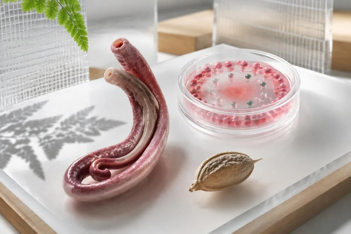
Dodatni wynik badania przeciwciał przeciwko Schistosoma może uzasadniać leczenie po wiarygodnym narażeniu na kontakt ze słodką wodą, jeśli nigdy nie zastosowano u Ciebie odpowiedniego leczenia, ale nie może potwierdzić aktywnego zakażenia ani wyleczenia. O dalszym postępowaniu decydują daty narażenia, wcześniejsze leczenie i dodatkowe badania.

Najważniejsze wnioski
- Dodatnie przeciwciała wskazują na odpowiedź immunologiczną na antygeny Schistosoma; 1 dodatni wynik nie pozwala odróżnić nieleczonego zakażenia od zakażenia leczonego wcześniej.
- Okres okna diagnostycznego ma znaczenie: CDC zaleca pobranie próbki do badania przeciwciał co najmniej 6–8 tygodni po podejrzeniu nowego zakażenia.
- Narażenie na kontakt ze słodką wodą oznacza kontakt skóry z potencjalnie skażonymi jeziorami, rzekami lub kanałami, a nie samo odwiedzenie kraju, w którym występuje choroba.
- Badanie mikroskopowe jest bardziej miarodajne, gdy lekarze zlecają zbadanie 3 próbek kału lub moczu pobranych w różne dni.
- Prazikwantel typowa całkowita dawka wynosi 40 mg/kg w ciągu 1 dnia w przypadku S. mansoni i S. haematobium, jednak wybór leczenia zależy od gatunku i okoliczności klinicznych.
- Czas leczenia zwykle oznacza odczekanie co najmniej 6–8 tygodni od ostatniego kontaktu ze słodką wodą u stabilnych klinicznie podróżnych, ponieważ prazikwantel jest najskuteczniejszy wobec dojrzałych robaków.
- Kontrolne badanie mikroskopowe może być zlecone 1–2 miesiące po leczeniu, gdy przed leczeniem obecne były jaja.
- Utrzymujące się IgG mogą pozostawać dodatnie przez lata po skutecznym leczeniu; kolejny dodatni wynik nie uzasadnia automatycznie kolejnego cyklu leczenia.
Czy dodatni wynik badania przeciwciał przeciwko Schistosoma wymaga leczenia?
Dodatni wynik testu na przeciwciała przeciwko Schistosoma nie oznacza automatycznie, że potrzebujesz leczenia. Lekarze często zalecają leczenie, gdy kontakt z wodą słodką jest wiarygodny, a wcześniej nie zastosowano odpowiedniego leczenia, nawet jeśli wynik badania mikroskopowego jest ujemny; po udokumentowanym leczeniu i bez ponownego narażenia 1 utrzymujący się dodatni wynik zwykle nie wystarcza, by uzasadnić ponowne leczenie.
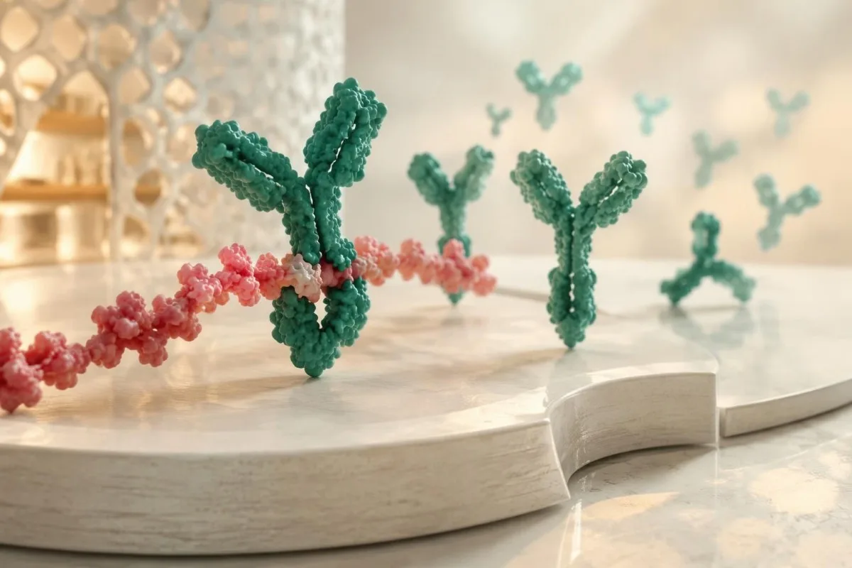
Znaczenie dodatniego wyniku Schistosoma IgG najlepiej rozumieć jako dowód rozpoznania przez układ odpornościowy, a nie pomiar liczby żywych robaków. Odpowiedź przeciwciał może utrzymywać się przez lata po zniknięciu pasożyta, a 2 osoby z takim samym zgłoszonym wynikiem mogą wymagać różnego postępowania — jedna po nieleczonym narażeniu, druga po skutecznym leczeniu, bez późniejszego kontaktu z wodą słodką.
Wcześniejsze narażenie i wcześniejsze leczenie to odrębne kwestie: dawna infekcja może pozostać nieleczona, natomiast dawny dodatni wynik testu może utrzymywać się po wyleczeniu. Przed zinterpretowaniem 1 wyniku jako oznaki aktywnej choroby lub niegroźnego śladu przebytej infekcji sprawdź nazwę leku, dawkę, datę i ewentualny późniejszy kontakt z wodą; nasze wyjaśnienie dodatnich wyników przeciwciał rozróżnia te różne rodzaje dowodów.
Kantesti to analizator badań krwi oparty na AI, który pomaga wyjaśnić, dlaczego 1 dodatni wynik Schistosoma IgG nie pozwala potwierdzić aktywnej infekcji, nawet gdy morfologia krwi jest prawidłowa. Naszą rolą jest objaśnianie wyników, a nie identyfikacja pasożytów ani przepisywanie leków; nasze organizacja i cel należy rozumieć odrębnie od opisanych tutaj decyzji podejmowanych pod kierunkiem lekarza.
Które narażenia na kontakt ze słodką wodą zwiększają wiarygodność wyniku?
Kontakt skóry z wodą słodką na obszarze występowania transmisji sprawia, że dodatni wynik ma większe znaczenie kliniczne. Pływanie, brodzenie, kąpiel, rafting lub mycie w jeziorach, rzekach i kanałach irygacyjnych mogą prowadzić do narażenia; zapisz 3 szczegóły dotyczące każdego zdarzenia: dokładne miejsce, rodzaj aktywności i datę ostatniego kontaktu.

Larwy Schistosoma uwalniane przez ślimaki słodkowodne wnikają przez skórę; połknięcie wody nie jest konieczne. Nawet krótkie brodzenie liczy się jako narażenie, a brak swędzącej wysypki go nie wyklucza, dlatego hipotetyczny podróżny, który wszedł do jeziora na 5 minut, powinien wspomnieć o tym zdarzeniu, zamiast odnotowywać wyłącznie dłuższe sesje pływania.
Woda morska i prawidłowo chlorowane baseny nie są typowymi źródłami narażenia na schistosomatozę. Kontakt z wodą słodką może również budzić obawy o inne choroby wymagające odmiennych badań i leczenia, w tym leptospirozę po kontakcie z wodą; gorączki rozpoczynającej się 1 tydzień po aktywności nad rzeką nie należy przypisywać przeciwciałom przeciwko Schistosoma bez szerszej oceny.
Sama nazwa kraju nie pozwala ustalić indywidualnego ryzyka, ponieważ transmisja różni się w obrębie krajów i zbiorników wodnych. Odtwórz nawet 1 zapomnianą aktywność — na przykład mycie sprzętu nad rzeką — i odróżnij narażenie na pasożyty od dokumentacji szczepień; badania przeciwciał podczas podróży odpowiadają na inne pytanie i nie zapewniają ochrony przed schistosomatozą.
Kiedy po podróży należy wykonać badania w kierunku schistosomatozy?
Badanie przeciwciał przeciwko schistosomatozie jest na ogół bardziej miarodajne co najmniej 6–8 tygodni po podejrzeniu nowej infekcji. Wytyczne CDC dotyczące badań klinicznych i diagnozowania schistosomatozy wyjaśniają, że taki odstęp pozwala na wytworzenie przeciwciał; ujemny wynik uzyskany wcześniej nie pozwala wiarygodnie wykluczyć niedawnej infekcji nabytej w wyniku kontaktu z wodą słodką.
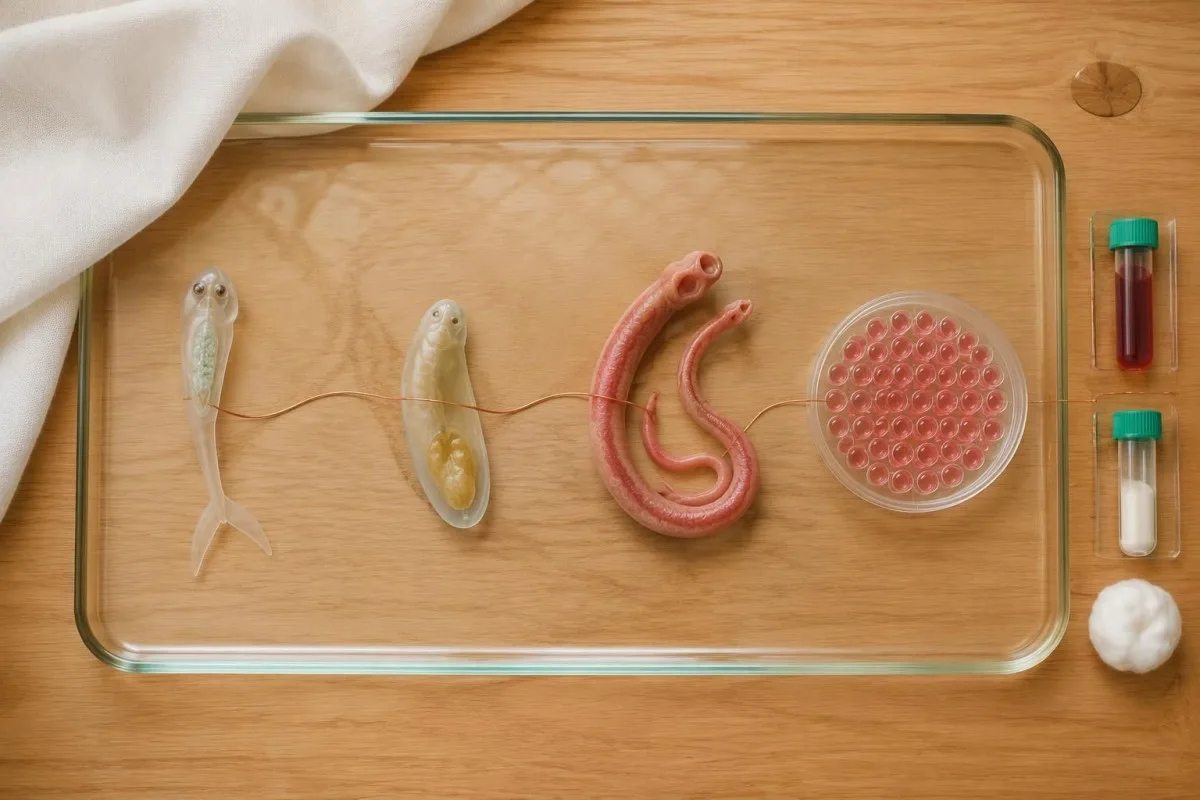
The last freshwater contact is a practical anchor when a trip involved several possible exposures. For example, a hypothetical traveler who swam on both the first and final day of a 3-week holiday should calculate the testing interval from the final swim, rather than assuming that the first exposure makes an immediate post-trip result conclusive.
Seroconversion is not identical across people or assays, so 6–8 weeks is a useful testing window rather than a guarantee of detection. When exposure is persuasive and an initial result is negative, a clinician may arrange another sample later, sometimes around 12 weeks after exposure; a second date should be agreed rather than inferred from a generic laboratory reference range.
Fever or significant symptoms after travel should be assessed without waiting for an antibody window to close, especially after a malaria-risk itinerary. At Kantesti, we encourage readers to keep 2 dates visible—last freshwater contact and sample collection—because czasie testowania infekcji can change the meaning of a negative report even though each organism requires its own diagnostic strategy.
Czy wynik badania przeciwciał przeciwko Schistosoma może być fałszywie dodatni?
Schistosoma antibody tests can produce false-positive or cross-reactive results, and their performance varies by assay. There is no universal IgG concentration or index that proves active schistosomiasis; 2 laboratories may use different antigen preparations, reporting units and thresholds, making direct numerical comparisons unreliable.
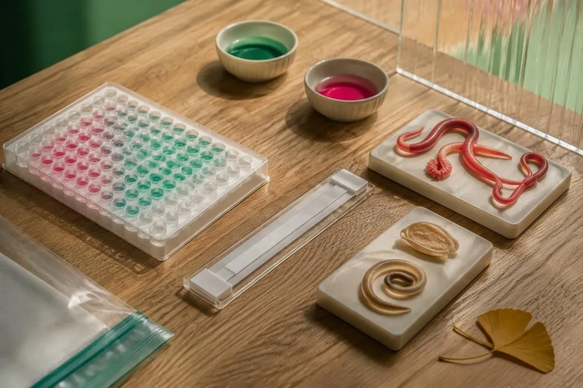
Cross-reactivity with other helminth antibodies depends on the test rather than occurring uniformly in every person. A laboratory may recommend an alternative assay or reference-laboratory review when the exposure history does not fit; 1 additional positive parasite antibody result also needs independent interpretation, as explained in our discussion of Toxocara antibody findings.
A hypothetical probability example shows why background risk matters: assume an imaginary test has 90% sensitivity and 95% specificity in 10,000 people, of whom 1% have the target infection. It would identify approximately 90 true positives and 495 false positives, so only about 15% of positive results would be true positives—these are teaching assumptions, not measured Schistosoma assay performance.
A high index should not be read as a high worm count, and a borderline index should not automatically be dismissed when exposure is strong. Ask for the assay name and the laboratory's interpretation of 1 equivocal or weakly positive result; confirmation with a different method can address assay uncertainty, but another antibody test still cannot reliably separate active infection from treated history.
Co można ustalić na podstawie mikroskopowego badania kału i moczu?
Finding characteristic Schistosoma eggs provides direct parasitological evidence, whereas negative microscopy does not exclude a light infection. CDC recommends examining 3 stool or urine samples collected on different days to improve sensitivity, because egg output can be sparse and intermittent, particularly in travelers with a low parasite burden.
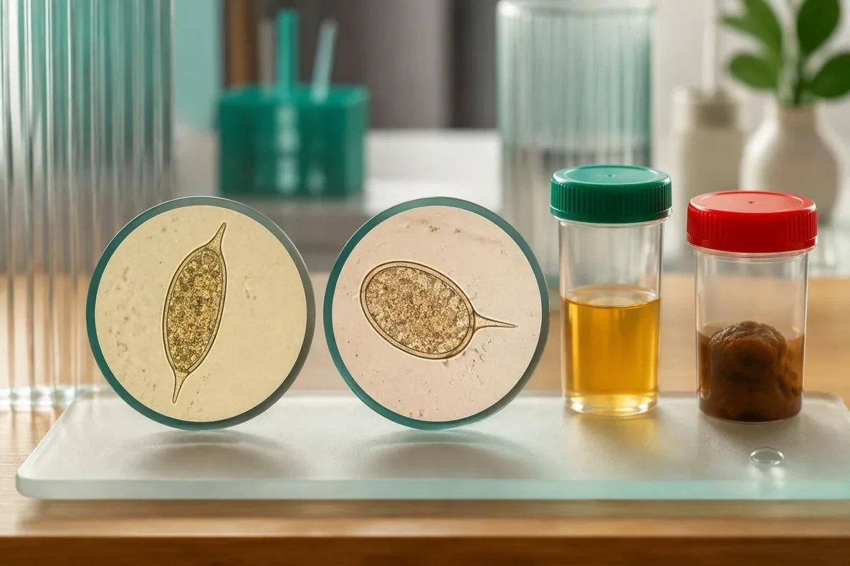
The suspected species determines specimen choice: S. mansoni and S. japonicum are usually investigated through stool, while S. haematobium is usually investigated through urine. Some exposure histories warrant both, so 1 negative stool examination does not address a possible urinary infection; interpretacji osadu moczu also explains why ordinary urinary findings are not equivalent to parasite identification.
Urinary egg excretion often peaks around midday, and laboratories may request a sample between approximately 12 noon and 3 pm. Urine filtration may use a measured volume such as 10 mL, but collection, transport and processing instructions vary; follow the receiving laboratory's protocol rather than improvising exercise, fluid restriction or a collection time.
Routine dipstick testing is not a substitute for a specific Schistosoma egg examination, and 1 routine stool pathogen panel may omit the parasite entirely. Check the exact tests ordered, including concentration or filtration methods when appropriate; our urinalysis method guide helps distinguish screening for cells and chemistry from targeted parasitology.
Czy testy antygenowe lub PCR wyraźniej wskazują na aktywne zakażenie?
Schistosoma antigen tests can provide evidence more closely linked to living worms than antibody tests, but access and performance differ. Circulating anodic antigen, or CAA, and circulating cathodic antigen, or CCA, are 2 distinct targets; PCR detects parasite DNA and needs interpretation according to specimen, timing and the laboratory method.
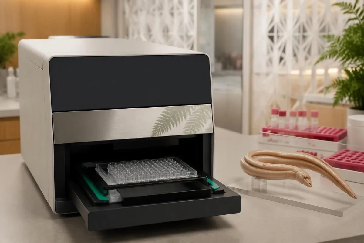
CAA assays can detect antigens released by living schistosomes, including some low-burden infections that microscopy misses. Urine CCA testing is used especially for S. mansoni, but trace readings and performance for other species complicate interpretation; 1 negative point-of-care CCA test should not be treated as a universal exclusion of urinary or intestinal schistosomiasis.
PCR may support diagnosis when microscopy is negative, but DNA detection is not identical to proving that every detected parasite component remains viable after treatment. The specimen matters—serum, stool and urine are not interchangeable—and 1 positive molecular result must be interpreted against the laboratory's validated application; antibodies versus direct detection illustrates this distinction in a different parasitic condition.
Kantesti is an AI lab test interpretation service that can explain the difference between 2 reports labeled Schistosoma IgG and Schistosoma PCR without treating them as interchangeable evidence. Our metodach interpretacji AI do not replace reference-laboratory expertise, and an antigen test's usefulness depends on whether the local service can obtain and validate it for your clinical question.
Które towarzyszące wyniki badań krwi i moczu mają znaczenie?
Eosinophilia, urinary abnormalities and signs of organ involvement can strengthen the case for further assessment, but none proves schistosomiasis alone. An absolute eosinophil count above approximately 0.5 × 10⁹/L is commonly considered elevated; a normal count does not exclude infection, especially outside the early immune-response phase.
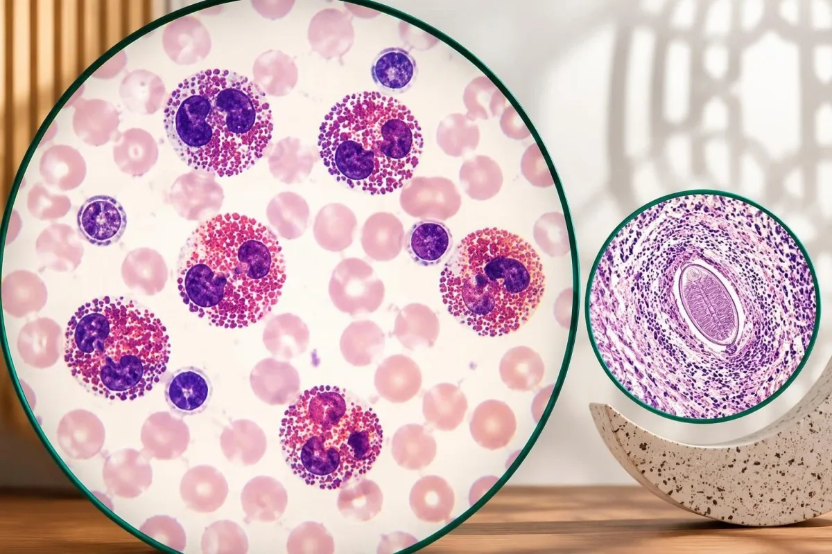
The absolute eosinophil count is usually more informative than the percentage because a percentage depends on the other white-cell populations. A hypothetical count of 0.8 × 10⁹/L may reflect schistosomiasis, another parasite, allergy or a medicine effect; persistent counts at or above 1.5 × 10⁹/L generally warrant broader evaluation, as discussed in our eosinophil count guide.
Normal liver enzymes do not reliably exclude hepatosplenic schistosomiasis because periportal fibrosis and portal hypertension can occur without the usual pattern of cirrhosis. For example, an ALT of 24 IU/L cannot overrule an enlarged spleen or suggestive ultrasound; interpretacją panelu wątrobowego explains why enzyme results answer different questions from structural imaging.
Blood in urine, protein in urine, persistent diarrhea or genital symptoms can change which investigations are appropriate. At Kantesti, we encourage keeping at least 3 evidence categories separate—exposure, parasite tests and organ findings—because a positive antibody result should not obscure a urinary obstruction, significant anemia or another explanation that requires its own assessment.
Dlaczego można zaproponować leczenie mimo ujemnego wyniku badania mikroskopowego?
Clinicians may treat a seropositive person with credible exposure and no adequate treatment history despite negative microscopy. This approach recognizes that light infections can escape egg detection and untreated adult worms can persist for years; the decision balances 3 factors: exposure probability, diagnostic limitations and the person's treatment risks.
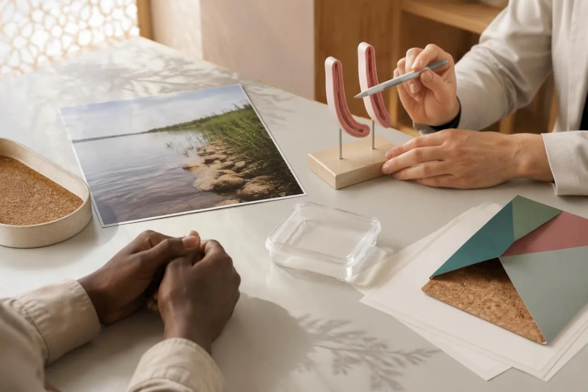
Absence of symptoms does not establish absence of infection or eliminate the potential benefit of treatment. A hypothetical traveler tested 10 weeks after a lake swim, with positive IgG and 3 negative stool examinations, may still be offered praziquantel if the exposure was credible and no earlier treatment occurred; negative microscopy lowers certainty but does not settle the question.
Documented earlier treatment changes the reasoning, particularly when no freshwater re-exposure has occurred. A person with a positive test 5 years after an appropriately timed course does not automatically need another course; the clinician should investigate whether treatment was complete, whether the initial infection was confirmed and whether any symptoms or direct tests now suggest persistence.
Visible blood in urine requires assessment even when Schistosoma antibodies are positive, because 1 parasite result does not exclude stones, other infections or cancer. Chronic urinary schistosomiasis is associated with increased bladder cancer risk, but risk is not diagnosis; our guide to bladder warning symptoms explains why persistent or recurrent urinary changes deserve separate attention.
Jak dobiera się dawkę prazikwantelu i termin leczenia?
Praziquantel is the standard medicine for schistosomiasis, but dose and timing depend on species and clinical circumstances. CDC's Clinical Care of Schistosomiasis guidance lists 40 mg/kg over 1 day in 2 divided doses for S. mansoni, S. haematobium and S. intercalatum, and 60 mg/kg in 3 divided doses for S. japonicum and S. mekongi.
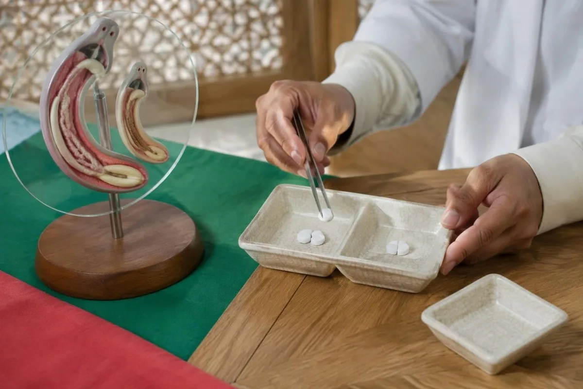
Praziquantel works best against mature worms, so otherwise stable travelers are generally treated at least 6–8 weeks after their last freshwater exposure. Taking it immediately after a swim is not reliable preventive treatment; if a course was given very early, the clinician may plan another course after maturation rather than assuming the initial prescription covered all developing worms.
A clearly hypothetical calculation illustrates a 40 mg/kg regimen: a 60 kg adult would receive 2,400 mg total over 1 day, split into 2 doses of 1,200 mg. Where 600 mg tablets are supplied, that arithmetic corresponds to 2 tablets per dose, but it is not an individual prescription—species, available formulation, rounding, swallowing ability and medical history must be checked.
Repeat treatment after 2–4 weeks may be appropriate in selected patients, especially with light infections or concerns about an incomplete response; it is not required solely because IgG remains positive. Medication review matters because rifampicin and enzyme-inducing antiseizure medicines can reduce praziquantel exposure, so patients taking carbamazepine should bring their regimen and any carbamazepine monitoring information rather than adjusting either medicine themselves.
Które objawy wymagają pilnej konsultacji lub pomocy doraźnej?
Fever after travel needs prompt assessment, and new weakness, seizures or bladder-control changes need emergency evaluation. Acute schistosomiasis can cause fever, cough, rash, diarrhea and eosinophilia around 2–8 weeks after exposure, but these symptoms overlap with other conditions and cannot be diagnosed from antibodies alone.
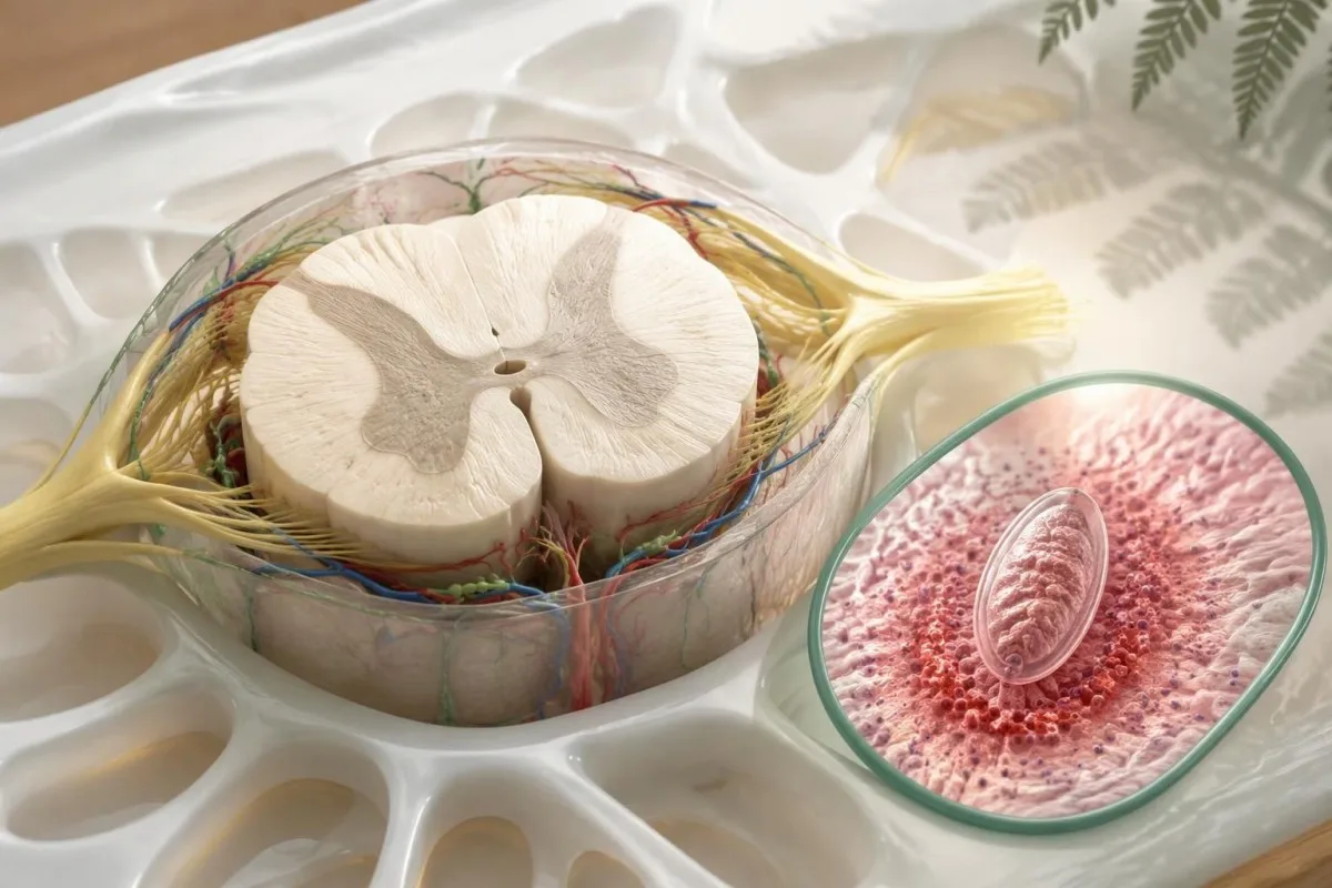
New leg weakness, difficulty walking, urinary retention or loss of bowel control can signal spinal cord involvement and should not wait for routine repeat serology. Schistosoma eggs can occasionally affect neurological tissue, but 1 positive antibody result does not establish the cause; emergency clinicians may need imaging and a broad differential diagnosis before specialist treatment is selected.
Acute systemic or neurological schistosomiasis may require specialist-directed corticosteroids alongside a carefully timed antiparasitic plan. If the exposure history also suggests Strongyloides, assess that possibility before immunosuppression when feasible, because steroids can precipitate dangerous hyperinfection; a Strongyloides IgG result needs its own management rather than assuming 1 antiparasitic medicine treats both organisms.
Fever after a malaria-risk itinerary should be assessed the same day, even if Schistosoma antibodies offer a plausible alternative explanation. Do not wait 6–8 weeks for repeat serology when there is persistent high fever, breathing difficulty, confusion, fainting, significant gastrointestinal bleeding or inability to pass urine; the immediate priority is identifying and treating the acute problem.
Jak ciąża, dzieciństwo i inne schorzenia wpływają na opiekę?
Pregnancy, young age and complex illness require individualized treatment decisions rather than automatically withholding or prescribing praziquantel. The WHO guideline on control and elimination of human schistosomiasis (2022) supports preventive treatment from age 2 years and includes pregnant women after the first trimester and lactating women in eligible programs.
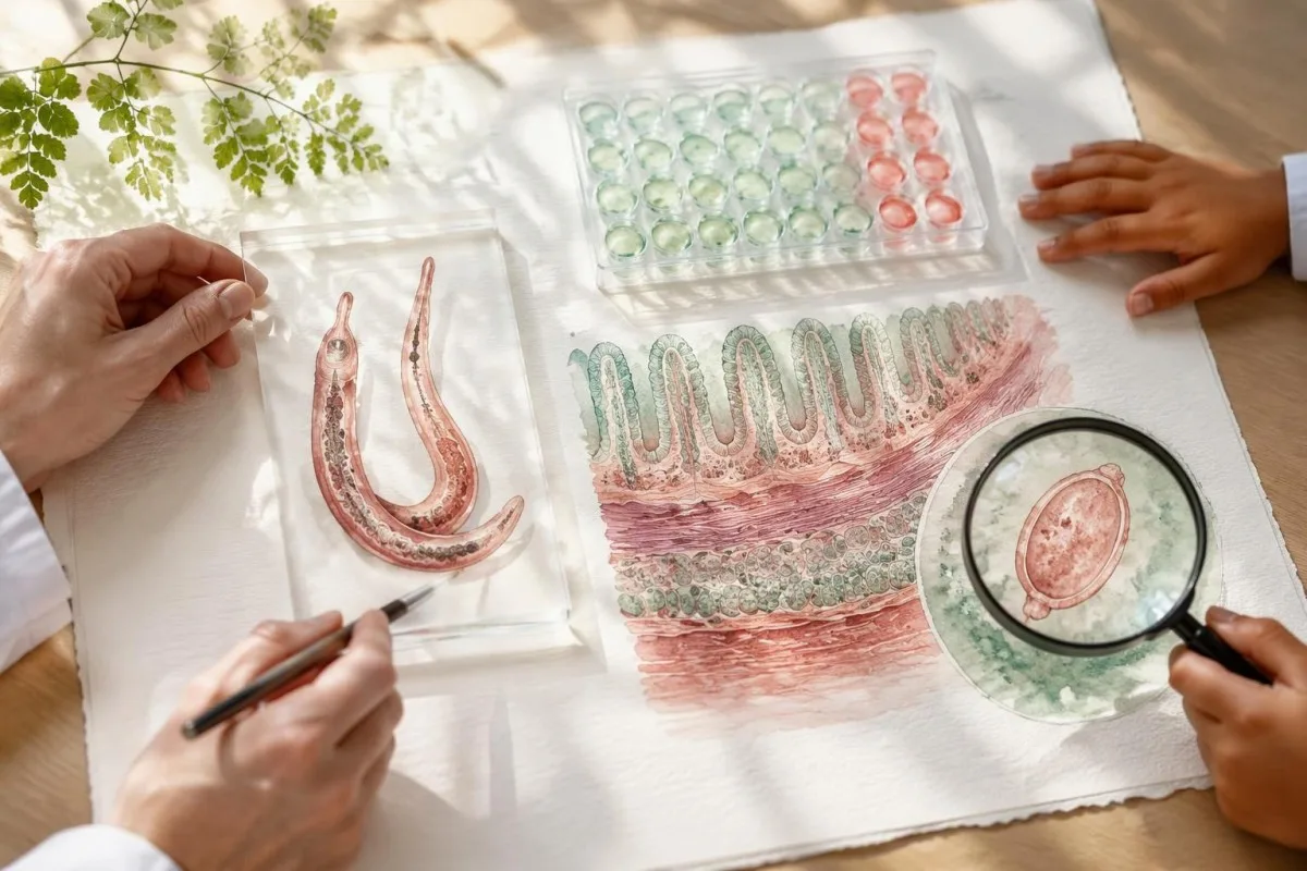
WHO program recommendations and treatment of an individual imported infection answer related but different questions. A hypothetical patient at 10 weeks of pregnancy needs a clinician-led balance of infection likelihood, severity and treatment timing; the recommendation to include pregnancy after the first trimester in preventive programs should not be turned into a blanket rule for every symptomatic patient.
Children's dosing requires an accurate weight and a formulation they can take safely; adult tablet arithmetic is not enough. WHO's inclusion of children aged 2 years and older in preventive chemotherapy does not make every product licensed for every younger age in every country, and our discussion of children's interpretation limits explains why pediatric results need age-specific clinical oversight.
A history of seizures, possible cysticercosis, significant liver disease or neurological symptoms should be reviewed before treatment. For 1 person with a straightforward travel-acquired infection, routine outpatient prescribing may be appropriate; another with possible central nervous system involvement may need specialist planning because the immune response to parasite treatment can itself affect symptoms.
Dlaczego wynik Schistosoma IgG może pozostać dodatni po skutecznym leczeniu?
Schistosoma IgG can remain positive for years after successful treatment and is not a reliable test of cure. CDC recommends considering repeat stool or urine examination 1–2 months after treatment when eggs were detected before treatment; follow-up must match the evidence used to establish infection and the person's subsequent clinical course.
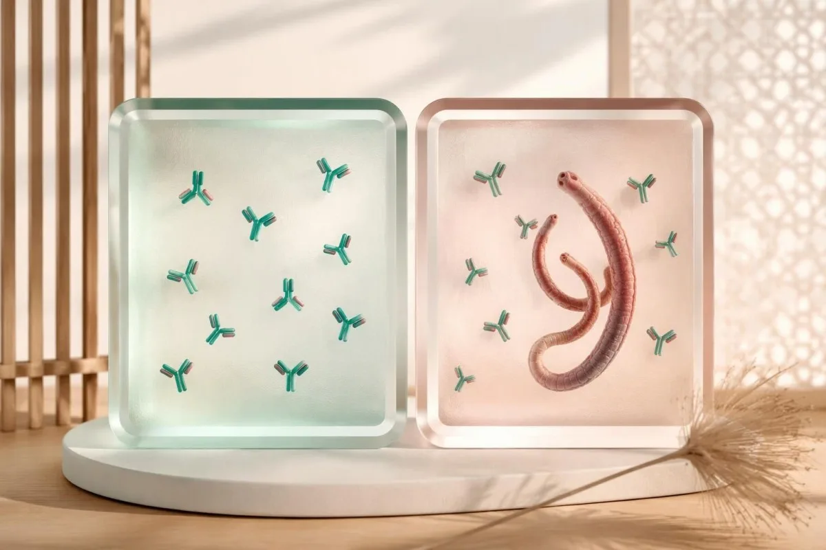
A positive IgG result 6 months after treatment does not, by itself, demonstrate treatment failure. Antibody decline varies, and a stable or temporarily higher signal cannot reliably quantify surviving worms; comparing results from 2 different laboratories adds assay differences, making numerical trend interpretation even less suitable for declaring cure.
Persistent symptoms or eosinophilia deserve reassessment, but neither automatically proves living Schistosoma remain. If eggs are still found at the 1–2 month follow-up, specialists may consider egg viability, collection details, treatment timing and whether another course is needed; PCR positivity can also require caution because detecting parasite DNA is not the same as identifying viable adult worms.
Kantesti can help organize 3 follow-up dates—treatment, specimen collection and any re-exposure—without labeling persistent IgG as treatment failure. Our vademecum analizy trendów lekowych explains why trends must be matched to the right clinical endpoint; an available validated antigen assay may be more useful than serial antibodies, but its follow-up schedule belongs to the treating service.
Jak ten sam dodatni wynik może prowadzić do różnych decyzji?
The same positive Schistosoma IgG result can lead to treatment, additional testing or observation because the surrounding evidence differs. The following 4 examples are hypothetical teaching scenarios—not patient encounters—and illustrate how exposure dates, prior therapy, assay uncertainty and organ findings can outweigh the apparent simplicity of a positive flag.
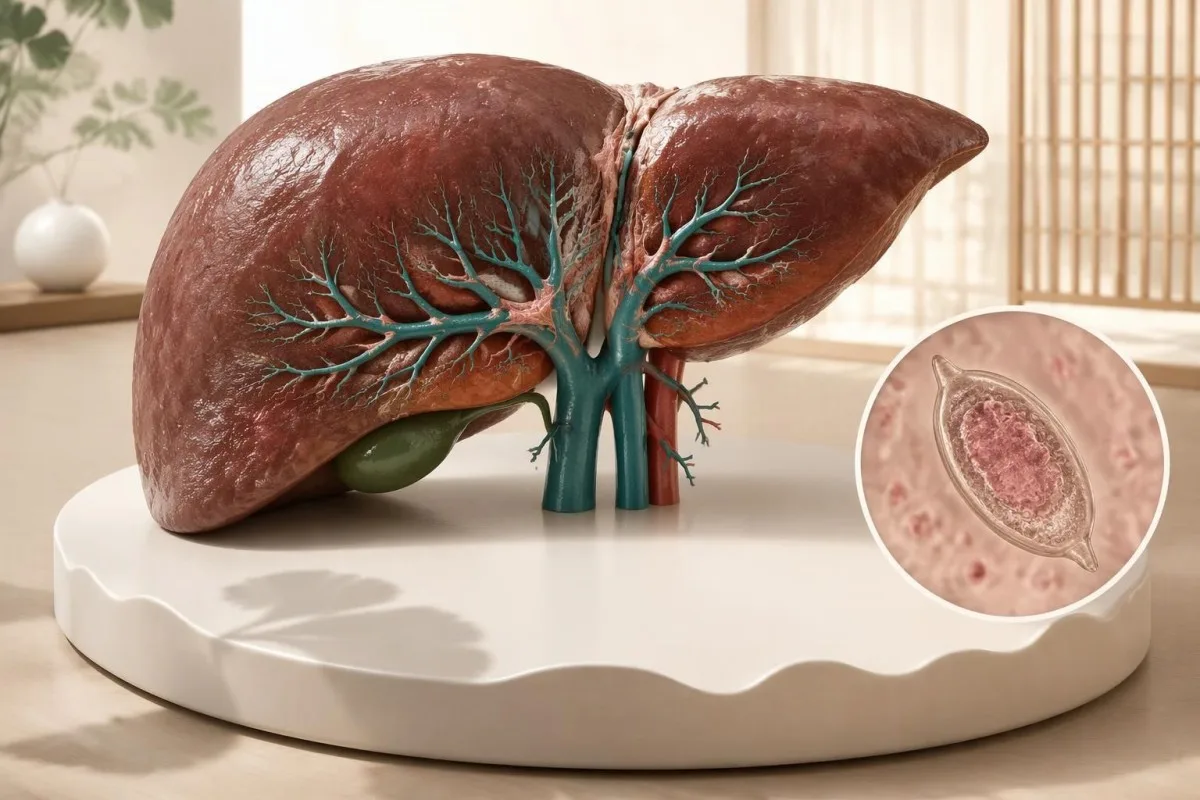
A hypothetical 28-year-old returns from a freshwater swim, tests positive 10 weeks later and has never received praziquantel; treatment may be reasonable despite negative microscopy. A hypothetical 44-year-old treated appropriately 4 years earlier, with no further exposure and no concerning findings, may not need retreatment; the timeline of laboratory results helps keep these histories distinct.
A hypothetical 35-year-old with no plausible freshwater exposure has a weakly positive screening assay and normal supporting investigations. The next step may be checking the original indication, assay and cross-reactivity rather than immediate treatment; changing a report from weak positive to strong positive on 1 repeat antibody assay still does not create a documented exposure or prove active infection.
A hypothetical 54-year-old who lived near endemic freshwater 20 years earlier has positive IgG, an enlarged spleen and an ALT of 24 IU/L within the local range. That combination merits assessment for portal and other organ disease despite normal enzymes; an old exposure is not automatically resolved, and common liver fibrosis scores should not be assumed validated for schistosomal periportal fibrosis.
Co zabrać na wizytę kontrolną i jakie źródła stanowią podstawę opieki?
Bring your exposure history, complete laboratory report and treatment record to a clinician experienced in travel medicine, infectious diseases or tropical medicine. Prepare 3 questions: whether the result fits your exposure, which direct tests could change the decision and how treatment response will be assessed without relying on antibody disappearance.
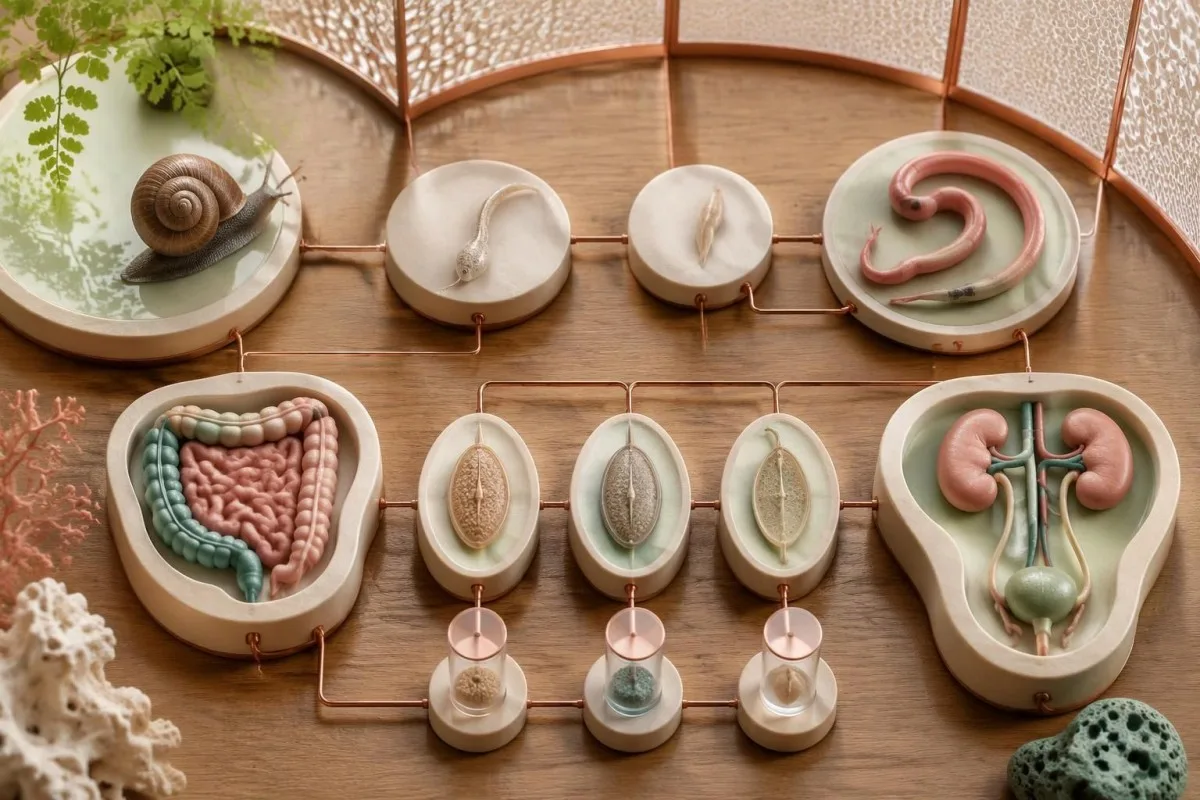
Your report should retain the assay name, units or index, reference interpretation and collection date, rather than only a cropped positive flag. Add 1 concise exposure timeline, previous praziquantel doses and dates, current medicines, pregnancy status where relevant and any urinary or neurological symptoms; this makes uncertainty visible and reduces the chance of repeating an uninformative antibody test.
Kantesti is an AI blood test interpretation platform that helps explain why 1 Schistosoma antibody result must be considered alongside exposure and treatment history, not used as a prescribing instruction. Our kliniczne ramy walidacji describes methodology and oversight, while our zasadami ochrony prywatności i przetwarzania danych information explains how to review data choices before sharing a travel or laboratory record.
The 2 Zenodo publications below describe Kantesti's validation framework and aggregate platform reporting; they are not clinical schistosomiasis guidelines and do not establish the sensitivity of a Schistosoma assay. Clinical recommendations in this article draw on CDC diagnostic and treatment guidance and the WHO guideline (2022), with our standardy treści medycznych distinguishing published evidence, hypothetical calculations and the limits of AI-supported explanation.
Często zadawane pytania
Czy dodatni wynik badania na obecność przeciwciał IgG przeciwko Schistosoma oznacza, że mam aktywne zakażenie?
Dodatni wynik badania IgG w kierunku Schistosoma nie pozwala wiarygodnie odróżnić aktywnego zakażenia od zakażenia wcześniej leczonego. Przeciwciała mogą pozostawać wykrywalne przez lata, dlatego 1 dodatni wynik należy interpretować z uwzględnieniem kontaktu z wodą słodką i historii leczenia. W odpowiednich przypadkach lekarze mogą wykonać badanie mikroskopowe kału lub moczu, badanie antygenowe albo PCR, aby uzyskać bardziej bezpośrednie dowody. Wiarygodny wywiad narażenia bez odpowiedniego wcześniejszego leczenia może uzasadniać leczenie, nawet gdy wynik badania mikroskopowego jest ujemny.
Czy mogę chorować na schistosomatozę, jeśli wyniki badań kału i moczu są ujemne?
Ujemny wynik badania mikroskopowego kału lub moczu nie wyklucza schistosomatozy, zwłaszcza po podróży, gdy obciążenie pasożytami może być niewielkie. CDC zaleca zbadanie 3 próbek pobranych w różne dni, ponieważ wydalanie jaj może być okresowe. Podejrzewany gatunek pasożyta również decyduje o tym, czy należy zbadać kał, mocz, czy obie próbki. Lekarz może mimo to zalecić leczenie, gdy wynik badania na obecność przeciwciał jest dodatni, narażenie na zakażenie jest wiarygodne i nie ma dokumentacji potwierdzającej wcześniejsze odpowiednie leczenie.
Po jakim czasie od kontaktu ze słodką wodą powinienem/powinnam się zbadać?
CDC zaleca pobieranie próbek do badania przeciwciał przeciwko Schistosoma co najmniej 6–8 tygodni po podejrzeniu nowego zakażenia. Jeśli podczas podróży doszło do kilku ekspozycji, jako praktyczny punkt odniesienia należy przyjąć ostatni kontakt ze słodką wodą. Wcześniejsze wyniki ujemne mogą wymagać powtórzenia, ponieważ przeciwciała mogą być jeszcze niewykrywalne. Gorączka lub inne istotne objawy wymagają pilnej oceny lekarskiej, zamiast czekania na upływ tego okresu przed badaniem.
Dlaczego po prazikwantelu nadal mam dodatni wynik przeciwciał przeciwko Schistosoma?
Przeciwciała przeciwko Schistosoma mogą pozostawać dodatnie przez lata po skutecznym leczeniu prazikwantelem, ponieważ odpowiedź immunologiczna utrzymuje się dłużej niż sam pasożyt. Dodatni wynik po 6 miesiącach od leczenia sam w sobie nie dowodzi niepowodzenia terapii ani nie uzasadnia kolejnego cyklu leczenia. Jeśli przed leczeniem wykryto jaja, lekarze mogą zlecić ponowne badanie kału lub moczu po 1–2 miesiącach. Nowa ekspozycja, utrzymujące się objawy i wyniki badań bezpośrednich są bardziej miarodajne niż samo utrzymywanie się przeciwciał.
Jaką dawkę prazikwantelu stosuje się w schistosomatozie?
CDC wymienia prazykwantel w dawce 40 mg/kg w ciągu 1 dnia, w 2 dawkach podzielonych, w przypadku S. mansoni, S. haematobium i S. intercalatum. Wymieniony schemat leczenia w przypadku S. japonicum i S. mekongi to 60 mg/kg w ciągu 1 dnia, w 3 dawkach podzielonych. Poza tym stabilni podróżni są zazwyczaj leczeni co najmniej 6–8 tygodni po ostatniej ekspozycji na wodę słodką, ponieważ prazykwantel działa najlepiej na dojrzałe robaki. Lekarz musi dobrać schemat leczenia i zweryfikować przyjmowane leki, ciążę, wiek oraz możliwe zajęcie układu nerwowego.
Czy dodatni wynik testu na przeciwciała przeciwko Schistosoma może być fałszywie dodatni?
0 Test na obecność przeciwciał przeciwko Schistosoma może dawać wyniki fałszywie dodatnie lub wykazywać reakcje krzyżowe z przeciwciałami przeciwko innym pasożytom, w zależności od zastosowanego testu. Nie istnieje uniwersalna wartość indeksu potwierdzająca aktywne zakażenie, a 2 laboratoria mogą stosować różne preparaty antygenowe i wartości odcięcia. Słabo dodatni wynik przy braku prawdopodobnego narażenia na kontakt ze słodką wodą może uzasadniać weryfikację laboratoryjną lub zastosowanie innej metody potwierdzającej. Samo ponowne badanie przeciwciał nadal nie pozwala wiarygodnie odróżnić przebytego i leczonego zakażenia od obecności żywych przywr.
Dowiedz się, jak czytać wyniki badań krwi
Prześlij wynik badania laboratoryjnego, a sztuczna inteligencja Kantesti w kilka sekund objaśni prostym językiem każdy biomarker. Z narzędzia korzysta ponad 2 miliony osób na całym świecie.
Odniesienia
- Klein, T., Mitchell, S. i Weber, H. (2026). Clinical Validation Framework v2.0 (strona walidacji medycznej). Badania medyczne z wykorzystaniem sztucznej inteligencji Kantesti. DOI
- Klein, T., Mitchell, S. i Weber, H. (2026). Analizator krwi AI: przeanalizowano 2,5 mln testów | Global Health Report 2026. Badania medyczne z wykorzystaniem sztucznej inteligencji Kantesti. DOI
- Centers for Disease Control and Prevention (2024). Clinical Testing and Diagnosis for Schistosomiasis. CDC Schistosomiasis Clinical Guidance.
- Centers for Disease Control and Prevention (2024). Clinical Care of Schistosomiasis. CDC Schistosomiasis Clinical Guidance.
- World Health Organization (2022). WHO guideline on control and elimination of human schistosomiasis. World Health Organization Guidelines.
- 2 mln+Analizowane badania
- 127+Kraje
- 100+Języki
O zespole medycznym


Sygnały zaufania E-E-A-T
Doświadczenie
Kliniczna weryfikacja procesów interpretacji przez lekarza.
Ekspertyza
Medycyna laboratoryjna skupiona na tym, jak zachowują się biomarkery w kontekście klinicznym.
Autorytatywność
Recenzja medyczna: dr Thomas Klein, przy współpracy dr Sarah Mitchell i prof. dr. Hansa Webera.
Solidność
Interpretacja oparta na dowodach, z jasnymi ścieżkami dalszego postępowania, aby ograniczyć alarm.
Zastrzeżenie medyczne
Ten artykuł ma wyłącznie charakter edukacyjny i nie stanowi porady medycznej. Zawsze konsultuj decyzje dotyczące diagnozy i leczenia z wykwalifikowanym pracownikiem ochrony zdrowia.
Opublikowano · Skontaktuj się z zespołem redakcyjnym
Opublikowano przez Kantesti Ltd, zarejestrowaną w Anglii i Walii, numer firmy. 17090423, Londyn, Wielka Brytania.
