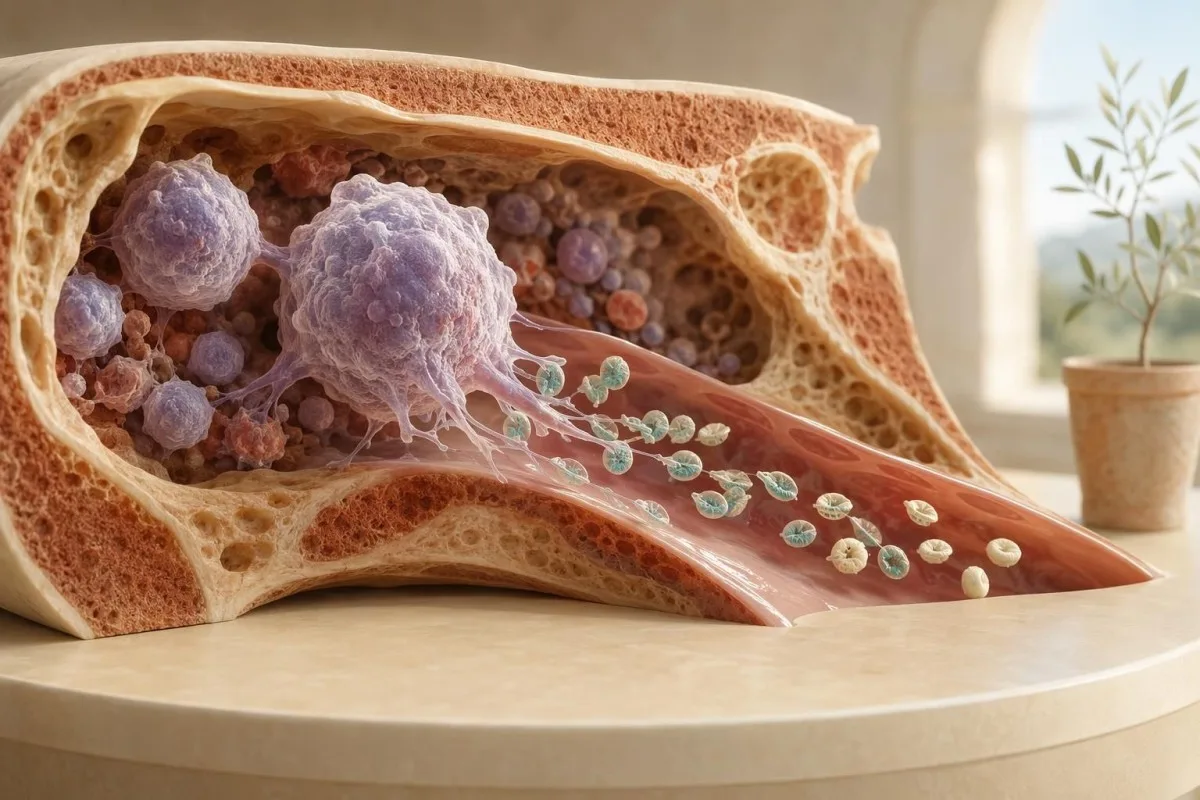
IPF estimates how many newly released platelets are circulating. When the platelet count is low, it helps clinicians decide whether the marrow is responding appropriately or whether production may be reduced.


- Immature platelet fraction (IPF) is the percentage of newly released, RNA-rich platelets in circulation; many adult laboratories use an approximate reference interval of 1.0% to 7.0%.
- High IPF meaning is usually faster platelet turnover, especially when a platelet count below 150 × 10^9/L suggests immune destruction, recovery after infection, or consumption.
- Low or normal IPF with thrombocytopenia can point toward inadequate marrow platelet production, medication suppression, severe nutritional deficiency, or marrow disease.
- Platelet count matters first: 150-450 × 10^9/L is the typical adult reference interval, while counts below 30 × 10^9/L generally need prompt clinical assessment.
- A peripheral smear is not optional in many cases because platelet clumping, giant platelets, blasts, schistocytes, and abnormal white cells can change the interpretation completely.
- IPF is not a diagnosis. A value of 10% can be reassuring in one person with immune thrombocytopenia and concerning in another person with sepsis or a falling platelet count.
- Trend beats one result: a rising IPF can precede platelet-count recovery by several days after marrow suppression or a viral illness.
- Urgent symptoms override the number: new neurological symptoms, black stools, uncontrolled bleeding, or widespread pinpoint purple spots need urgent medical care.
What an immature platelet fraction result tells you
Immature platelet fraction (IPF) helps distinguish low platelet production from increased platelet loss or destruction. A low platelet count with a high IPF usually means the bone marrow is releasing extra young platelets in response to peripheral loss; a low count with a low or inappropriately normal IPF raises concern that production is not keeping pace. Kantesti is an AI blood test analyzer that reads IPF beside the platelet count, CBC pattern, and reported laboratory range rather than treating one percentage as a diagnosis.
Platelets are tiny circulating cell fragments that help form an initial plug after vessel injury, and most survive roughly 7 to 10 days. Newly released platelets contain more residual RNA and are generally larger and more reactive than older platelets; automated analyzers identify that feature to calculate IPF. A standard CBC overview shows the platelet number, but not necessarily whether the marrow is responding.
In my clinical work, the most useful IPF question is not “is this high?” but “is it high enough for this platelet count?” An IPF of 9% with platelets of 45 × 10^9/L may fit an active marrow response, whereas 9% with platelets of 280 × 10^9/L is often much less informative. The percentage can rise simply because the total platelet denominator has fallen.
Thomas Klein, MD, Chief Medical Officer at Kantesti, advises patients not to use IPF alone to self-diagnose immune thrombocytopenia, cancer, or a marrow disorder. The practical next step is to check the platelet trend, symptoms, medicines, and whether a clinician or laboratory has reviewed a peripheral cell sample slide. That combination prevents a surprising number of false alarms.
How the IPF blood test measures young platelets
An IPF blood test uses fluorescent technology to detect residual RNA in recently produced platelets. The result is usually reported as a percentage of total platelets, and some laboratories also report an absolute immature platelet count, often called IPF#.
Modern hematology analyzers expose a laboratory sample to a fluorescent dye that binds nucleic-acid material, then use light scatter and fluorescence intensity to distinguish platelet populations. Because immature platelets retain more RNA, they create a stronger fluorescence signal than older platelets. The method is quick, but the exact gate settings and reference interval differ among analyzer manufacturers and laboratories.
An IPF% answers, “what share of circulating platelets is young?” An absolute IPF, commonly reported in × 10^9/L, answers, “how many young platelets are present?” In severe thrombocytopenia, the absolute value can prevent a misleading impression created by a percentage alone; 12% of 20 × 10^9/L is a very different platelet output from 12% of 250 × 10^9/L.
Kantesti AI interprets immature platelet fraction results by identifying whether the report contains IPF%, IPF#, platelet count, mean platelet volume, and a laboratory-specific range. Readers who want the broader context can use our 15,000-plus biomarker guide, particularly when their report uses unfamiliar abbreviations.
Normal IPF range and why laboratories differ
Many laboratories place the adult IPF reference interval around 1.0% to 7.0%, but there is no universal cutoff. A result should be compared with the range printed beside it because analyzer method, local validation, age, and platelet count distribution all affect the expected value.
A laboratory may flag IPF above 7%, another above 8.0%, and a few use a lower upper limit near 6.1%. This variation is not trivial: a result of 7.4% can be mildly above range at one laboratory and ordinary at another. If your report has an H or L flag, our guide to out-of-range blood test results explains why flags are screening prompts, not diagnoses.
A high IPF does not have a recognised “critical value” in the same way that a platelet count below 10 × 10^9/L may require immediate action. Clinicians react to the bleeding risk, the rate of platelet decline, and the suspected cause. A stable IPF of 11% in known immune thrombocytopenia is a different situation from a new IPF of 11% with fever, confusion, kidney injury, and fragmented red cells.
Absolute immature platelet counts are less widely standardised, but published adult intervals often sit roughly near 3 to 16 × 10^9/L on platforms that report them. Ask the laboratory or clinician which measure was used before comparing results from two facilities. This is one reason a percentage should never be copied into a generic online calculator without the original report.
High IPF meaning when the platelet count is low
A high IPF with thrombocytopenia usually supports increased peripheral platelet destruction, consumption, or recovery. The marrow appears to be responding by releasing a larger share of young platelets, although this pattern cannot by itself prove immune thrombocytopenia.
When platelets fall below 150 × 10^9/L and IPF rises above the local upper limit, common possibilities include immune thrombocytopenia (ITP), recovery after a viral illness, medication-related immune destruction, bleeding, and consumptive illness. Pons et al. (2010) found IPF useful for separating peripheral platelet destruction from decreased production, but the authors did not present it as a standalone diagnostic test. That distinction gets lost in casual internet explanations.
I recently reviewed a patient with platelets of 58 × 10^9/L and IPF of 15.2% after a self-limited viral syndrome; her smear showed larger young platelets and no platelet clumps, and the count improved over 2 weeks. That is a reassuring direction, not a guarantee. A falling count over hours or days needs a different level of attention than a stable count over months, as our post-viral platelet recovery guide explains.
High IPF can also occur in thrombotic microangiopathy, disseminated intravascular coagulation, major bleeding, and some inflammatory states where platelets are being consumed rapidly. Those conditions are assessed with symptoms, haemoglobin, creatinine, LDH, bilirubin, coagulation studies, and the smear. The reason we worry about high IPF plus schistocytes is that together they suggest platelet consumption and red-cell injury, whereas high IPF alone usually does not.
Low IPF with low platelets: reduced production clues
A low or normal IPF in someone with a low platelet count can indicate insufficient platelet production. This pattern may occur when marrow megakaryocytes are suppressed, depleted, infiltrated, or unable to respond appropriately to the degree of thrombocytopenia.
Causes of reduced platelet production include chemotherapy, heavy alcohol exposure, severe acute illness, aplastic anaemia, myelodysplastic syndromes, marrow infiltration, and selected viral infections. Profound vitamin B12 or folate deficiency can suppress multiple blood cell lines, although isolated thrombocytopenia is less typical. A low IPF is most useful when it is “inappropriately low” for a platelet count such as 35 × 10^9/L.
The rest of the CBC changes the probability substantially. Low platelets plus low haemoglobin and low neutrophils suggests a broader marrow issue more strongly than isolated thrombocytopenia does; a low reticulocyte count can reinforce that concern, as outlined in our article on low reticulocyte patterns. Conversely, an isolated low platelet count with normal red and white cell lines can still have several benign or reversible explanations.
Briggs et al. (2006) described rising IPF as an early marker of platelet recovery after stem-cell transplantation, sometimes before the platelet count rises. That practical insight applies more broadly: a stable low IPF may justify further investigation, while a rising IPF after a temporary marrow insult can be an encouraging early sign. A haematologist may still recommend marrow testing if there are blasts, persistent cytopenias, or unexplained abnormalities.
How clinicians read platelet count and IPF together
The platelet count establishes severity, while IPF describes the marrow response. The most informative pattern is usually low platelets with a high IPF, which favours peripheral loss, versus low platelets with a low IPF, which favours impaired production.
A platelet count of 120 × 10^9/L with IPF 8% may require only repeat testing if the person is well and prior counts were similar. A count of 22 × 10^9/L with IPF 2% is more concerning for inadequate production, particularly if the haemoglobin or white-cell count is also low. No single matrix replaces examination, but this pairing directs the next question efficiently.
Mean platelet volume (MPV) may provide a supporting clue because younger platelets tend to be larger, yet MPV is much less reliable when the sample is delayed or platelet clumps are present. Kantesti AI flags discordant platelet patterns rather than assuming that high MPV and high IPF always mean ITP. Our explanation of full blood panel clusters shows why related results should be interpreted as a group.
Do not overinterpret small changes. A shift from IPF 6.8% to 7.5% may be ordinary analytical and biological variation, especially if the platelet count is unchanged. A shift from platelets 180 to 74 × 10^9/L over 48 hours is clinically meaningful even if IPF barely moves; our delta-check guide explains why sudden changes should first be verified.
A practical four-pattern framework
Normal platelets plus normal IPF generally indicates steady platelet turnover. Low platelets plus high IPF supports peripheral loss or recovery; low platelets plus low IPF supports reduced production; and normal platelets plus high IPF is often nonspecific but may prompt a look at recent illness, bleeding, inflammation, or a changing trend.
Why a blood smear can change the IPF interpretation
A peripheral blood smear can confirm whether a low platelet count is real and reveal clues that IPF cannot provide. It can identify platelet clumping, giant platelets, fragmented red cells, blasts, dysplasia, parasites, and abnormal white-cell populations.
EDTA-dependent platelet clumping is a classic cause of pseudothrombocytopenia: the analyzer records a low platelet count because platelets have stuck together in the collection tube, not because the body lacks platelets. A smear shows the clumps, and repeating the count in a citrate tube often clarifies the issue. IPF in a clumped specimen may be uninterpretable or misleading.
Large platelets on a smear can support active turnover, inherited macrothrombocytopenia, or ITP, while schistocytes require prompt consideration of a microangiopathic process. The 2019 International Consensus Report for ITP recommends reviewing the peripheral smear as part of excluding alternative causes of thrombocytopenia (Provan et al., 2019). Patients can read more about the broader CBC, smear, and flow-cytometry pathway when multiple cell lines are abnormal.
Smear review is especially valuable when the platelet result is unexpectedly low in a person with no bruising or prior history. In practice, I have seen an apparent platelet count of 38 × 10^9/L become 196 × 10^9/L on a promptly repeated citrate sample after clumping was recognised. That is why a frightening number should be confirmed before anyone starts treatment—unless active bleeding or severe illness makes the situation urgent.
IPF limitations, sample errors, and false signals
IPF can be distorted by platelet clumping, delayed processing, very low platelet counts, and differences between analyzer methods. It is a useful response marker, not an infallible measurement of marrow function.
A sample that sits too long before analysis can alter platelet size and activation characteristics, while microclots can lower the reported platelet count. Laboratories commonly inspect analyser flags and may repeat a count by an alternative method when results conflict with prior measurements. An unexpected isolated platelet result should be repeated rather than explained away.
Kantesti is an AI lab test interpretation service designed to identify results that need verification, not to override laboratory quality control or a clinician's judgement. Our methodology follows pattern-based safeguards described in the AI technology guide, including checks for implausible unit combinations, missing reference ranges, and abrupt changes between reports.
IPF can look relatively high at very low platelet counts because the denominator has become small. Some analyzers also have reduced precision when platelets are under 20 × 10^9/L, precisely the range where clinical decisions are most consequential. If the result could affect admission, transfusion, anticoagulant decisions, or steroid treatment, a manual review and repeat sample are sensible safeguards.
Conditions that can cause a high immature platelet fraction
High IPF most often reflects accelerated platelet turnover, but the cause ranges from benign recovery after infection to serious platelet-consuming illness. Clinical history and accompanying CBC findings separate these possibilities better than the IPF number alone.
ITP is a common consideration when thrombocytopenia is isolated, IPF is elevated, the smear lacks schistocytes or blasts, and the patient is otherwise well. In adults, ITP remains a diagnosis of exclusion rather than a test result; platelet counts can range from less than 10 to more than 100 × 10^9/L. Provan et al. (2019) emphasise history, examination, CBC, and smear review rather than a single biomarker.
Viral infections can transiently lower platelets through immune effects, splenic sequestration, and marrow suppression; a rising IPF during recovery is common. New medications matter as well, including heparin, quinine-containing products, trimethoprim-sulfamethoxazole, linezolid, valproate, and some immune therapies. Our guide to infection blood-test results explains why symptoms and timing are more useful than guessing from a CBC alone.
Autoimmune disease, liver disease with splenic enlargement, severe infection, and pregnancy-related conditions can produce mixed mechanisms: destruction, sequestration, and reduced production can coexist. That is one reason the evidence for a single diagnostic IPF cutoff is honestly mixed. High IPF is a directional clue, not a shortcut around careful clinical assessment.
IPF in pregnancy: when thrombocytopenia needs extra care
Pregnancy can lower platelet counts modestly, but IPF does not by itself distinguish harmless gestational thrombocytopenia from pre-eclampsia, HELLP syndrome, or immune thrombocytopenia. A platelet trend, blood pressure, liver tests, haemolysis markers, and symptoms are more decisive.
Gestational thrombocytopenia is usually mild, appears later in pregnancy, and often leaves the platelet count above 100 × 10^9/L. A count below 100 × 10^9/L, an abrupt decline, or thrombocytopenia in early pregnancy warrants a more deliberate work-up. Our trimester-specific platelet range guide gives useful context, though the treating maternity team should set the plan.
IPF may rise when platelet turnover accelerates in pre-eclampsia or HELLP syndrome, but it is not a validated screening test that can rule these conditions in or out. Headache with visual disturbance, upper abdominal pain, shortness of breath, new swelling, high blood pressure, or reduced fetal movements should prompt urgent maternity assessment. Waiting for a repeat IPF is the wrong move in that scenario.
For delivery planning, clinicians focus on platelet count trajectory, bleeding history, medications, and the possibility of neuraxial anaesthesia rather than IPF alone. A count of 70 × 10^9/L may be managed very differently depending on whether it has been stable for weeks or fell from 180 in 48 hours. Review our same-day pregnancy lab red flags if you are unsure which symptoms should accelerate follow-up.
When low IPF raises concern about bone marrow production
Low IPF becomes more concerning when thrombocytopenia occurs with anaemia, neutropenia, abnormal cells on a smear, or persistent unexplained fatigue and infection. This combination can indicate a broader production problem and often merits haematology input.
A low platelet count plus haemoglobin below the laboratory range and low neutrophils is called bicytopenia or pancytopenia, depending on how many cell lines are affected. These patterns can result from nutritional deficiency, medications, infection, autoimmune disease, marrow failure, or marrow infiltration. A smear may identify blasts or dysplastic features that make the evaluation more urgent.
Kantesti AI classifies low platelets with concurrent low haemoglobin or white cells as a higher-priority pattern for clinician review; it does not diagnose leukaemia or myelodysplasia from routine blood test results. Our clinical approach is described in the medical validation framework, which emphasises source-report confirmation, reference-range matching, and escalation for red-flag clusters.
A marrow problem is not the most likely explanation for every low IPF. Iron, B12, folate, liver tests, infection history, alcohol use, prescription medicines, and recent chemotherapy often clarify the picture before invasive testing is considered. Our article on anaemia pattern clues can help patients understand why haemoglobin indices are reviewed alongside platelet production.
Using IPF trends to spot platelet recovery
A rising IPF can appear several days before the platelet count recovers after temporary marrow suppression or peripheral platelet loss. The most meaningful trend compares samples from the same laboratory, collected under similar conditions, with the platelet count shown beside each IPF result.
Following chemotherapy, severe infection, or marrow recovery, an IPF rise may precede a platelet-count increase by roughly 2 to 5 days, though the interval varies widely by cause and treatment. Briggs et al. (2006) reported its value as an early recovery marker after stem-cell transplantation. That does not mean every small IPF rise predicts a safe recovery, especially if bleeding symptoms are present.
Kantesti is an AI-powered blood test analysis tool that compares platelet count, IPF, haemoglobin, white cells, and prior dates to identify meaningful direction of change. A longitudinal lab trend review is particularly helpful when a “normal” result is actually falling quickly from a person's usual baseline.
Try to compare results from the same laboratory when possible. One clinic may report IPF as 6.5% with an upper limit of 7.0%, while another may use different fluorescence gating and flag 6.5% differently. Saving the collection date, illness status, medications, and whether a smear was reviewed adds more clinical value than repeatedly testing IPF without context.
When platelet and IPF blood test results need urgent review
Urgent assessment is needed for active bleeding, black stools, severe headache, confusion, new weakness, chest symptoms, or platelet counts below about 10-20 × 10^9/L. IPF does not reduce the urgency of a dangerously low count or concerning symptoms.
Spontaneous major bleeding risk rises as platelet counts fall, although the exact threshold differs with age, fever, anticoagulants, kidney disease, trauma, and the underlying cause. Counts below 10 × 10^9/L are generally treated as a medical urgency even in people without obvious bleeding. New pinpoint purple spots, gum bleeding, nosebleeds that do not stop, or blood in urine should be reported promptly.
If the result appeared in a portal before a doctor called, do not assume the delay means it is harmless. Our guide to seeing lab results before clinician review explains why results may release automatically and why a repeat test is often the first step. Avoid aspirin, ibuprofen, and new supplements that affect platelets unless a clinician says they are safe for you.
As of July 22, 2026, there is no guideline-supported IPF percentage that tells a patient to stay home or go to emergency care. Thomas Klein, MD, recommends basing urgency on the platelet count, bleeding, neurological symptoms, rapid change, pregnancy status, and whether there are signs of serious systemic illness. An IPF value is supporting evidence, not a triage score.
Questions to ask after an abnormal IPF result
The best follow-up questions clarify whether thrombocytopenia is genuine, whether the marrow response fits the count, and what needs repeating. Ask for the exact platelet count, the laboratory IPF reference interval, whether the smear was reviewed, and when the result should be rechecked.
Useful questions include: “Could platelet clumping have affected this count?”, “Is my IPF high for this laboratory?”, “Are my haemoglobin and white-cell results also abnormal?”, and “Should I have a citrate-tube repeat or smear review?” Bring a dated list of medications, supplements, recent infections, vaccines, alcohol intake, and prior blood counts. These details often identify the cause faster than another isolated test.
If your platelets are mildly low, for example 110 to 149 × 10^9/L, repeat timing often depends on the trend and clinical setting rather than IPF alone. A repeat may occur within days when the count is falling or within weeks when it is stable and asymptomatic. A concise doctor-visit lab summary can make that conversation much more efficient.
Kantesti's medical team, including the clinicians on our Medical Advisory Board, treats AI interpretation as an educational layer rather than a substitute for examination, smear review, or emergency care. Most patients find that understanding the production-versus-destruction question makes the follow-up plan less frightening. The answer, though, must still be tailored to the actual report and the person in front of us.
Bottom line: what IPF adds to blood test results
IPF is most valuable when it is read with platelet count, a peripheral smear, the rest of the CBC, and the clinical story. High IPF with low platelets generally supports increased turnover; low IPF with low platelets raises the possibility of reduced production.
The number answers a biological question: is the marrow releasing an unusually high share of young platelets? It does not answer the diagnostic question by itself: why are platelets low, and is the person at risk today? That distinction is the safest way to understand a high IPF meaning without either dismissing or catastrophising it.
In a straightforward case of isolated thrombocytopenia, IPF may help prevent unnecessary marrow investigations by supporting peripheral destruction or recovery. In a complex case with low haemoglobin, abnormal white cells, kidney injury, fever, or smear abnormalities, it adds only one piece to a much larger clinical puzzle. A well-interpreted CBC is a pattern-recognition exercise, not a collection of independent flags.
Keep your original report, including units and laboratory range, and compare it with earlier values before drawing conclusions. If the count is markedly low or you have bleeding symptoms, seek care rather than waiting for a trend. That is the sensible clinical line I would want a member of my own family to follow.
Frequently Asked Questions
What does a high immature platelet fraction mean?
A high immature platelet fraction usually means the bone marrow is releasing a larger proportion of newly made platelets because platelets are being destroyed, consumed, lost, or are recovering after a temporary fall. Many laboratories flag IPF above about 7% to 8%, but the reported laboratory range is the correct comparator. High IPF is especially informative when the platelet count is below 150 × 10^9/L. It cannot diagnose ITP, infection, or another cause without the platelet trend, symptoms, and a peripheral smear.
Is an IPF of 10% high?
An IPF of 10% is above the upper reference limit in many adult laboratories, where a common interval is approximately 1.0% to 7.0%. Its meaning depends heavily on the platelet count: 10% with platelets of 40 × 10^9/L supports active marrow response, while 10% with platelets of 250 × 10^9/L may be nonspecific. Some analyzers use different validated cutoffs, so the number should be interpreted against the range on the report. A clinician may repeat the CBC and request a smear if the result is new or unexpected.
Can IPF diagnose immune thrombocytopenia or ITP?
IPF cannot diagnose immune thrombocytopenia, but an elevated IPF with isolated low platelets can support increased peripheral platelet destruction, which is compatible with ITP. ITP is a diagnosis of exclusion that requires review of medical history, medications, examination, CBC findings, and a peripheral smear. The 2019 International Consensus Report for ITP recommends excluding alternative causes rather than relying on one biomarker. Platelet counts in ITP may range from below 10 × 10^9/L to over 100 × 10^9/L, so severity cannot be inferred from IPF alone.
What does low IPF with low platelets mean?
Low IPF with low platelets can mean the bone marrow is not producing enough platelets for the degree of thrombocytopenia. Potential causes include chemotherapy, medications, severe illness, alcohol-related marrow suppression, vitamin B12 or folate deficiency, aplastic anaemia, and marrow disorders. The pattern is more concerning when haemoglobin and neutrophils are also low or when the smear shows abnormal cells. A platelet count below 30 × 10^9/L, especially with low IPF or bleeding, needs prompt clinical review.
Can platelet clumping cause a false low platelet count and abnormal IPF?
Yes, platelet clumping in an EDTA collection tube can create pseudothrombocytopenia, where the analyzer reports too few platelets even though the circulating platelet count is normal. A peripheral smear can show the clumps, and a repeat sample collected in citrate often gives a more accurate count. IPF can be unreliable in a clumped or partially clotted specimen because the platelet population is not being measured cleanly. This is why an unexpected platelet count such as 35 × 10^9/L should often be confirmed before treatment decisions are made, unless urgent symptoms are present.
Does a high IPF mean platelet count will recover?
A rising IPF can precede platelet-count recovery by approximately 2 to 5 days after temporary marrow suppression or platelet loss, but it does not guarantee recovery. Studies in stem-cell transplantation, including Briggs et al. (2006), found that IPF can rise before platelet counts increase. Recovery timing depends on the cause, such as infection, chemotherapy, immune destruction, bleeding, or ongoing consumption. A falling platelet count, active bleeding, fever, kidney injury, or neurological symptoms needs medical assessment even if IPF is high.
Can alcohol use affect my platelet count and IPF result?
Alcohol can lower platelet counts by temporarily suppressing marrow production and, with liver disease, by contributing to platelet pooling in the spleen. IPF alone cannot identify alcohol as the cause, so tell your clinician about recent alcohol intake rather than assuming it explains a low count.
Get AI-Powered Blood Test Analysis Today
Join over 2 million users worldwide who trust Kantesti for instant, accurate lab test analysis. Upload your blood test results and receive comprehensive interpretation of 15,000+ biomarkers in seconds.
📚 Referenced Research Publications
Klein, T., Mitchell, S., & Weber, H. (2026). AI Blood Test Analyzer: 2.5M Tests Analyzed | Global Health Report 2026. Kantesti AI Medical Research.
Klein, T., Mitchell, S., & Weber, H. (2026). RDW Blood Test: Complete Guide to RDW-CV, MCV & MCHC. Kantesti AI Medical Research.
📖 External Medical References
Pons I et al. (2010). Correlation between immature platelet fraction and reticulated platelets. Usefulness in the etiology diagnosis of thrombocytopenia.. European Journal of Haematology.
Briggs C et al. (2006). Immature platelet fraction measurement: a future guide to platelet transfusion requirement after haematopoietic stem cell transplantation.. Transfusion Medicine.
⚕️ Medical Disclaimer
This article is for educational purposes only and does not constitute medical advice. Always consult a qualified healthcare provider for diagnosis and treatment decisions.
E-E-A-T Trust Signals
Experience
Physician-led clinical review of lab interpretation workflows.
Expertise
Laboratory medicine focus on how biomarkers behave in clinical context.
Authoritativeness
Written by Dr. Thomas Klein with review by Dr. Sarah Mitchell and Prof. Dr. Hans Weber.
Trustworthiness
Evidence-based interpretation with clear follow-up pathways to reduce alarm.