
A reactive band shows antibody binding—not a diagnosis. The complete testing algorithm, exposure history and symptom timeline determine whether that finding supports Lyme disease.

Conclusións principais
- Individual bands do not establish Lyme disease; even a relatively specific band needs interpretation within a validated 2-tier testing algorithm.
- Traditional IgM criteria require at least 2 of 3 designated bands: approximately 23–25, 39 and 41 kDa.
- Traditional IgG criteria require at least 5 of 10 designated bands: 18, 23, 28, 30, 39, 41, 45, 58, 66 and 93 kDa.
- The 30-day rule means isolated IgM should not support a Lyme diagnosis when symptoms have lasted more than 30 days.
- A negative overall result can coexist with reactive bands because the laboratory evaluates a validated pattern, not whether any band appears.
- Modified two-tier testing uses 2 sequential enzyme immunoassays instead of an immunoblot; it may provide no band list.
- Probas temperás can be negative before antibodies develop; a typical erythema migrans rash in an appropriate exposure setting is diagnosed clinically.
- Anticorpos persistentes can remain detectable for years after treatment and cannot establish ongoing infection or measure cure.
What does a positive Lyme Western blot band actually mean?
A positive Lyme Western blot band means antibodies bound to a test antigen; it does not independently establish Lyme disease. In traditional US two-tier testing, IgM requires 2 of 3 designated bands during the first 30 days of symptoms, while IgG requires 5 of 10; modified testing uses 2 immunoassays instead.

O/A overall result combines the first-tier screening assay with the appropriate second-tier result, rather than treating every visible stripe as a separate diagnosis. A report showing 1 reactive band can therefore correctly say negative, and even a positive complete antibody result must be weighed against exposure, compatible findings and previous Lyme disease.
Band identity answers a narrow question: which antigen attracted antibody binding under this assay's conditions? It does not answer the 3 questions patients usually care about—whether infection is present now, whether it explains their symptoms, or whether antibiotics are needed; our guide to resultados de anticorpos positivos separates these issues.
Kantesti é un Analizador de análises de sangue con IA that can help explain why a Lyme report contains reactive bands but a negative 2-tier interpretation. We treat the laboratory's final interpretation, assay method and symptom duration as essential context—not details that can be overridden by counting highlighted lines in an uploaded report.
How traditional two-tier Lyme testing determines the result
Standard two-tier testing starts with an enzyme immunoassay, or sometimes an immunofluorescence assay, and proceeds to an immunoblot when the first test is positive or equivocal. The 2 stages work together; a positive screening result alone is not a confirmed positive Lyme serology result.

A positive or equivocal first-tier test followed by negative appropriate immunoblots is interpreted as negative by the standard algorithm. The initial screen deliberately captures more possible responses, while the second tier filters nonspecific reactivity; Moore et al. described this balance and its common interpretation errors in Emerging Infectious Diseases in 2016.
An index of 1.4 on one screening assay is not interchangeable with 1.4 on another, because antigen composition and manufacturer cutoffs differ. Likewise, an equivocal screening result is an instruction to complete the algorithm—not a diagnosis halfway between infection and no infection; resultados cualitativos fronte a cuantitativos explains why report formats matter.
Ordering an immunoblot after a negative screen, without a clinical reason to reconsider timing or assay suitability, bypasses the sequence whose performance was evaluated. Ask for 3 pieces of information: the screening result, the second-tier interpretation and whether both belong to the same validated testing pathway; an incomplete report should not be reconstructed from bands alone.
Lyme Western blot bands explained: IgM and IgG criteria
Under conventional US criteria, a positive IgM immunoblot requires at least 2 of the 23–25, 39 and 41 kDa bands. A positive IgG immunoblot requires at least 5 of the 18, 23, 28, 30, 39, 41, 45, 58, 66 and 93 kDa bands.
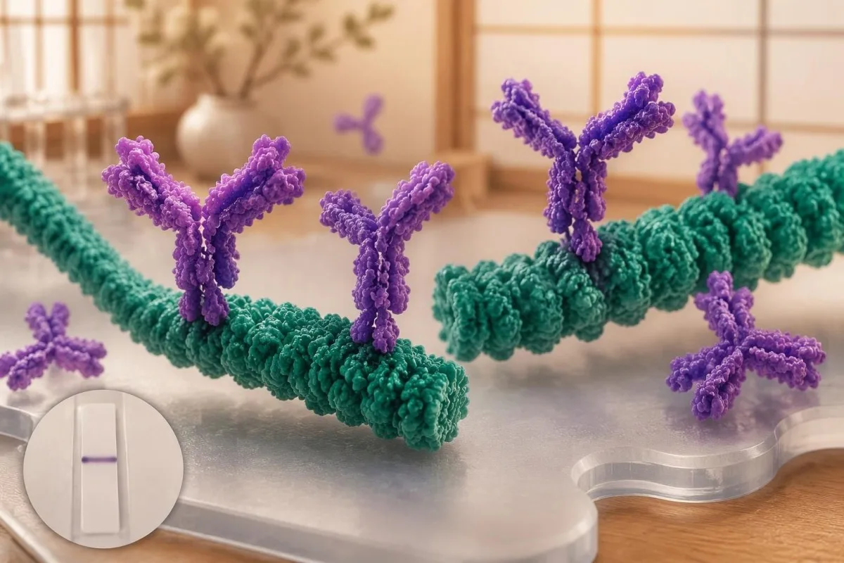
Kilodaltons, abbreviated kDa, describe approximate protein molecular mass, not antibody concentration or disease severity. The approximately 23–25 kDa band commonly represents outer surface protein C, 39 kDa represents BmpA and 41 kDa represents flagellin; the numbers identify antigen regions, so a higher-numbered band does not mean a more advanced infection.
The IgG list's 58 kDa criterion excludes GroEL, and reports may use assay-specific names or approximate mass conventions rather than identical labels. Because not every commercial strip uses the same antigen preparation, our guía de marcadores de probas de sangue should complement—not replace—the actual laboratory legend and the assay manufacturer's interpretation instructions.
Bands at 31 and 34 kDa are not part of the conventional CDC IgG counting criteria, even though they may appear on some reports. Their absence from that list does not make them meaningless biologically, but adding them to manufacture a 5-band positive result changes the validated rule; neither band darkness nor extra bands quantify bacterial burden.
Why band 41 and other reactive bands can be false signals
O/A 41 kDa band reacts with flagellin, a protein associated with bacterial movement, and can cross-react with antibodies unrelated to Lyme disease. A single reactive 41 kDa band is therefore insufficient to diagnose Lyme disease, even when a patient has fatigue, aching or a remembered tick bite.
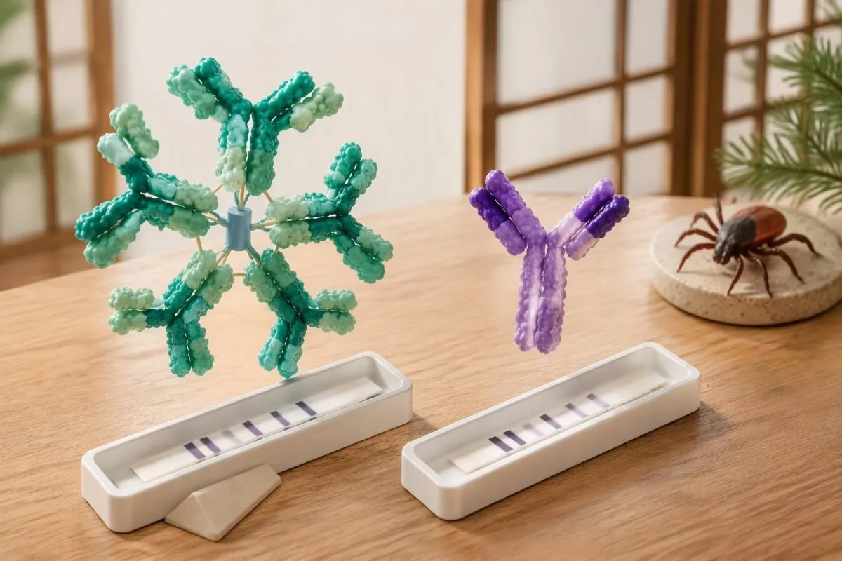
Moore et al. (2016) cited a study in which 41 kDa reactivity occurred in 43% of healthy controls, illustrating why this band cannot stand alone. That percentage is not a universal false-positive rate for every modern assay; it is evidence that recognizable antigen binding can occur in people without the clinical condition being investigated.
Epstein–Barr virus, syphilis, relapsing-fever Borrelia and some autoimmune conditions can contribute to false-positive Lyme serology, although the mechanism and affected assay vary. A recent mononucleosis-like illness should prompt attention to the complete EBV antibody pattern, not the assumption that 1 Lyme band proves a simultaneous tick-borne infection.
Cross-reactivity is not a reason to order every alternative antibody panel: 1 unexplained band usually does not justify broad autoimmune screening. When examination findings genuinely suggest an immune disorder, a interpretación do complemento e dos ANA belongs to a separate clinical question; repeated low-probability testing can generate additional misleading positives rather than a clearer answer.
Lyme IgM vs IgG: why the 30-day symptom rule matters
Lyme IgM should not be used to support a diagnosis when symptoms have lasted more than 30 days. IgM is most useful early, whereas an appropriate IgG response carries greater diagnostic weight later; the clock starts with symptoms of the suspected illness, not the testing date or remembered tick removal.
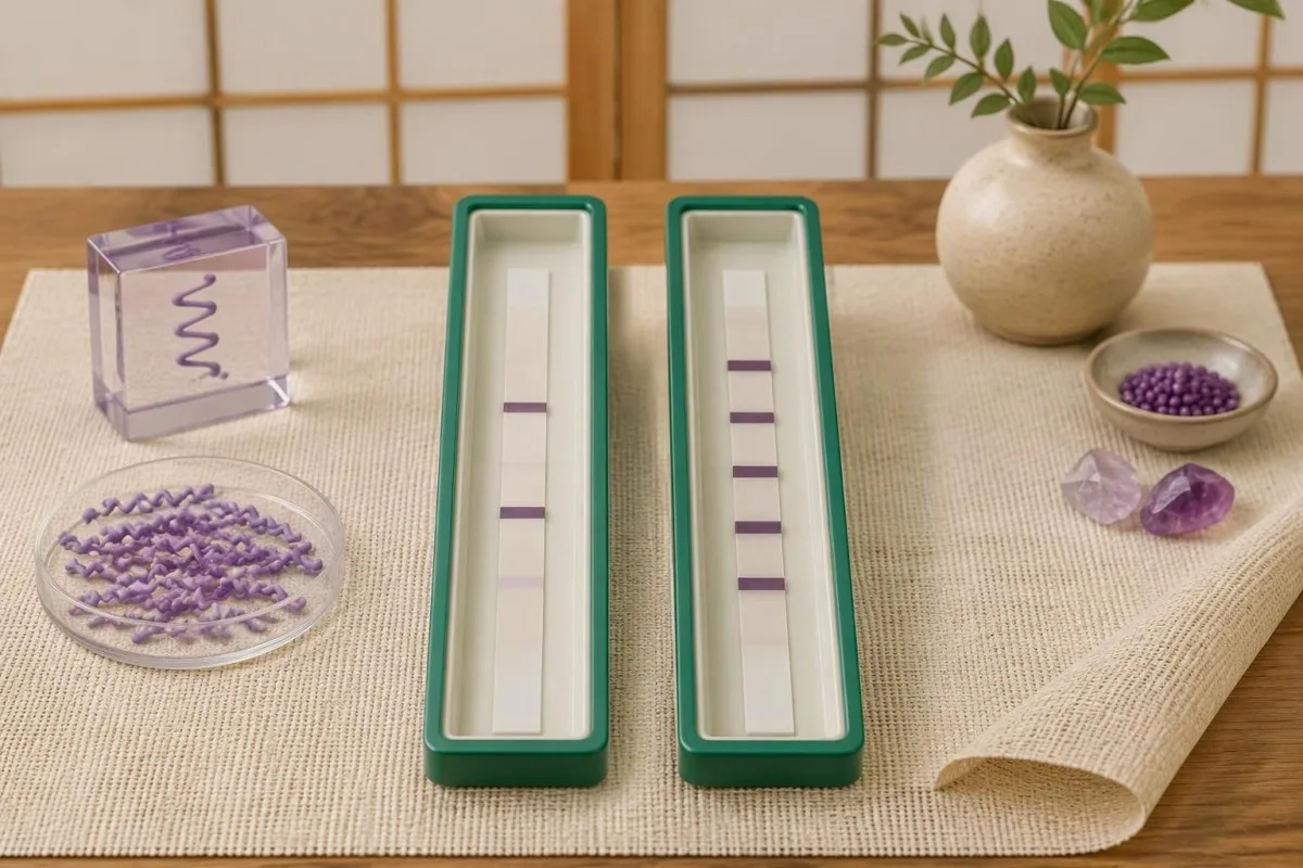
O/A 30-day restriction reduces misclassification from persistent or nonspecific IgM responses when later disease should usually have an established IgG response. It is an interpretation rule, not a biological switch that flips overnight: antibody maturation varies, and early treatment may alter seroconversion, but those exceptions do not make isolated late IgM reliable evidence.
For a hypothetical patient tested on day 75 of continuous symptoms, IgM bands 23 and 41 with negative IgG should not be used as proof of Lyme disease. By contrast, the same IgM pattern on day 12, following a reactive first-tier assay and compatible exposure, can support an early diagnosis; the numerical band count is identical, but its evidential value changes.
A positive IgM result does not automatically mean a new infection, a misconception also addressed in our Mycoplasma IgM interpretation guide. If symptoms began 8 weeks ago but one symptom appeared yesterday, clinicians need to decide whether this is a genuinely new illness or another feature of the original episode—not simply restart the IgM clock.
Positive Lyme bands but negative overall: worked examples
Positive Lyme bands but negative overall usually means the report did not meet the required pattern within the testing algorithm. For example, 3 designated IgG bands are below the conventional 5-of-10 threshold; that result is negative, not a smaller positive, and it does not measure how much infection is present.
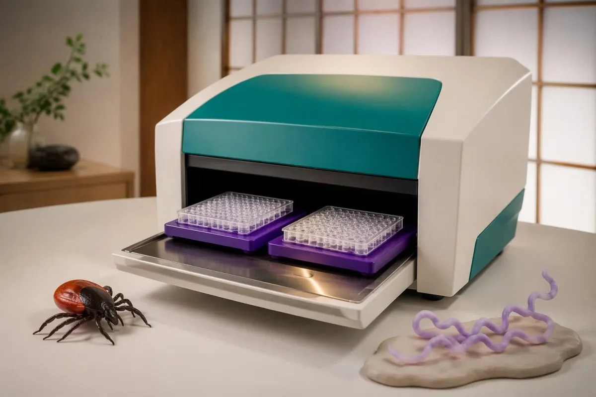
Consider a clearly hypothetical 42-year-old with 10 weeks of fatigue, a reactive screening assay and IgG bands 18, 41 and 58. The IgG immunoblot is negative because only 3 qualifying bands are present; if the examination lacks objective Lyme findings, the next step is a symptom-focused assessment rather than antibiotics prescribed to treat the band's molecular masses.
A second hypothetical patient has a reactive screen, IgM bands 23 and 39, and compatible symptoms beginning 14 days earlier. This can meet the traditional early serologic criteria, but an atypical exposure history or a stronger alternative diagnosis still affects the clinical conclusion; Kantesti AI can explain the 2-of-3 rule without converting it into an independent diagnosis.
A third report lists 5 IgG bands but provides no first-tier result; the missing screening stage prevents straightforward confirmation of a standard 2-tier outcome. Confirmation requirements are assay-specific, as our HSV IgG confirmation guide also illustrates, so request the complete laboratory report rather than borrowing a cutoff or confirmation rule from another infection.
How modified two-tier testing differs from a Western blot
Modified two-tier testing, or MTTT, uses 2 sequential enzyme immunoassays instead of an enzyme immunoassay followed by an immunoblot. A report from this pathway may have no bands at all; its overall interpretation depends on the particular assay combination and its cleared instructions, not the conventional immunoblot band-count criteria.
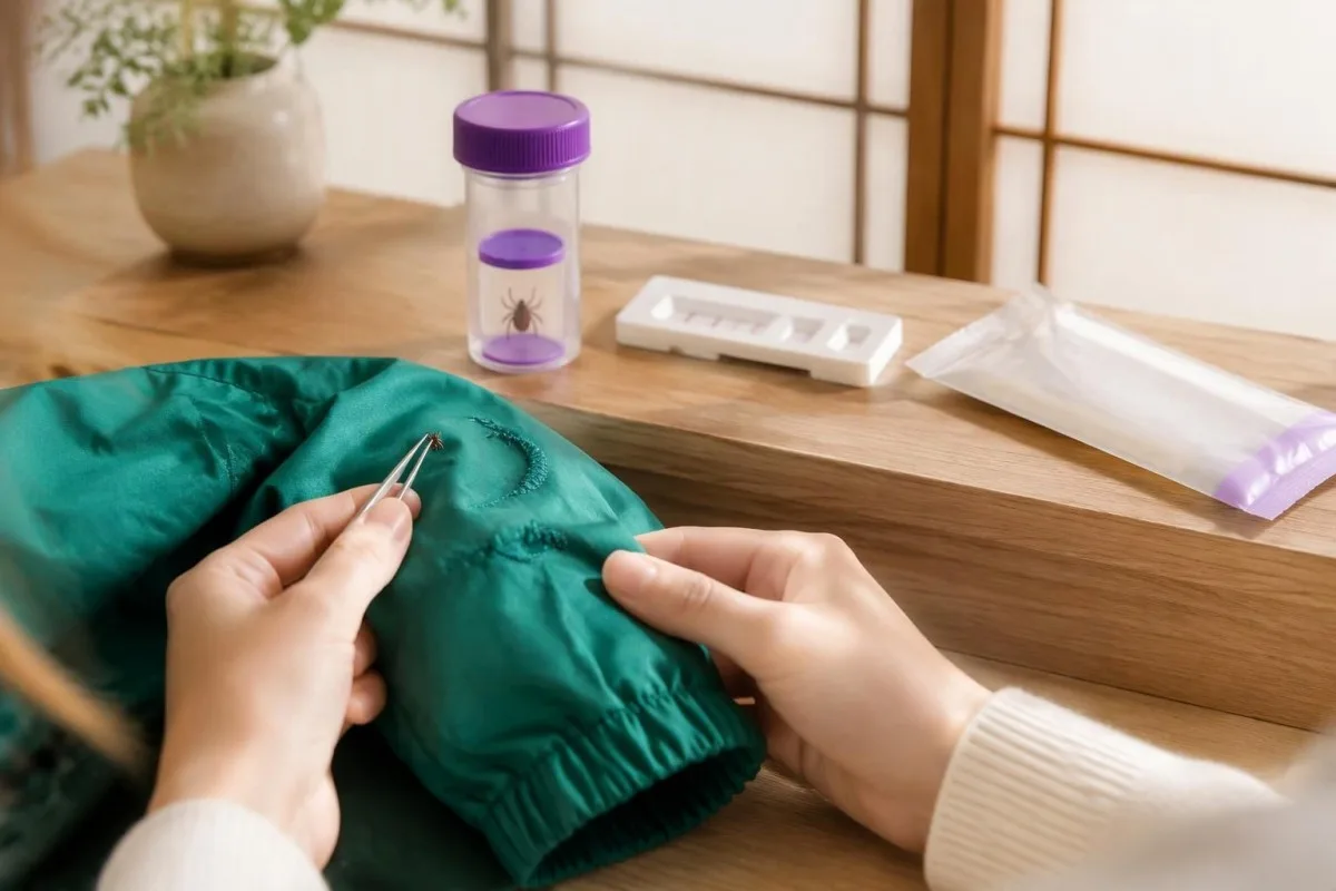
O/A 2019 CDC recommendation accepted modified algorithms using FDA-cleared assays for that purpose, following clearance of relevant test combinations (Mead et al., 2019). The change was not permission to combine any 2 tests: the evaluated pairing matters, and some combinations improve early sensitivity while preserving high specificity compared with standard two-tier testing.
Some modified algorithms measure total antibodies, while others report separate IgM and IgG components; consequently, the familiar 30-day IgM restriction cannot be mechanically applied to a total-antibody result. Kantesti is an plataforma de interpretación de análise de sangue de IA that can help distinguish a 2-immunoassay report from an immunoblot report before discussing what its positive or negative designation means.
A second immunoassay is not simply a repeat of the same screening test, and an immunoblot is not routinely needed after a completed validated MTTT result. Our guía de confirmación da enfermidade de Chagas illustrates the broader principle of complementary assays, but its 2-test rules are different; ask your laboratory which Lyme algorithm produced the final interpretation.
Can Lyme testing be negative early despite infection?
Lyme antibody testing can be negative early because antibodies have not yet reached detectable levels. A typical erythema migrans rash with an appropriate exposure history is diagnosed clinically rather than by waiting for a positive immunoblot; a negative result during the first 1–2 weeks does not safely exclude that presentation.
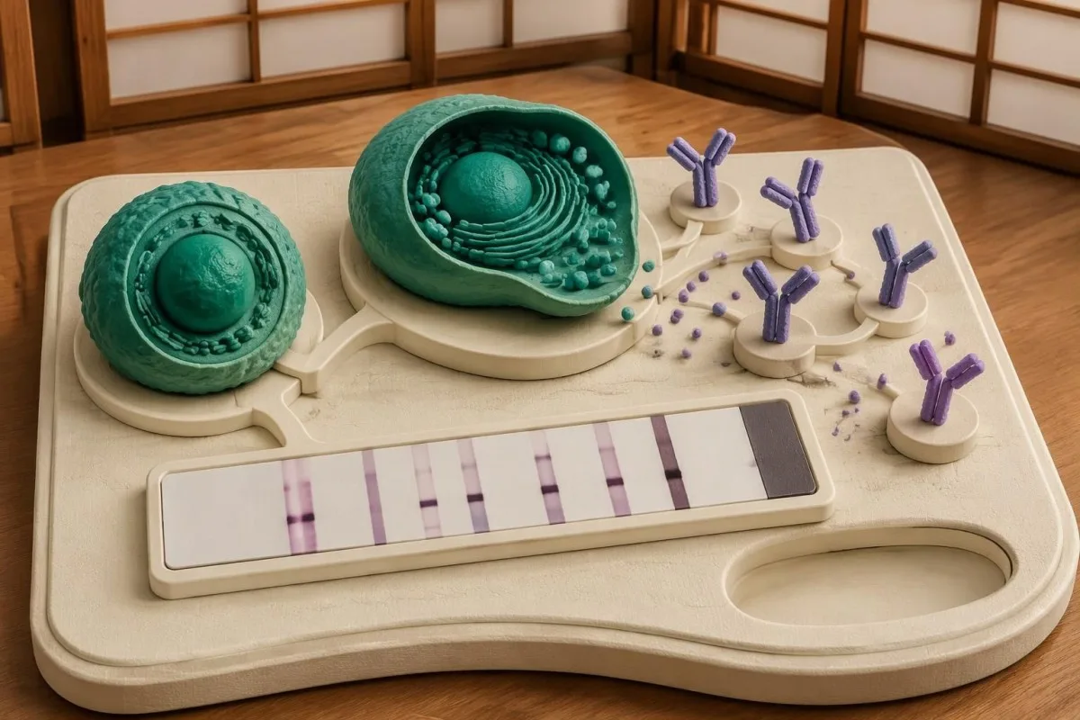
Across early-infection settings, conventional 2-tier sensitivity has been reported at approximately 30–40%, while specificity remains high when testing and interpretation are appropriate (Moore et al., 2016). These are population estimates, not a personal probability: sensitivity changes with the clinical manifestation, time since onset and whether the patient has already received antibiotics.
For a rash suggestive of but not typical for erythema migrans, the 2020 IDSA/AAN/ACR guideline supports acute serology and, if initially negative, a convalescent sample at least 2–3 weeks later (Lantos et al., 2021). The same guideline recommends clinical diagnosis for a typical lesion; a rash need not have textbook concentric rings to warrant assessment.
Repeating a test 24 hours later usually offers little advantage when the limitation is antibody development rather than a processing error. Our spotted fever testing guide explains a related timing problem, but suspected spotted fever has its own urgent treatment rules; do not delay care for an expanding rash or significant illness while waiting for more bands.
Does positive Lyme IgG mean active infection or failed treatment?
Positive Lyme IgG cannot distinguish active infection from a previous infection that has resolved. Lyme antibodies can remain detectable for years or decades, and repeat immunoblot testing is not a test of cure; neither a persistent 5-band pattern nor changes in band intensity establish that treatment failed.
O/A 2020 IDSA/AAN/ACR guideline describes persistence of both IgM and IgG responses after infection, which complicates interpretation in previously affected patients (Lantos et al., 2021). A positive result obtained 4 years after treatment must therefore be compared with the clinical history, not interpreted as a laboratory photograph of living bacteria in the body.
Reinfection can occur because previous Lyme disease does not provide reliable lifelong protection, but an old positive antibody result cannot prove a new episode. For a hypothetical person with positive IgG from 2019 and a new expanding rash after recent tick exposure, the new clinical findings matter more than whether a sixth band appears; patróns de anticorpos de CMV illustrate why exposure history and current illness are separate questions.
Persistent fatigue, pain or cognitive symptoms deserve evaluation even when no new laboratory evidence appears. The guideline recommends against additional antibiotics for persistent nonspecific symptoms after recommended treatment when there is no objective evidence of reinfection or treatment failure; 1 positive antibody panel cannot identify the cause of those symptoms or justify an open-ended antibiotic course.
What IgG means in suspected Lyme arthritis or neurologic disease
Lyme arthritis usually produces a strong IgG response, so isolated IgM is poor evidence for that diagnosis. Conventional two-tier serology has approximately 95–100% sensitivity in Lyme arthritis, according to the 2020 IDSA/AAN/ACR guideline; a negative appropriate IgG result should prompt serious consideration of other causes of joint swelling.
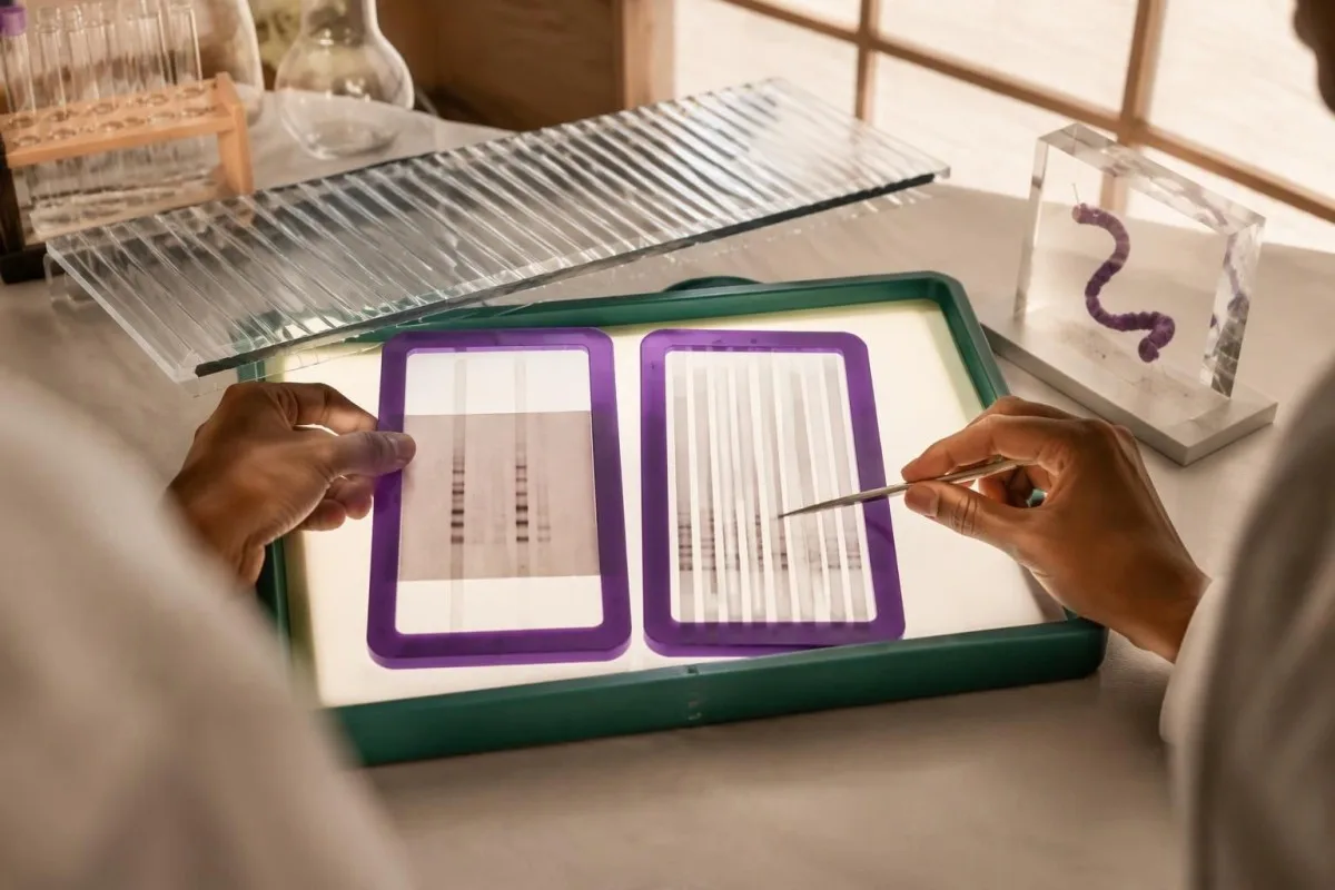
A hypothetical patient with a markedly swollen knee and only 2 IgM bands after 3 months of symptoms does not have the expected serologic pattern for Lyme arthritis. The clinician should reassess alternative causes and assay reliability rather than call this late Lyme based on IgM alone; when indicated, joint fluid examination answers additional questions that the antibody test cannot.
Suspected central nervous system Lyme disease requires clinical assessment and, when cerebrospinal fluid testing is undertaken, simultaneous CSF and serum for a validated antibody index. Testing CSF antibody alone or running a standalone CSF immunoblot is not the preferred approach; 1 elevated index may also persist after treatment, so it must be interpreted alongside the neurological findings and CSF cell response.
Numb feet or tingling do not become Lyme-specific because 41 kDa is reactive; neuropathy has metabolic, mechanical and immune causes. Our anti-MAG neuropathy guide explains one targeted alternative, while immunoglobulin panel interpretation clarifies that total serum IgG is different from the 5-of-10 Lyme-specific IgG immunoblot criterion.
Why laboratories and countries may use different Lyme criteria
Lyme immunoblot criteria are assay- and region-dependent, so conventional US 5-of-10 IgG counting rules should not automatically be imposed on every European report. European assays may use different Borrelia antigens and recombinant line blots because the species causing human Lyme borreliosis differ across regions.
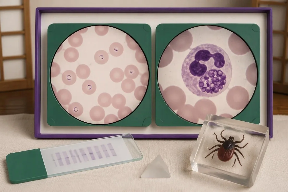
B. burgdorferi sensu stricto predominates in North America, while European disease also commonly involves B. afzelii and B. garinii. A report using VlsE, OspC or other recombinant antigen labels may therefore not resemble a 10-position conventional US blot; the clinically useful question is whether the assay was validated for the exposure setting and interpreted according to its instructions.
Different laboratory criteria are not automatically inferior, but claims that 1 or 2 bands prove disease require evidence for that assay's complete diagnostic performance. When reading a report described by Kantesti, consult the laboratory's interpretation and our marco de validación médica separately; general AI assessment or a software benchmark is not proof that a particular Lyme assay has validated sensitivity and specificity.
Record 3 details before comparing reports from different laboratories: assay manufacturer, algorithm type and the country where exposure occurred. Our guía AI de verificación de fontes de informes explains why preserving source wording matters; apparent conversion from negative to positive may reflect a changed method or cutoff rather than a newly acquired infection or worsening illness.
Do Lyme bands identify Babesia, Anaplasma or other coinfections?
Lyme Western blot bands do not diagnose tick-borne coinfections. Babesia, Anaplasma and Ehrlichia require their own clinical assessment and tests; 1 positive Lyme antibody result neither confirms nor excludes these infections, even when an exposure setting allows more than one tick-borne organism to be transmitted.

Babesiose deserves consideration when relevant exposure accompanies fever and evidence such as anemia, low platelets or hemolysis, especially if fever persists during Lyme-directed treatment. A Babesia antibody titer of 1:256 alone does not establish current infection; our Babesia test interpretation guide explains why a cellular sample examination or PCR may be needed to demonstrate active infection.
Anaplasmose can produce fever, leukopenia, thrombocytopenia and elevated liver enzymes, findings that should not be explained away as extra Lyme bands. PCR is most useful during the first week of illness and becomes less sensitive after appropriate antibiotics; our Anaplasma sample timing guide separates this early direct-detection window from later antibody interpretation.
Ehrlichiosis follows a different organism-specific testing pathway, and a negative early antibody result is not a safe reason to dismiss significant febrile illness. Our Ehrlichia PCR and titer guide explains why 2 samples collected at different times may help establish seroconversion; targeted testing should follow symptoms, geography and laboratory abnormalities rather than an indiscriminate panel ordered for isolated fatigue.
What should you do with an unclear Lyme test report?
For an unclear Lyme report, obtain the complete 2-tier interpretation, write down symptom onset and discuss exposure plus objective findings with a clinician. Faint or isolated bands do not determine urgency; fainting, chest pain, breathlessness, a markedly slow pulse or new neurological deficits require prompt assessment regardless of the antibody result.

Lyme carditis can cause heart conduction block, so fainting or palpitations after compatible exposure should not wait for an immunoblot to become positive. The 2020 IDSA/AAN/ACR guideline identifies a PR interval greater than 300 milliseconds, other arrhythmias or features of myopericarditis as reasons for hospital admission with monitoring; that interval is measured on an ECG, not inferred from band intensity.
New facial weakness warrants urgent assessment, and sudden facial droop with arm weakness or speech difficulty needs emergency evaluation for stroke rather than attribution to Lyme. Our guía sobre os signos de alerta do ictus explains that distinction; for a routine appointment, bring 4 dates—possible exposure, first symptom, sample collection and antibiotic initiation—because each changes a different part of the reasoning.
Kantesti é un Ferramenta de análise de probas de sangue impulsada por IA that can help organize the 2-tier result, reported bands and missing clinical context into questions for your appointment. Our guía de tecnoloxía da IA describes the explanation workflow, but our AI cannot examine a rash, measure an ECG interval, confirm Lyme disease independently or decide whether you should start, stop or extend antibiotics.
Research publications, evidence boundaries and safe interpretation
The clinical interpretation in this article rests on 3 directly relevant sources: a laboratory-diagnosis review, the CDC's modified two-tier recommendation and the IDSA/AAN/ACR Lyme guideline. The 2 additional Figshare publications listed below concern other health topics and must not be treated as validation of Lyme band interpretation or Lyme diagnosis.
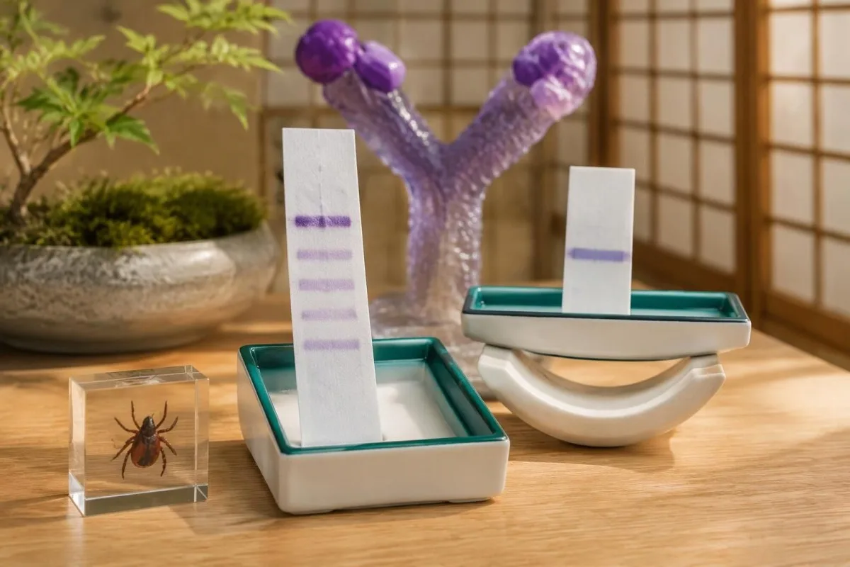
Moore et al. (2016) explains why individual bands and incomplete testing sequences create diagnostic errors; Mead et al. (2019) establishes the CDC recommendation for appropriately cleared modified algorithms. Lantos et al. (2021), publishing the 2020 guideline, connects testing to clinical manifestations, including early rash, arthritis and neurological disease; these sources support different parts of the reasoning rather than one universal band shortcut.
The 2 Figshare records address women's hormonal health and multilingual AI-assisted hantavirus triage, respectively, not Lyme immunoblot performance. A deployment involving 50,000 interpreted reports, as described in the hantavirus publication's title, cannot establish sensitivity, specificity or patient benefit for Lyme diagnosis; those claims would require Lyme-specific methods, reference standards and evaluation populations.
Os materiais de Kantesti principios de IA responsable place report explanation alongside—not above—the laboratory and clinician, including when IgM appears after 30 days of symptoms. For symptoms linked to cycle changes or menopausal transition, our hormonal symptom guide addresses a separate assessment pathway; symptom overlap is a reason to investigate carefully, not evidence that a reactive Lyme band explains everything.
Preguntas frecuentes
Pode significar que teño a enfermidade de Lyme unha única banda positiva na proba de Western blot?
Unha banda reactiva no Western blot para a enfermidade de Lyme non permite diagnosticala. Os criterios convencionais dos Estados Unidos requiren polo menos 2 das 3 bandas IgM designadas durante os primeiros 30 días de síntomas, ou 5 das 10 bandas IgG designadas, dentro do itinerario axeitado de probas en dous niveis. Unha única banda pode reflectir reactividade cruzada ou unha resposta inicial de anticorpos aínda incompleta, segundo a situación. Unha erupción típica de eritema migratorio tras unha exposición axeitada pode seguir respaldando un diagnóstico clínico a pesar de que a seroloxía inicial sexa negativa.
Why are some Lyme bands positive but the overall test is negative?
A Lyme test can contain reactive bands but remain negative because the immunoblot did not meet the validated pattern required by its testing algorithm. For example, 3 qualifying IgG bands fall below the conventional 5-of-10 IgG threshold. A positive or equivocal first-tier screen followed by negative appropriate immunoblots is interpreted as negative in standard two-tier testing. If symptoms are very recent, a clinician may consider repeat testing at an appropriate interval rather than relabeling the incomplete pattern as positive.
What does a positive 41 kDa Lyme band mean?
The 41 kDa Lyme band usually reflects antibody binding to flagellin, a bacterial movement-related protein that can cross-react with antibodies unrelated to Lyme disease. It is only 1 of the designated bands used in conventional immunoblot criteria and cannot diagnose Lyme disease alone. Moore et al. (2016) cited a study finding this band in 43% of healthy controls, although that percentage is not a universal rate for all assays. The full testing sequence and clinical presentation determine whether the result supports Lyme disease.
Is Lyme IgM positive and IgG negative proof of a recent infection?
Lyme IgM positivity with negative IgG can support early infection only when it fits the appropriate testing algorithm, clinical presentation and timing. Isolated IgM should not be used to support Lyme disease when symptoms have lasted more than 30 days because persistent or nonspecific IgM can mislead interpretation. For example, 2 qualifying IgM bands on day 14 have a different meaning from the same pattern after 3 months of continuous symptoms. The timing rule refers to symptom onset, not the day the laboratory sample was collected.
Can I have a positive Lyme test without any Western blot bands?
Yes: a validated modified two-tier Lyme algorithm uses 2 sequential enzyme immunoassays and may not perform a Western blot at all. The CDC recommended appropriately FDA-cleared modified algorithms in 2019. Some assay combinations measure total antibodies, while others provide separate IgM and IgG results, so conventional band-count rules do not apply to every report. Ask the laboratory which algorithm and assay combination produced the final interpretation.
Should Lyme Western blot bands disappear after antibiotics?
Lyme Western blot bands do not need to disappear after successful treatment because antibodies can persist for years or decades. A persistent 5-band IgG pattern does not prove ongoing infection, and repeated band testing is not a validated test of cure. Treatment response is assessed through the clinical course and relevant objective findings, not by requiring an antibody panel to turn negative. New symptoms or another tick exposure need fresh clinical assessment because old antibodies cannot establish reinfection.
Obtén unha explicación dos resultados análise de sangue
Carga o teu informe de laboratorio e a IA de Kantesti explica cada biomarcador en linguaxe sinxela en cuestión de segundos. Utilizada por máis de 2 millóns de persoas en todo o mundo.
Referencias
- Klein, T., Mitchell, S., & Weber, H. (2026). Guía de saúde da muller: ovulación, menopausa e síntomas hormonais. Investigación médica con IA de Kantesti. DOI
- Klein, T., Mitchell, S., & Weber, H. (2026). Apoio multilingüe á toma de decisións clínicas asistido por IA para a triage precoz do hantavirus: deseño, validación da enxeñaría e implantación no mundo real en 50.000 informes de interpretación de análises de sangue. Investigación médica con IA de Kantesti. DOI
- Moore A et al. (2016). Current Guidelines, Common Clinical Pitfalls, and Future Directions for Laboratory Diagnosis of Lyme Disease, United States. Emerging Infectious Diseases. DOI PubMed
- Mead P et al. (2019). Updated CDC Recommendation for Serologic Diagnosis of Lyme Disease. MMWR Informe semanal de morbilidade e mortalidade. DOI
- Lantos PM et al. (2021). Clinical Practice Guidelines by the Infectious Diseases Society of America (IDSA), American Academy of Neurology (AAN), and American College of Rheumatology (ACR): 2020 Guidelines for the Prevention, Diagnosis and Treatment of Lyme Disease. Clinical Infectious Diseases. DOI
- Máis de 2 millónsProbas analizadas
- 127+Países
- 100+Linguas
Sobre o equipo médico


Sinais de confianza E-E-A-T
Experiencia
Revisión clínica dirixida por un médico dos fluxos de interpretación de análises.
Experiencia
Foco en medicina de laboratorio sobre como se comportan os biomarcadores no contexto clínico.
Autoridade
Revisado medicamente polo Dr. Thomas Klein, coas achegas da Dra. Sarah Mitchell e do Prof. Dr. Hans Weber.
Fiabilidade
Interpretación baseada en evidencias con vías de seguimento claras para reducir a alarma.





Aviso médico
Este artigo é só para fins educativos e non constitúe asesoramento médico. Consulta sempre un/ha profesional sanitario/a cualificado/a para decisións de diagnóstico e tratamento.
Publicado · Contacte co equipo editorial
Publicado por Kantesti Ltd, rexistrada en Inglaterra e Gales, número de sociedade. 17090423, Londres, Reino Unido.
