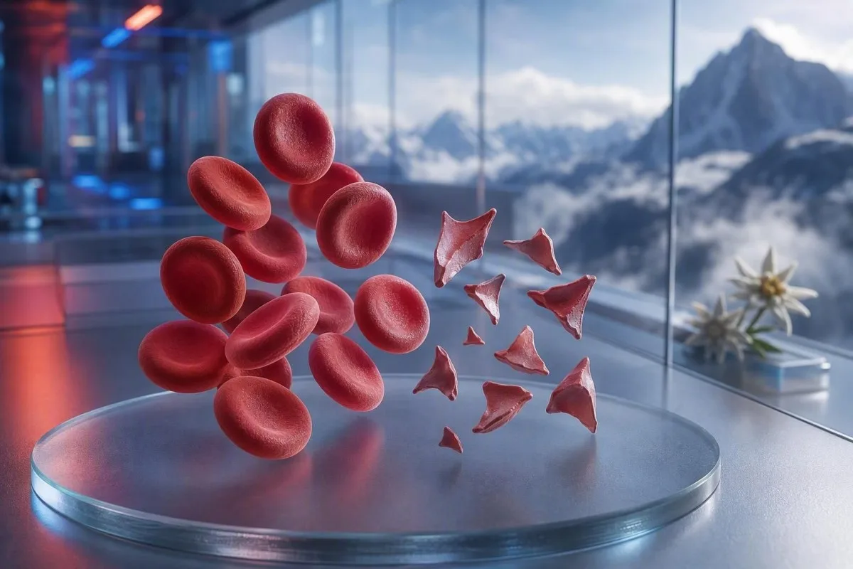
Schistocyty jsou fragmentované červené krvinky, které mohou signalizovat nebezpečné srážení v malých cévách, ale jediná poznámka z nátěru není diagnóza. Naléhavost závisí na procentu fragmentů, počtu trombocytů, trendu hemoglobinu, funkci ledvin, markerů hemolýzy a příznaků.


- Prahová hodnota schistocytů: Více než 1% schistocytů na dobře připraveném periferním nátěru dospělého je podezřelé z trombotické mikroangiopatie, pokud chybí jiné změny tvaru červených krvinek.
- Urgentní shluk: Schistocyty plus trombocyty pod 150 × 10^9/L, klesající hemoglobin, zvýšené LDH a nízký haptoglobin vyžadují vyšetření lékařem ještě tentýž den.
- Indicie TTP: Závažná aktivita ADAMTS13 pod 10% podporuje imunitní trombotickou trombocytopenickou purpuru, stav léčený ještě před návratem výsledků potvrzujících vyšetření, pokud je klinické podezření vysoké.
- Ne každý fragment je nebezpečný: Mechanické srdeční chlopně, těžká hypertenze, dialyzační okruhy, popáleniny a poškození vzorku mohou způsobit fragmentované červené krvinky.
- Nátěr je manuální důkaz: Automatizovaný analyzátor nedokáže spolehlivě diagnostikovat schistocyty; zkušený morfolog by měl vyšetřit preparát.
- Těhotenství zvyšuje sázky: Schistocyty s vysokým krevním tlakem, bílkovinami v moči nebo nízkými trombocyty po 20. týdnu vyžadují okamžité vyšetření porodníkem.
- Opakované testování pomáhá: Čerstvě odebraný vzorek v EDTA a opakovaný nátěr mohou odlišit artefakt z odběru od skutečně probíhající fragmentace červených krvinek.
- Neléčte se sami: Železo, kyselina listová nebo doplňky neléčí mikroangiopatickou hemolýzu a mohou odvádět pozornost od urgentního vyšetření.
Když fragmentované červené krvinky vyžadují urgentní lékařské vyšetření
Schistocyty vyžadují urgentní kontrolu ještě týž den, objeví-li se spolu s nízkým počtem trombocytů, anémií, biochemickou hemolýzou, poškozením ledvin, neurologickými příznaky, horečkou nebo těžkou hypertenzí. Samotná poznámka k nátěru může být benigní nebo arteficiální; nebezpečný je vzorec. Ke dni 7. srpna 2026 bych nové schistocyty spolu s trombocyty pod 100 × 10^9/L považoval za „zavolat lékaři hned“ výsledek, nikoli za rutinní kontrolu.
Pokles trombocytů je často nejrychlejší vodítko urgentnosti. Hemoglobin 92 g/L při trombocytech 68 × 10^9/L a nově hlášených schistocytech je podstatně znepokojivější než 1% fragmentů s hemoglobinem 145 g/L a stabilními trombocyty 260 × 10^9/L. Kantesti je analyzátor krevních testů s AI, který čte tento shluk v rámci krevního obrazu (complete blood count) místo toho, aby izolovaně zvyšoval poznámku k morfologii.
Za 15 let klinické praxe jsou moje rychlé „call“ pro pacienty, jejichž výsledky se změnily během dnů: klesající hemoglobin, stoupající kreatinin a stoupající laktátdehydrogenáza, tedy LDH. Praktické pravidlo doktora Thomase Kleina je jednoduché: zmatenost, nová slabost, bolest na hrudi, dušnost, velmi málo moči nebo silná bolest hlavy spolu s tímto vzorcem znamenají dnes urgentní vyšetření.
Schistocyty nejsou důvod čekat na opakovaný test, pokud je daná osoba nemocná. Normální draslík nebo normální počet leukocytů nezruší riziko trombotické mikroangiopatie. Pacienti, kteří se snaží porozumět nízkému počtu, si mohou také prostudovat naši příručku k příznakům nízkých červených krvinek, ale akutní příznaky vždy mají přednost před interpretací na internetu.
Okamžitě volejte záchrannou službu pro
Novou jednostrannou slabost, omdlení, záchvat, těžkou dušnost, svíravý diskomfort na hrudi, černou stolici nebo rychle se zhoršující zmatenost vyžadují záchrannou službu, nikoli zprávu do ordinace. Tyto příznaky mohou odrážet postižení orgánů a žádné procento schistocytů to bezpečně nevylučuje.
Co schistocyty na periferním nátěru ve skutečnosti znamenají
Schistocyty jsou nepravidelné fragmenty červených krvinek vznikající, když jsou cirkulující buňky fyzicky „natrženy“—nejčastěji fibrinovými vlákny nebo abnormálními povrchy s vysokým smykovým napětím v malých cévách. Typicky mají ostré úhly, formu „přilby“ nebo malé trojúhelníkovité tvary a postrádají centrální bledost neporušené červené krvinky.
Pravé schistocyty jsou nález v morfologii, nikoli hodnota vytvořená standardním analyzátorem v rámci CBC. Mezinárodní rada pro standardizaci v hematologii doporučuje při kvantifikaci, kdy na ní záleží, spočítat alespoň 1 000 červených krvinek v optimální zóně nátěru (Zini et al., 2021). Tento detail vysvětluje, proč jedna laboratoř může uvádět “vzácné fragmenty”, zatímco jiná uvádí procento.
Klasická obava je mikroangiopatická hemolytická anémie, kde se červené krvinky setkávají s trombocyty bohatými mikrothromby a fibrinem v oběhu. Přesto však těžká aortální stenóza, mechanická chlopeň, mimotělní oběhové okruhy a závažné tepelné poranění mohou vytvářet skutečné fragmenty jiným mechanismem. Otázka tedy není “Jsou fragmenty skutečné?”, ale “Co způsobuje fragmentaci?”
Kantesti je platforma pro rozbor krevních výsledků pomocí AI, která dokáže nahraný kompletní krevní obraz umístit vedle co zahrnuje CBC a zvýraznit chybějící doprovodné výsledky, jako je bilirubin, retikulocyty nebo haptoglobin. Nedokáže však zkontrolovat samotný nátěr, což zůstává úkolem vyškoleného pracovníka klinické laboratoře.
Jak moc je schistocytóza znepokojivá u dospělého nátěru
Počet schistocytů nad 1% na periferním nátěru dospělého je podezřelý z trombotické mikroangiopatie, pokud jsou fragmenty převládající abnormitou červených krvinek. Počty pod 1% nevylučují onemocnění, zejména pokud byl film vyhotoven po podání tekutin, transfúzi nebo při probíhajícím onemocnění.
Hraniční hodnota 1% je diagnostický podnět, nikoli škála závažnosti. Doporučení ICSH používají referenční interval pod 1% pro zdravé dospělé a donošené novorozence; předčasně narození novorozenci mohou mít až 5% fragmentů bez stejné implikace (Zini et al., 2021). Výsledek 2% při těžké trombocytopenii je znepokojivější než 4% po zavedení mechanického zařízení pro podporu oběhu.
Laboratoře mohou uvádět “ojedinělé”, “několik” nebo “1–2 na pole vysokého zvětšení” místo procent. Tyto formulace nejsou zaměnitelné, protože velikost pole, tloušťka nátěru a pravidla hlášení se liší. Otázka, zda laboratoř provedla procentuální stanovení ve stylu ICSH, je často užitečnější než pokoušet se převést popisnou formulaci sami.
Vysoké hodnoty RDW neměří schistocyty. RDW odráží variabilitu velikosti buněk a může stoupat při retikulocytóze, nedostatku železa, transfúzi nebo fragmentaci. Naše podrobné RDW a vodítko pro indexy červených krvinek vysvětluje, proč vysoké RDW samo o sobě nepodporuje ani TTP, ani artefakt.
Vzor výsledků krevních testů, který podporuje pravou hemolýzu
Skutečná fragmentace červených krvinek obvykle vede k vysokému LDH, nízkému haptoglobinu, zvýšenému nepřímému bilirubinu a retikulocytární odpovědi spolu s anémií. Žádný jediný marker není definitivní, protože onemocnění jater, zánět, nedávná transfuze a onemocnění kostní dřeně mohou obraz zkreslit.
LDH je citlivé, ale není specifické. LDH nad 600 IU/L často podporuje aktivní hemolýzu ve správném kontextu, ale může se zvýšit i při poranění svalů, poškození jater, infekci a rakovině. Haptoglobin pod dolní hranicí laboratoře, běžně pod 0,3 g/L, přidává na váze, protože váže volný hemoglobin uvolněný do plazmy.
Počet retikulocytů by obvykle měl během periferní destrukce stoupat, ale může zůstat nízký při nedostatku folátu, nedostatku železa, onemocnění ledvin nebo akutní supresi kostní dřeně. Tuto zdánlivou protichůdnost lze snadno přehlédnout: pacient může mít skutečnou hemolýzu a zároveň nepřiměřeně nízkou retikulocytární odpověď. výsledek haptoglobinu vede k pokrytí běžných interpretačních pastí.
Kantesti AI interpretuje vzorce fragmentovaných červených krvinek porovnáním hemoglobinu, MCV, trombocytů, LDH, bilirubinu, kreatininu a retikulocytů ze stejného časového bodu. Pokles hemoglobinu o více než 20 g/L během 48 až 72 hodin je klinicky informativnější než mírně abnormální hodnota bez předchozího srovnání.
Proč může být bilirubin normální
Nepřímý bilirubin může být časně při hemolýze normální, po naředění intravenózní tekutinou, nebo když játra bilirubin účinně odstraňuje. Normální bilirubin by proto neměl přebít přesvědčivé fragmenty, nízký haptoglobin a klesající hemoglobin.
Proč se TTP léčí jako hematologická pohotovost
Trombocytopenická trombotická purpura neboli TTP se předpokládá, když schistocyty doprovázejí trombocytopenii a mikroangiopatickou hemolytickou anémii bez jasnější alternativy. Neléčená imunitní TTP může rychle progredovat, takže klinici mohou zahájit plazmaferézu a imunitní léčbu ještě před návratem výsledků ADAMTS13.
Aktivita ADAMTS13 pod 10% silně podporuje TTP, ale výsledek může trvat hodiny nebo dny. Diagnostická doporučení ISTH uvádějí, že je vhodné odhadnout pretest pravděpodobnost pomocí klinických nástrojů, jako je PLASMIC skóre, při odběru vzorku před zahájením plazmatické léčby (Zheng et al., 2020). Skóre je most k akci, nikoli náhrada za odborný úsudek.
Známá “pentáda” horečky, neurologických příznaků, renálního postižení, trombocytopenie a hemolytické anémie je u mnoha reálných pacientů neúplná. George a Nester popsali trombotickou mikroangiopatii jako syndrom s překrývajícími se příčinami spíše než pět povinných „krabic“ (George & Nester, 2014). V praxi je čekání na všechny pět rysů nebezpečný starý zvyk.
Trombocyty pod 30 × 10^9/L při hemolýze si zaslouží zvlášť urgentní diskuzi s hematologií. Transfuze trombocytů se při suspektní TTP obvykle vyhýbá, pokud nejde o život ohrožující krvácení nebo invazivní výkon, který nelze odložit. Pro související testování naše průvodce koagulačními testy vysvětluje, proč mohou být PT a aPTT v TTP normální.
Jiné nebezpečné příčiny schistocytů, které zvažují lékaři
Schistocyty se také vyskytují při diseminované intravaskulární koagulaci, hemolyticko-uremickém syndromu vyvolaném Shiga-toxinem, těžké hypertenzi, mikroangiopatii související s těhotenstvím, některých lécích a trombotické mikroangiopatii spojené s transplantací. Příslušné nálezy na trombocytech, koagulaci, ledvinách a klinické nálezy obvykle tyto stavy odliší.
DIC často kombinuje fragmenty s prodlouženým PT, prodlouženým aPTT, nízkým nebo klesajícím fibrinogenem a výrazně zvýšeným D-dimerem. TTP má častěji relativně normální rutinní časy srážení. Fibrinogen pod 1,5 g/L u nemocného pacienta přesouvá obavy směrem ke konzumpční koagulopatii, i když fibrinogen může zpočátku vypadat normálně, protože při akutním zánětu stoupá.
Těžká hypertenze může „stříhat“ červené krvinky, když tlak poškodí mikrocirkulaci; hodnoty na úrovni nebo nad 180/120 mmHg s bolestí hlavy, změnou vidění, bolestí na hrudi nebo dysfunkcí ledvin jsou urgentní stav. Proto patří měření krevního tlaku vedle zprávy z krevního nátěru. Pacienti mohou zkontrolovat sekundární hypertenze – laboratorní vodítka poté, co byla řešena bezprostřední hrozba.
Průjem následovaný sníženým výdejem moči, anémií a trombocytopenií vyvolává obavy z hemolyticko-uremického syndromu. Vyšetření stolice může pomoci identifikovat Shiga toxin, ale stav hydratace, kreatinin, draslík a neurologické zhodnocení by měly nejprve řídit urgentní péči. Vysoký laktát nebo nestabilní vitální funkce vyžadují vyšetření na sepsi a DIC spíše než přístup „vyčkat a uvidět“; viz tyto vzorcům markerů sepse.
Mechanické příčiny fragmentovaných červených krvinek
Mechanická destrukce může vytvořit skutečné schistocyty i bez TTP, zejména při použití protetických srdečních chlopní, těžké aortální stenózy, ventrikulárních asistenčních zařízení, dialyzačních okruhů nebo mimotělní oxygenace (ECMO). Tyto příčiny si stále zaslouží lékařské zhodnocení, ale léčba cílí na mechanický zdroj spíše než na imunitní TTP.
Vzor u mechanické chlopně často zahrnuje chronické mírné zvýšení LDH a nízký haptoglobin při stabilních trombocytech. Nová anémie, zhoršující se dušnost, tmavá moč nebo změna zvuku chlopně mohou naznačovat paravalvulární leak nebo dysfunkci zařízení a měly by vést k revizi kardiologem. Krevní nátěr poskytuje vodítko; echokardiografie najde zdroj.
Sportovci se někdy ptají, zda může maraton vysvětlit fragmenty. Hemolýza při došlapu může po dlouhodobém běhu způsobit přechodně volný hemoglobin a nízký haptoglobin, ale přesvědčivá schistocytóza není něco, co bych bez dalšího přisuzoval cvičení—zejména pokud trombocyty klesají. Porovnejte s širším laboratorním průvodcem pro maratonské běžce.
Těžké popáleniny a velké cévní operace mohou vyvolat fragmentované buňky prostřednictvím fyzického a endoteliálního poškození. V těchto situacích může být trend laboratorních hodnot každých 6 až 24 hodin výmluvnější než jeden nátěr. Stabilní CBC po vyvolávající události je uklidňující; kontinuálně klesající hemoglobin není.
Jak chyby při odběru a artefakty na preparátu napodobují schistocyty
Špatný odběr vzorku, opožděná příprava nátěru, nedonaplněné zkumavky s antikoagulanciem a poškozený okraj nátěru mohou vytvářet tvary připomínající schistocyty, aniž by odrážely hemolýzu na úrovni cirkulace. Opakovaný vzorek odebraný čistě a zhodnocený v monovrstvě je obvykle první kontrola, když je klinický obraz klidný.
Zkrenované buňky, deformace na okraji nátěru, artefakt při vysychání a překrývající se buňky jsou časté falešné napodobeniny. Často se objevují difuzně nebo pouze na „pírkovité“ hraně spíše než rovnoměrně v ideální čtecí zóně. Zkušený morfolog odliší ostrou geometrii skutečného fragmentu od buňky, která je jen deformovaná při přípravě sklíčka.
Obtížný odběr může hemolyzovat zkumavku a falešně zvýšit draslík nebo LDH, ale automaticky to nevede ke skutečným cirkulujícím schistocytům. Pokud výsledek zahrnuje hemolýzní index nebo poznámku “vzorek kompromitován”, lékaři obvykle odeberou znovu vzorek dříve, než stanoví diagnózu onemocnění. Náš článek o chyby při odběru draslíku vysvětluje, proč stejný preanalytický problém může zavést v omyl několik výsledků najednou.
Shlukování trombocytů závislé na EDTA může falešně snížit počet trombocytů a vytvořit alarmující kombinaci vzhledu. Počet trombocytů ve zkumavce s citrátem, korigovaný na ředění, a čerstvý film to mohou odhalit. Nuance je důležitá: artefakt může koexistovat se skutečným onemocněním, takže opakovaný test by nikdy neměl oddalovat péči u někoho, kdo má příznaky.
Proč mohou upozornění analyzátoru a formulace v laboratoři zavádět
Automatizované hematologické analyzátory mohou upozornit na abnormální populace erytrocytů, ale nedokážou samostatně diagnostikovat schistocyty s dostatečnou spolehlivostí k vyloučení trombotické mikroangiopatie. Ruční revize periferního krevního nátěru zůstává nutná, když je klinická otázka hemolýza nebo TTP.
Komentář “přítomny fragmentované červené krvinky” by měl vyvolat otázky ohledně množství, kvality nátěru a souběžné morfologie. Výrazná anisopoikilocytóza při nedostatku železa, megaloblastové anémii nebo hereditárních poruchách červených krvinek činí pravidlo 1% méně specifickým. Fragmenty jsou nejvíce diagnosticky užitečné tehdy, když dominují abnormální morfologii, nikoli když se objevují mezi mnoha nesouvisejícími typy tvarů.
Kantesti je služba interpretace laboratorních výsledků pomocí AI navržená k odhalování nesouladných kombinací, jako je nízký počet trombocytů spolu s komentářem, že jsou přítomny shluky trombocytů. Nenahrazuje laboratorní mikroskopickou kontrolu ani klinika, který může požádat o opakovaný nátěr. Náš přístup ke kontrole kvality je popsán v přehled lékařské validace.
Delta check porovnává aktuální výsledek s předchozím výsledkem a může odhalit náhlou nepravděpodobnou změnu. Pokles hemoglobinu z 138 g/l na 87 g/l během 24 hodin je klinicky významný, ale ředění intravenózními tekutinami nebo chybně označený vzorek zůstává možný. Přečtěte si více o náhlých změnách v laboratoři.
Co dělat po výsledcích krevních testů, které zmiňují schistocyty
Pokud jsou schistocyty nově hlášeny, kontaktujte klinika nebo službu, která test objednala, ještě tentýž den, a okamžitě vyhledejte urgentní péči, pokud máte neurologické příznaky, dušnost, bolest na hrudi, tmavou moč, silnou bolest hlavy, horečku nebo sníženou tvorbu moči. Neinterpretujte komentář k nátěru jako diagnózu samotnou.
Mějte k dispozici skutečnou zprávu včetně hemoglobinu, trombocytů, kreatininu, bilirubinu, LDH a jakéhokoli indexu hemolýzy. Zaznamenejte také nedávné těhotenství, průjmové onemocnění, závažně zvýšené hodnoty krevního tlaku, nové léky, operaci chlopní, dialýzu, transfúzi a autoimunitní onemocnění. Tyto údaje mění diferenciální diagnostiku více než jedno jediné slovo v poli morfologie.
Vidím, že pacienti ztrácejí cenný čas tím, že začnou užívat železo, protože si přečtou “anémii”, než kdokoli identifikuje příčinu. Nedostatek železa může koexistovat, ale nevysvětluje rychlý pokles trombocytů s fragmenty. Dr. Thomas Klein doporučuje pořídit fotografii celé zprávy místo přepisování pouze zvýrazněných čísel; přepis často vynechá laboratorní komentář, který dává vzor do souvislostí.
Vyhněte se aspirinu, ibuprofenu a antikoagulanciím bez předpisu, dokud klinik neporadí jinak, pokud jsou trombocyty velmi nízké nebo je přítomno krvácení. Nepřerušujte předepsané antikoagulans bez urgentního lékařského pokynu ani tak. Lidé s únavou nebo motáním hlavy mohou použít naše krevní testy pro závratě vedou k přípravě otázek poté, co jsou vyloučeny urgentní příčiny.
Schistocyty v těhotenství, u novorozenců a u dětí
Schistocyty během těhotenství vyžadují urgentní vyšetření, když se vyskytují spolu s hypertenzí, proteinurií, zvýšenými jaterními enzymy, nízkými trombocyty, bolestí hlavy, zrakovými příznaky nebo bolestí v nadbřišku. HELLP syndrom, preeklampsie, TTP, atypický HUS a DIC se mohou překrývat a načasování kolem porodu pomáhá, ale diagnózu nevyjasní.
HELLP syndrom je klasicky definován hemolýzou, zvýšenými jaterními enzymy a nízkými trombocyty, často po 20. týdnu těhotenství nebo brzy po porodu. Počet trombocytů pod 100 × 10^9/l nebo AST nad 70 IU/l může podpořit obavy, ale lokální porodnické týmy interpretují celý klinický obraz. S touto kombinací nečekejte na rutinní prenatální návštěvu.
Term novorozenci mohou mít méně než 1% schistocytů, zatímco předčasně narození mohou mít až 5%, protože morfologie erytrocytů u novorozenců je odlišná (Zini et al., 2021). U dítěte s průjmem, bledostí, sníženou tvorbou moči a trombocytopenií však mohou fragmenty ukazovat na HUS a vyžadují okamžité pediatrické vyšetření.
Referenční intervaly pro trombocyty a hemoglobin se významně mění podle věku a stadia těhotenství. Výsledek označený jako “normální” v obecné aplikaci může být v kontextu stále klinicky znepokojivý. Naše zdroje o rozmezích trombocytů v těhotenství a upozorněních na laboratorní vyšetření v těhotenství tentýž den poskytuje užitečné pozadí a není náhradou za třídění (triáž) v těhotenské pohotovosti.
Vyšetření, která klinici objednávají k objasnění pravých schistocytů
První sada vyšetření při podezření na mikroangiopatickou hemolýzu obvykle zahrnuje opakované vyšetření CBC a nátěru, počet retikulocytů, LDH, haptoglobin, frakce bilirubinu, kreatinin, vyšetření moči, PT, aPTT, fibrinogen a D-dimer. Vyšetření aktivity ADAMTS13 a inhibitorů by mělo být odebráno před plazmaferézou, pokud je podezření na TTP, ale léčba nesmí čekat.
Přímý antiglobulinový test, také nazývaný DAT nebo Coombsův test, pomáhá odlišit imunitní hemolýzu od neimunitní fragmentace. Pozitivní DAT nevylučuje druhý probíhající proces, ale klasická TTP je typicky DAT-negativní. Vyšetření moči mikroskopicky může odhalit proteinové nebo buněčné válce, pokud je aktivní poranění mikrovasculatury ledvin; viz naše vysvětlení z granulární močové cylindry.
Testování na Shiga-toxin ve stolici se považuje za vhodné po kompatibilním gastrointestinálním onemocnění, zatímco doplňkové testy komplementu, revize medikace a genetické nebo funkční studie mohou být použity pro atypický HUS. Pořadí se liší podle země a nemocnice. Zvýšení kreatininu o 26,5 µmol/l během 48 hodin splňuje jedno kritérium akutního poškození ledvin a zaslouží si pozornost i dřív, než se z toho stane dramatické číslo.
Index produkce retikulocytů pod 2 při významné anémii naznačuje nedostatečnou odpověď kostní dřeně, i když se zdá, že procento retikulocytů je vysoké. Tato kalkulace koriguje nižší hmotnost červených krvinek při anémii. To vodítko pro retikulocyty a hematologické ukazatele pomáhá vysvětlit, proč mohou být procenta zavádějící.
Kdy je rozumné nátěr zopakovat a kdy ne
Opakování nátěru během několika hodin až do 24 hodin dává smysl, pokud jsou fragmenty vzácné, pacient se cítí dobře, trombocyty a hemoglobin jsou stabilní a je zpochybněna kvalita vzorku. Opakování není bezpečné odkládat, pokud jsou přítomny trombocytopenie, aktivní hemolýza, poškození orgánů nebo znepokojivé příznaky.
Dobře odebraný opakovaný vzorek je čerstvě odebrán do správně naplněné zkumavky s EDTA a zhotoven do nátěru neprodleně. Žádost by měla výslovně požadovat zhodnocení podezřelých schistocytů a shlukování trombocytů, pokud je kterékoli z toho pravděpodobné. Laboratoř může porovnat oba nátěry vedle sebe, což je mnohem užitečnější než pouze znovu spustit automatizované CBC.
Směr trendu záleží. Pokles počtu trombocytů z 210 na 165 na 98 × 10^9/l během tří dnů je znepokojivý, i když první dvě hodnoty mohou spadat do lokálního referenčního rozmezí. Kantesti AI používá podélný kontext k odhalení těchto trajektorií, což je princip probíraný v našem průvodce analýzou trendů krevních testů.
Transfuze může dočasně naředit nebo zamlžit původní morfologii. Pokud jste dostal/a erytrocyty před druhým vzorkem, sdělte týmu datum a počet jednotek. Náš průvodce k změnám hodnot po propuštění z nemocnice vysvětluje, proč post léčebné trendy vyžadují odlišný výchozí stav (baseline).
Stavy, které mohou vypadat podobně, aniž by šlo o onemocnění schistocytů
Nedostatek železa, deficit vitaminu B12, onemocnění jater, dědičné poruchy červených krvinek a závažná infekce mohou vytvářet abnormální tvary červených krvinek bez specifického fragmentačního vzoru trombotické mikroangiopatie. Celková morfologie nátěru a klinická časová osa zabraňují nadhodnocení fragmentů.
Závažný deficit vitaminu B12 může způsobit pseudo-trombotickou mikroangiopatii s anémií, vysokým LDH, nízkými trombocyty a občasnými fragmenty. Často vede k výrazné makrocytóze, nízké odpovědi retikulocytů a hodnotám LDH, které někdy přesahují 2 500 IU/l—znaky, které by měly spustit vyšetření B12 a methylmalonové kyseliny, spíše než automatickou léčbu TTP. Náš průvodci B12 vs folát vysvětluje tento důležitý „mimiker“.
Nedostatek železa obvykle způsobuje mikrocytózu, hypochromii, „pencil cells“ a zvýšené hodnoty RDW; fragmenty mohou být v závažných případech uvedeny, ale jen zřídka jsou dominantním rysem. Ferritin pod 15 µg/L silně podporuje vyčerpané zásoby železa u jinak nezkomplikovaného dospělého, i když zánět může nedostatek maskovat zvýšením ferritinu.
Z mé zkušenosti je nejvíce uklidňující výsledek ten, že to není jen “žádné schistocyty”, ale ucelené alternativní vysvětlení se stabilními trombocyty, stabilní funkcí ledvin a bez biochemické hemolýzy. Pokud zjištění zůstávají nesouladná, může hematolog zkontrolovat skutečný preparát na sklíčku spíše než se spoléhat na znění finální zprávy.
Použití interpretace pomocí AI bez přehlédnutí urgentní situace na nátěru
AI může pomoci uspořádat výsledky krevních testů a identifikovat nebezpečné kombinace, ale nedokáže ověřit diagnózu schistocytů z PDF CBC ani nahradit urgentní klinické vyšetření. Periferní nátěr je vizuální mikroskopie a původní laboratorní sklíčko zůstává zdrojem pravdy.
Kantesti je nástroj pro analýzu krevních testů poháněný AI, který mapuje související výsledky napříč zprávami, včetně vývoje trombocytů, změn hemoglobinu, kreatininu, bilirubinu a LDH. To může snížit častou chybu, kdy se každý abnormální marker čte odděleně. Nedokáže však zjistit, zda je tvar označený jako schistocyt skutečný fragment, artefakt na okraji nátěru, nebo konvence uvádění.
Záleží na kvalitě nahrání, protože vynechaná jednotka nebo desetinná tečka může význam výsledku zcela obrátit. Před sdílením zprávy zkontrolujte identifikátory pacienta, datum odběru, jednotky, referenční rozmezí a zda zpráva uvádí “estimated”, “pending” nebo “sample haemolysed”. Naše checklist přesnosti AI reportu je užitečné pro tu finální lidskou kontrolu.
Klinické metody Kantesti jsou revidovány s přispěním naší Lékařská poradní rada, a naše technologie je popsána v vysvětluje, jak Kantesti váží pomocí neuronové sítě přilehlé biomarkery, směr trendu a vnitřní konzistenci. Z mé zkušenosti je to nejdůležitější právě u hraničních hodnot BUN mezi. Jako Dr. Thomas Klein bych si však stále přál, aby klinik zavolal přímo do laboratoře u pacienta s novými fragmenty, trombocyty pod 100 × 10^9/L a příznaky—protože právě tehdy je rychlost a klinické vyšetření nejdůležitější.
Často kladené otázky
Jaká úroveň schistocytů je nebezpečná?
Více než 1% schistocytů na pečlivě zhodnoceném nátěru dospělého periferního nátěru je podezřelé z trombotické mikroangiopatie, když jsou fragmentované buňky hlavní abnormalitou červených krvinek. Jakékoli procento schistocytů se stává urgentním, když se vyskytuje při počtu trombocytů pod 150 × 10^9/l, při klesajícím hemoglobinu, vysokém LDH, nízkém haptoglobinu, poškození ledvin nebo neurologických příznacích. Výsledek pod 1% spolehlivě nevylučuje probíhající stav. Klinický vzorec určuje nebezpečí více než samotné procento.
Mohou být schistocyty způsobeny špatným odběrem krve?
Obtížná odběrová souprava, zpožděná příprava, nedonaplněná zkumavka s EDTA nebo špatně vyrobený nátěr mohou vytvářet tvary buněk, které napodobují schistocyty, i když nevytvářejí skutečnou cirkulující fragmentaci. Laboratoře to mohou upřesnit pomocí čerstvě odebraného vzorku a manuální revize optimální oblasti nátěru. Hemolyzovaný vzorek může také falešně zvýšit draslík a LDH, což může zprávu učinit nápadnější. Opakované testování by nemělo oddalovat urgentní vyšetření, pokud jsou přítomny příznaky, nízké trombocyty nebo anémie.
Znamenají schistocyty vždy TTP?
Schistocyty ne vždy znamenají trombotickou trombocytopenickou purpuru, tedy TTP. Mohou se vyskytovat při DIC, těžké hypertenzi, HUS vyvolaném shiga-toxinem, poruchách souvisejících s těhotenstvím, mechanických srdečních chlopních, dialyzačních okruzích a při závažném poranění tkání. TTP se stává vysoce prioritním problémem, když se schistocyty vyskytují současně s trombocytopenií a neimunitní hemolytickou anémií, zejména když je aktivita ADAMTS13 nižší než 10%. Protože vyšetření ADAMTS13 může trvat, u suspektní TTP je nutné urgentní odborné vyšetření ještě před návratem výsledku.
Jaké krevní testy jsou zvýšené při hemolýze se schistocyty?
Pravá fragmentační hemolýza běžně zvyšuje LDH a nepřímý bilirubin, snižuje haptoglobin a vyvolává zvýšený počet retikulocytů, zatímco hemoglobin klesá. LDH může při aktivní hemolýze překročit 600 IU/l, ale není specifické, protože zvýšení vyvolává i poranění svalů, jater a dalších tkání. Haptoglobin pod přibližně 0,3 g/l podporuje intravaskulární hemolýzu, pokud je jaterní produkce dostatečná. Kreatinin, počet trombocytů, PT, aPTT, fibrinogen a D-dimer pomáhají určit základní příčinu.
Jsou schistocyty v těhotenství normální?
Schistocyty se nepovažují za běžný normální nález v těhotenství, pokud se vyskytují spolu s hypertenzí, proteinurií, zvýšenými jaterními enzymy, klesajícími trombocyty, bolestí hlavy, poruchami vidění nebo bolestí v nadbřišku. Tato kombinace může signalizovat HELLP syndrom, preeklampsii, TTP, atypický HUS nebo DIC a vyžaduje okamžité porodnické vyšetření. Počet trombocytů pod 100 × 10^9/l v tomto kontextu je obzvlášť znepokojivý. Těhotenství specifické referenční rozmezí nečiní hemolytický vzorec bezpečným k ignorování.
Může mechanická srdeční chlopeň způsobit schistocyty?
Mechanické srdeční chlopně a těžká aortální stenóza mohou způsobit skutečné schistocyty, protože vysokorychlostní proudění krve mechanicky fragmentuje červené krvinky. Často to vede k mírné chronické hemolýze se zvýšeným LDH a sníženým haptoglobinem, zatímco počet trombocytů zůstává stabilní. Nová anémie, tmavá moč, dušnost nebo změna funkce chlopně vyžadují kardiologické vyšetření, protože za to může být zodpovědný únik nebo problém se zařízením. Nález by neměl být automaticky označen jako TTP bez zohlednění kardiální anamnézy.
Získejte analýzu krevních testů poháněnou AI ještě dnes
Přidejte se k více než 2 milionům uživatelů po celém světě, kteří důvěřují Kantesti pro okamžitou a přesnou analýzu laboratorních testů. Nahrajte své výsledky krevních testů a během několika sekund získejte komplexní interpretaci biomarkerů 15,000+.
📚 Odkazované publikace výzkumu
Klein, T., Mitchell, S., & Weber, H. (2026). Předregistrované, rubrikou řízené automatizované technické benchmarkování interpretačního enginu pro krevní testy Kantesti na 100 000 syntetických testovacích případech. Kantesti AI Medical Research.
Klein, T., Mitchell, S., & Weber, H. (2026). Rámec klinické validace v2.0 (stránka lékařské validace). Kantesti AI Medical Research.
📖 Externí lékařské reference
Zheng XL a kol. (2020). Doporučení ISTH pro diagnostiku trombotické trombocytopenické purpury.
⚕️ Lékařské prohlášení
Tento článek je pouze pro vzdělávací účely a nepředstavuje lékařské poradenství. Pro rozhodnutí o diagnóze a léčbě se vždy poraďte s kvalifikovaným poskytovatelem zdravotních služeb.
Signály důvěry E-E-A-T
Zažít
Lékařem vedená klinická revize pracovních postupů pro interpretaci laboratorních výsledků.
Odbornost
Zaměření laboratorní medicíny na to, jak se biomarkery chovají v klinickém kontextu.
Autoritativnost
Napsal Dr. Thomas Klein, recenze: Dr. Sarah Mitchell a Prof. Dr. Hans Weber.
Důvěryhodnost
Interpretace založená na důkazech s jasnými následnými kroky pro snížení poplachu.