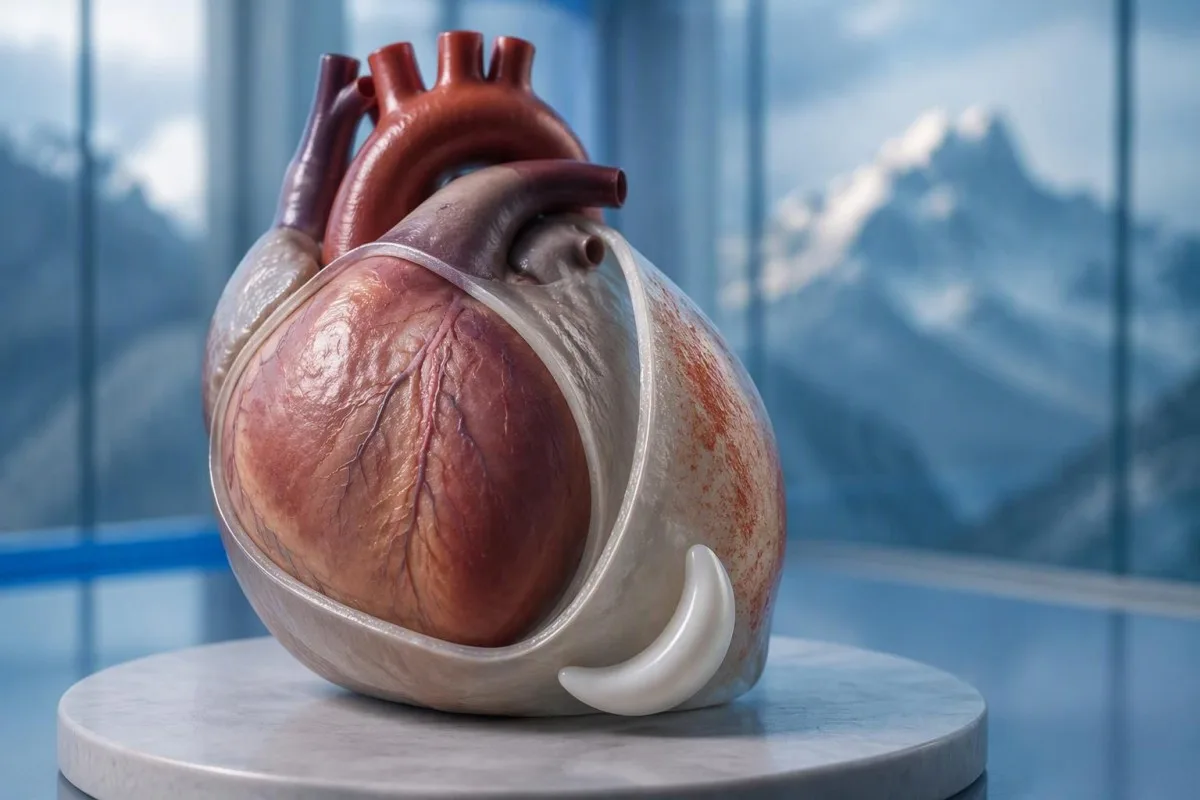
Da — bol u grudima kod perikarditisa često se pogoršava ležeći ravno, a poboljšava sjedeći ili naginjući se naprijed. Ali taj obrazac ne može isključiti srčani udar; nova neobjašnjiva bol u grudima zahtijeva hitnu medicinsku procjenu.


- Bol povezana s položajem često se pogoršava ležeći ravno i poboljšava se naginjanjem naprijed, ali nijedan odgovor ne potvrđuje perikarditis niti isključuje srčani udar.
- Bol povezana s disanjem može se pogoršati dubokim udahom ili kašljanjem; plućna embolija i druga stanja pluća mogu proizvesti isti obrazac.
- Hitni simptomi uključuju jaku ili upornu bol u grudima, kratak dah, nesvjesticu, hladno znojenje ili širenje boli u ruku, vilicu ili leđa. Nazovite svoj lokalni broj hitne pomoći bez čekanja na sve simptome.
- Promjene EKG-a tipične za akutni perikarditis prijavljene su u do 36% slučajeva u smjernici ESC-a iz 2015.; normalan EKG ne isključuje stanje.
- Ehokardiografija procjenjuje tekućinu oko srca i njen utjecaj na punjenje. Izljev veći od 20 mm je faktor visokog rizika, a ne prag ispod kojeg je sva tekućina bezopasna.
- Troponin iznad 99. percentila specifičnog za analizu ukazuje na ozljedu srčanog mišića; sam rezultat ne može razlikovati srčani udar od miokarditisa.
- Testovi na zapaljenje poput CRP i ESR pomažu pri procjeni i praćenju, ali normalni rezultati sami po sebi ne isključuju perikarditis.
- Praćenje obično se javlja u roku od otprilike 1 sedmice za odgovarajuće odabrane ambulantne pacijente; pogoršanje simptoma zahtijeva ranije ponovno procjenjivanje.
Zašto se bol u grudima kod perikarditisa pogoršava ležeći?
Bol u grudima kod perikarditisa se često pogoršava pri ležanju, a smanjuje se pri sjedenju ili naginjanju naprijed. Promjene u položaju mijenjaju mehanički odnos između iritirane srčane ovojnice i obližnjih struktura, ali olakšanje u jednom položaju je dijagnostički trag—ne dokaz da je srčani udar nemoguć.
The perikard je dvoslojna vrećica koja okružuje srce, a njena vanjska ovojnica ima osjetljive na bol veze koje sam srčani mišić ne dijeli na isti način. Poznato objašnjenje da se slojevi jednostavno “trljaju jedni o druge” je nepotpuno: kretanje, iritacija okolnog tkiva i individualni senzorni odgovori također utiču na bol.
Hipotetski 42-godišnjak koji razvije oštar bol nakon respiratorne bolesti i ne može udobno spavati ravno, ima obrazac koji je kompatibilan sa perikarditisom. Još uvijek bih pitao o tačnom početku, naporu, nedostatku daha i kardiovaskularnoj istoriji prije postavljanja dijagnoze; dob i nedavna prehlada ne čine da koronarna bolest ili plućna embolija nestanu.
Kantesti je AI analizator krvi koji može pomoći u objašnjavanju prijavljenih CRP i troponin rezultata nakon kliničke procjene; ne može odlučiti da li je bol u grudima povezan s položajem bezbedan. Kao Thomas Klein, jasno formulišem ovu razliku: 1 ohrabrujući simptom nikada ne bi trebao nadjačati neobjašnjivu akutnu bol u grudima; informacije o našoj organizaciji objašnjava našu obrazovnu ulogu.
Pogoršava li se bol kod perikarditisa kod dubokog disanja?
Bol kod perikarditisa se obično pogoršava pri dubokom udahu, kašljanju ili ponekad gutanju. Kliničari bol povezan s disanjem nazivaju pleuritičnim, ali taj opis identificira obrazac bola, a ne 1 specifičnu bolest; pluća, grudni koš i perikard ga mogu generirati.
Duboko udisanje pomiče dijafragmu i mijenja odnose unutar grudnog koša, što može pogoršati iritaciju srčane ovojnice. Veze koje uključuju frenične nerve, koji potiču uglavnom iz C3–C5, pomažu objasniti zašto neki pacijenti osjećaju prenesenu nelagodu u blizini ramena ili grebena trapeziusa, a ne samo iza grudne kosti.
Bol koji se javlja pri svakom udisaju zahtijeva širu procjenu kada počne iznenada, posebno uz nedostatak daha ili oticanje noge. Plućna embolija može proizvesti istu tegobu bez klasičnog osjećaja pritiska; čak ni zasićenost kisikom od 98% ne može samostalno isključiti.
Osjetljivost pri pritisku kliničara na 1 dio grudi čini izvor u grudnom zidu vjerovatnijim, ali ne pruža apsolutno isključenje koronarne bolesti. Naše objašnjenje testova za neobjašnjivu bol razlikuje podržavajuće laboratorijske tragove od stanja koja zahtijevaju pregled i snimanje.
Koji drugi simptomi perikarditisa su važni?
Simptomi perikarditisa mogu uključivati groznici, umor, nedostatak daha i ubrzan rad srca, kao i bol u grudima. Nijedan nije obavezan, i njihova kombinacija je važnija od bilo kog 1 simptoma; izrazita slabost, nesvjestica ili pogoršanje disanja mijenjaju hitnost procjene.
Temperatura iznad 38°C is a higher-risk feature in the 2015 ESC pericardial guideline, particularly when the cause is uncertain. A patient with 37.4°C and considerable pain may still need urgent assessment, however; temperature thresholds help clinicians organize risk rather than define a safe home-care boundary.
A fast pulse can reflect pain, fever, dehydration, an arrhythmia, or impaired cardiac filling. I would interpret a resting pulse of 110 beats/min differently in someone comfortable and well hydrated than in someone pale and faint; our vodič za testiranje palpitacija explains why laboratory results are only part of that assessment.
Breathlessness when lying flat is not automatically the same thing as chest pain when lying flat. Someone who needs 3 pillows because they cannot breathe comfortably may have congestion, an effusion, or another cardiopulmonary problem; the distinction is covered in our rezultati za nedostatak daha vode, but active breathing difficulty should not wait for online interpretation.
Kada bol u grudima povezana s položajem zahtijeva hitnu pomoć?
Call emergency services for severe, persistent, or rapidly worsening chest pain, especially with breathlessness, fainting, cold sweating, or pain spreading to the arm, jaw, or back. In the UK call 999 or 112; elsewhere use your local emergency number, and do not drive yourself.
Sudden maximal pain, collapse, new confusion, or coughing up blood also warrants emergency assessment because the differential includes pulmonary embolism and aortic emergencies. Do not wait 5 or 15 minutes to see whether leaning forward fixes the problem; concerning accompanying symptoms make a waiting experiment unsafe.
New unexplained chest pain that is not severe still deserves prompt, usually same-day, medical assessment rather than a routine appointment several weeks away. The 2021 AHA/ACC chest pain guideline emphasizes structured assessment and discourages using supposedly “atypical” symptoms to dismiss acute coronary syndrome (Gulati et al., 2021).
Stop exertion while arranging help, and follow the emergency dispatcher’s instructions about any medication rather than starting treatment for presumed pericarditis. Our hitnih krvnih testova za bol u grudima article explains why 1 laboratory result cannot replace an emergency ECG, examination, and appropriately timed testing.
Kako kliničari procjenjuju sumnju na perikarditis?
Clinicians assess suspected pericarditis by combining the history, examination, ECG, echocardiography, and selected blood tests. The widely taught 2015 ESC framework requires at least 2 of 4 findings: typical pain, a pericardial rub, characteristic ECG changes, or a new or worsening pericardial effusion (Adler et al., 2015).
Sharp pain, pain worse lying down, and relief leaning forward are not 3 independent diagnostic criteria; they are different features of the same symptom. This distinction prevents a persuasive story from being mistaken for confirmation, especially when the ECG and imaging have not yet been assessed.
A pericardial friction rub may be transient, so a clinician may listen again after an initial examination rather than treating 1 quiet examination as decisive. Vital signs, signs of poor circulation, heart failure, and the history of recent procedures or systemic illness can matter more urgently than whether the rub is audible.
The 2025 ESC myocarditis and pericarditis guideline gives contemporary imaging and overlapping myocardial involvement a central place in assessment, so the older checklist is not a complete care pathway (Schulz-Menger et al., 2025). Our clinical standards information should be read as documentation about laboratory interpretation—not evidence that software can reproduce this bedside evaluation.
Šta EKG pokazuje kod perikarditisa?
An ECG may show widespread ST-segment elevation and PR-segment depression in acute pericarditis, but these changes are not always present. The 2015 ESC guideline reports characteristic ECG changes in up to 60% of cases, so a normal tracing does not exclude pericarditis (Adler et al., 2015).
A EKG sa 12 odvoda records electrical activity from several perspectives; clinicians assess which leads are affected, reciprocal changes, rhythm, and comparison with previous tracings. “The ST segment looks elevated” is not enough to separate pericarditis, early repolarization, myocardial infarction, and other causes without the clinical setting.
A heart attack more often produces changes corresponding to a coronary territory, whereas pericarditis may produce a broader pattern—but neither rule is absolute. ECG abnormalities can evolve over sati, and repeating the tracing during recurrent or changing symptoms may reveal information absent from the first recording.
Electrolyte disturbances can add another layer of confusion, particularly when kidney disease or medication effects are present. A potassium concentration around 6,0 mmol/L can be clinically urgent depending on context, and our potassium and ECG guide explains why ECG interpretation should never be detached from the rest of the assessment.
Zašto se ehokardiografija koristi za perikarditis?
Echocardiography checks for fluid around the heart, ventricular function, and signs that fluid is impairing cardiac filling. A normal echocardiogram does not exclude uncomplicated pericarditis; an effusion greater than 20 mm is a recognized high-risk feature, not the definition of every dangerous effusion.
The 2015 ESC guideline describes effusions as small below 10 mm, moderate at 10–20 mm, and large above 20 mm, using the echo-free space on imaging. These measurements help communicate size, but the distribution of fluid and its hemodynamic effects still require interpretation rather than a number-only decision.
Cardiac tamponade occurs when pericardial pressure compromises filling; how quickly fluid accumulates can matter more than its absolute volume. A collection that develops over sati may be less well tolerated than a larger, slowly developing collection, and the classic low-pressure, raised-neck-vein, quiet-heart-sound triad is not reliably present in every patient.
The 2025 ESC guideline supports integrating echocardiography with other imaging when the diagnosis or extent of involvement remains uncertain (Schulz-Menger et al., 2025). An elevated NT-proBNP may add information about cardiac strain, but 1 value cannot diagnose tamponade; our NT-proBNP interpretation guide explains several alternative reasons for elevation.
Može li troponin razlikovati perikarditis od srčanog udara?
Troponin identifies heart muscle injury, but an elevated result alone cannot distinguish a heart attack from myocarditis or overlapping myopericarditis. The relevant threshold is the assay-specific gornje referentne granice 99. percentila, interpreted alongside symptoms, ECG findings, serial measurements, and sometimes imaging.
There is no universal normal troponin number for every laboratory: troponina I i troponina T use different assays, and some laboratories apply sex-specific reference limits. If a hypothetical report lists an upper limit of 14 ng/L, a result of 20 ng/L exceeds that particular limit; it does not independently identify the cause, as our troponin I versus T explanation details.
An early normal troponin may precede a detectable rise, which is why emergency departments use validated serial pathways, often with repeat sampling at 1 or 2 hours for suitable high-sensitivity assays. Selected patients can meet validated single-sample rule-out criteria, but that decision depends on assay, symptom timing, ECG, and risk—not a home interpretation of “normal.”
Pericardial symptoms plus raised troponin prompt assessment for myocardial involvement, especially with arrhythmia, breathlessness, or reduced ventricular function. A 30-year-old with a recent viral illness still needs a proper differential; our myocarditis troponin guide explains why neither youth nor the size of the rise settles the diagnosis.
Šta dodaju CRP, ESR i krvne slike?
CRP, ESR, and a full blood count can support evidence of inflammation, but they do not confirm pericarditis or exclude a heart attack. Many laboratories use a standard CRP upper reference limit around 5 mg/L, while others use different limits; the printed laboratory range takes precedence.
CRP od 45 mg/L indicates a substantial inflammatory response in a laboratory with an upper limit of 5 mg/L, but it cannot say whether the source is pericardial, pulmonary, autoimmune, or elsewhere. High-sensitivity CRP measures the same protein at low concentrations; acute illness values should not be interpreted using stable cardiovascular-risk categories.
Kantesti je AI platforma za tumačenje krvne slike that can explain a CRP result against the supplied reference range and help organize its trend alongside other reported results. If CRP changes from 45 to 12 mg/L, the direction may support improvement, but persistent symptoms still matter; our WBC and CRP comparison discusses why these markers can disagree.
ESR often changes more slowly than CRP and is influenced by age, anemia, pregnancy, and protein concentrations, so comparing 2 different markers as though they were interchangeable can mislead follow-up. The 2015 ESC guideline treats inflammatory tests as supportive evidence rather than standalone diagnostic criteria (Adler et al., 2015); our causes of elevated ESR adds context for an unexpectedly persistent elevation.
Koji uzroci i faktori rizika mijenjaju plan liječenja?
Pericarditis may follow a viral illness, occur after cardiac injury, or accompany autoimmune disease, severe kidney disease, tuberculosis, or other conditions. Temperatura iznad 38°C, a large effusion, tamponade, or failure to improve with initial treatment are among the features that can justify hospital assessment and a broader search for causes.
The 2015 ESC guideline lists lack of response to anti-inflammatory treatment within 7 dana as a major adverse prognostic feature, alongside subacute onset and the imaging findings above (Adler et al., 2015). This is a clinician’s reassessment trigger—not permission to wait a week through worsening pain, fever, or breathlessness.
Autoimmune testing is most useful when the history supplies a reason: recurrent episodes, joint symptoms, rashes, mouth ulcers, or known systemic disease. 1 positive antibody does not prove autoimmune pericarditis; our vodič za rezultat anti-dsDNA explains why antibody findings need compatible clinical evidence.
Severe kidney dysfunction can change both the cause of pericarditis and medication safety; an eGFR below 15 mL/min/1.73 m² identifies kidney failure-range function but does not itself diagnose uremic pericarditis. Our urgent low-eGFR symptoms guide is relevant to that background, while chest pain in this setting requires direct medical assessment.
Da li poboljšanje lijekovima potvrđuje perikarditis?
Improvement with ibuprofen, aspirin, or a change in posture does not confirm pericarditis. For appropriately diagnosed uncomplicated cases, clinicians commonly use an anti-inflammatory medicine with colchicine, often for approximately 3 mjeseca after a first episode, but the regimen must account for kidney function, interactions, and the underlying cause.
A reduction in pain from 8 out of 10 to 3 out of 10 is useful clinical information, but several chest-pain causes can respond to analgesia. Do not use medication response as a reason to cancel assessment, and do not start high-dose anti-inflammatory treatment for undiagnosed chest pain without professional advice.
Colchicine requires particular care with kidney or liver impairment and certain interacting medicines, including some antibiotics; taking 2 medicines together can alter exposure even when each prescription is familiar. Corticosteroids are not routine first-line treatment for every uncomplicated case, and selected bacterial or systemic causes need a different strategy rather than simply more pain relief.
Kantesti can help organize laboratory follow-up for a clinician through our vodič za trendove sigurnosti lijekova, but it should not select the treatment or tapering schedule. A plan that includes 1 named prescriber, clear interaction checks, and instructions for worsening symptoms is more useful than a generic dosing chart.
Kada je vježbanje sigurno nakon poboljšanja simptoma perikarditisa?
Exercise should be restricted during active pericarditis, and return should follow clinical reassessment rather than pain relief alone. The decision changes substantially if troponin, rhythm testing, or imaging suggests myocardial involvement; 1 symptom-free day is not evidence that strenuous activity is safe.
The older 2015 ESC guideline used a minimum 3-mjesečnog restriction for athletes with isolated pericarditis, while contemporary guidance places greater emphasis on individualized remission and risk assessment. Patients should not borrow an athlete’s timetable—or a myocarditis timetable—without knowing which diagnosis their clinician has established.
For a hypothetical 28-year-old runner, the difference between isolated pericarditis and myocardial involvement can change follow-up testing and return-to-training decisions. I would ask whether symptoms, inflammatory markers, ventricular function, and any relevant rhythm concerns have resolved; a normal resting pulse of 65 beats/min is only one small part of that picture.
Daily activity, physical work, and competitive sport are 3 different workloads, so a written return plan should specify what the patient can actually do. Our vodič za krvne biomarkere can explain reported measurements, but no biomarker page, wearable reading, or AI result constitutes exercise clearance.
Šta ako se bol u grudima vrati ili testovi izgledaju normalno?
Persistent or recurrent chest pain needs reassessment even when an earlier ECG, echocardiogram, or CRP result was normal. Appropriately selected low-risk outpatients are commonly reviewed within about 1 sedmica, but recurrence with faintness, breathlessness, or worsening pain warrants earlier—and sometimes emergency—assessment.
Residual discomfort, recurrent inflammation, chest-wall pain, and a new unrelated problem can feel similar to the patient, so 2 episodes should not automatically receive the same explanation. Cardiac MRI can sometimes identify pericardial or myocardial involvement when first-line tests leave uncertainty; it is selected for a clinical question, not ordered for every ache.
A follow-up note should record symptom timing, temperature, activity tolerance, medication changes, and the actual laboratory reference ranges. CRP values of 8 and 6 mg/L from different laboratories may not represent a meaningful improvement without knowing the methods and context; our post-discharge result changes guide explains why small numerical changes deserve restraint.
Kantesti je Alat za analizu krvnih nalaza uz pomoć AI-a that can help organize reported results for discussion, but it cannot verify the cause of renewed chest pain from a PDF. My advice here, as Thomas Klein, is to agree on 3 things before leaving follow-up: the next review date, permitted activity, and the symptoms that require immediate help.
Kliničke granice i povezane istraživačke publikacije
Symptoms and laboratory interpretation cannot safely replace an acute chest-pain assessment. Od 3. oktobar 2026., the clinical references below support the distinction between pericardial clues, myocardial injury, and emergency risk; the 2 Zenodo resources are background laboratory guides, not trials validating a pericarditis diagnosis.
Kantesti-ove medical advisory information describes the professional expertise behind our educational work, but readers should not equate a medical title with an examination of their own chest pain. A personal care plan still needs 1 accountable treating clinician or team who can examine the patient, review the ECG, and act on imaging or troponin findings.
The Normalni raspon aPTT-a: D-dimer, protein C, vodič za zgrušavanje krvi is available as a Zenodo educational publication with DOI 10.5281/zenodo.18262555. Its associated clotting-test interpretation guide is relevant when clinicians consider coagulation questions, but a normal aPTT cannot exclude pulmonary embolism and D-dimer must be used within an appropriate clinical probability pathway.
The Vodič za serumske proteine: Test krvi za globuline, albumine i odnos A/G is a second Zenodo educational publication with DOI 10.5281/zenodo.18316300. The associated serum protein interpretation guide supplies background on systemic laboratory findings; neither albumin nor the A/G ratio independently diagnoses pericarditis, and the discovery links below are searches rather than verified copies of these publications.
Često postavljana pitanja
Da li je bol u grudima kod perikarditisa gori kada legnete?
Bol u grudima kod perikarditisa često se pogoršava ležanjem na ravnom, a poboljšava sjedenjem ili naginjanjem unaprijed. Taj obrazac je sugestivan, ali predstavlja samo 1 komponentu kliničke procjene i ne može isključiti srčani udar. Kliničari kombinuju anamnezu sa pregledom, EKG-om, ehokardiografijom i odabranim krvnim pretragama. Nova neobjašnjiva bol u grudima zahtijeva hitnu procjenu, uz hitnu pomoć kod jake boli, otežanog disanja, nesvjestice ili hladnog znojenja.
Da li bol u grudima koji se ublažava naginjanjem unaprijed isključuje srčani udar?
Bol u grudima olakšan naginjanjem naprijed ne isključuje srčani udar. Smjernice AHA/ACC iz 2021. za bol u grudima podržavaju strukturiranu procjenu umjesto odbacivanja simptoma jer zvuče neklasično. Možda će biti potreban EKG i pravovremeno testiranje na visokoosjetljivi troponin, a rani normalan rezultat nije automatski konačan. Pozovite hitnu pomoć kod jakog ili upornog bola ili povezanog nedostatka daha, nesvjestice ili hladnog znojenja.
Može li se javiti perikarditis sa normalnim EKG-om?
Da, perikarditis se može javiti uz normalan EKG. Smjernice ESC-a iz 2015. o perikardu izvijestile su o karakterističnim promjenama EKG-a kod do 60% akutnih slučajeva, što znači da značajan dio nije pokazao klasični obrazac. Nalazi EKG-a također se mogu mijenjati, pa kliničari mogu ponoviti EKG snimanje od 12 odvoda kada se simptomi promijene. Samo normalan EKG ne može isključiti perikarditis ili svaki srčani udar.
Hoće li ehokardiogram uvijek pokazati perikarditis?
Ehokardiogram ne pokazuje uvijek nekomplikovani perikarditis jer se upala može dogoditi bez vidljive kolekcije tečnosti. Ehokardiografija je posebno korisna za mjerenje perikardne tečnosti, procjenu ventrikularne funkcije i traženje oštećenog punjenja. Izliv veći od 20 mm smatra se velikim prema klasifikaciji ESC iz 2015. godine, ali manje brzo nakupljajuće kolekcije i dalje mogu biti opasne. Dalje snimanje može se razmotriti kada kliničko pitanje ostane neriješeno.
Znači li normalan troponin da bol u grudima nije ozbiljna?
Normalan rezultat troponina ne znači automatski da je bol u grudima bezopasna. Vrijeme, 99. percentil granica specifična za test, EKG nalazi i ukupna procjena rizika određuju šta rezultat može isključiti. Validirani putevi visokoosjetljivog troponina često koriste ponovljena mjerenja nakon 1 ili 2 sata, iako odabrani pacijenti mogu ispunjavati uslove za jednosmjerni put isključenja. Troponin također ne isključuje samostalno plućnu emboliju, akutne aortalne probleme ili perikarditis.
Koliko dugo traju simptomi perikarditisa?
Simptomi perikarditisa mogu se poboljšati tokom dana do sedmica uz odgovarajući tretman, ali vremenski okvir varira u zavisnosti od uzroka, recidiva i uključenosti miokarda. Kolhicin se obično propisuje otprilike 3 mjeseca nakon prve nekomplikovane epizode, što ne znači da bol treba da traje tokom tog perioda. Nedostatak poboljšanja u roku od 7 dana od antiinflamatornog tretmana je prepoznati okidač za ponovnu procjenu u smjernicama ESC iz 2015. godine. Pogoršanje bola, kratkog daha ili nesvjestice zahtijeva raniju procjenu, a ne čekanje zakazanog pregleda.
Nabavite analizu krvne slike uz AI već danas
Pridružite se više od 2 miliona korisnika širom svijeta koji vjeruju Kantesti-u za trenutnu i tačnu analizu laboratorijskih testova. Otpremite svoje rezultate krvne slike i dobijte sveobuhvatno tumačenje 15,000+ biomarkera za nekoliko sekundi.
📚 Referisane naučne publikacije
Klein, T., Mitchell, S., & Weber, H. (2026). Normalni raspon aPTT-a: D-dimer, protein C, vodič za zgrušavanje krvi. Kantesti AI Medical Research.
Klein, T., Mitchell, S., & Weber, H. (2026). Vodič za serumske proteine: Test krvi za globuline, albumine i odnos A/G. Kantesti AI Medical Research.
📖 Eksterne medicinske reference
Gulati M i sur. (2021). Smjernice AHA/ACC/ASE/CHEST/SAEM/SCCT/SCMR za procjenu i dijagnostiku bola u prsima iz 2021. godine. Circulation.
Schulz-Menger J et al. (2025). 2025 ESC Guidelines for the management of myocarditis and pericarditis. European Heart Journal.
⚕️ Medicinska izjava o odricanju odgovornosti
Ovaj članak je samo u edukativne svrhe i ne predstavlja medicinski savjet. Za odluke o dijagnozi i liječenju uvijek se posavjetujte s kvalifikovanim zdravstvenim radnikom.
E-E-A-T signal(i) povjerenja
Iskustvo
Klinička revizija radnih tokova tumačenja laboratorijskih nalaza koju vodi ljekar.
Stručnost
Fokus laboratorijske medicine na to kako se biomarkeri ponašaju u kliničkom kontekstu.
Autoritativnost
Napisao dr. Thomas Klein, uz recenziju dr. Sarah Mitchell i prof. dr. Hans Weber.
Pouzdanost
Tumačenje zasnovano na dokazima, s jasnim sljedećim koracima kako bi se smanjila uzbuna.