Ano — bolest na hrudi při perikarditidě se často zhoršuje vleže a zlepšuje v sedě nebo předklonu. Ale tento vzorec nemůže vyloučit infarkt myokardu; nová nevysvětlená bolest na hrudi vyžaduje rychlé lékařské vyšetření.


- Bolest spojená s polohou se často zhoršuje vleže a zlepšuje v předklonu, ale ani jedna reakce nepotvrzuje perikarditidu ani nevylučuje infarkt myokardu.
- Bolest spojená s dýcháním se může zhoršit při hlubokém nádechu nebo kašli; plicní embolie a jiná plicní onemocnění mohou vyvolat stejný vzorec.
- Nouzové příznaky zahrnují silnou nebo přetrvávající bolest na hrudi, dušnost, mdloby, studený pot nebo bolest šířící se do paže, čelisti nebo zad. Okamžitě zavolejte své místní tísňové číslo, aniž byste čekali na všechny příznaky.
- Změny EKG typické pro akutní perikarditidu byly hlášeny až u 36 % případů v doporučení ESC z roku 2015; normální EKG nevylučuje toto onemocnění.
- Echokardiografie hodnotí tekutinu kolem srdce a její vliv na plnění. Výpotek větší než 20 mm je vysoce rizikový faktor, nikoli prahová hodnota, pod kterou je veškerá tekutina neškodná.
- Troponin nad 99. percentilem specifickým pro test indikuje poškození srdečního svalu; samotný výsledek nemůže rozlišit infarkt myokardu od myokarditidy.
- Zánětlivé testy jako CRP a ESR podporují hodnocení a sledování, ale normální výsledky samy o sobě nemohou vyloučit perikarditidu.
- Následná kontrola obvykle dochází během přibližně 1 týdne u vhodně vybraných ambulantních pacientů; zhoršující se příznaky vyžadují dřívější přehodnocení.
Proč se bolest na hrudi při perikarditidě zhoršuje vleže?
Bolest na hrudi při perikarditidě se často zhoršuje vleže a méně intenzivní vsedě nebo v předklonu. Změny postavení mění mechanický vztah mezi podrážděným osrdečníkem a blízkými strukturami, ale úleva v jedné poloze je diagnostickým vodítkem – nikoli důkazem, že infarkt myokardu je nemožný.
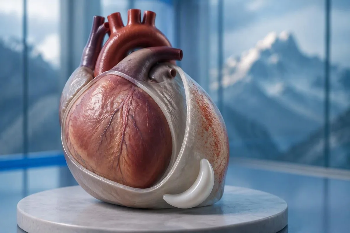
The osrdečník je dvouvrstvý vak obklopující srdce a jeho vnější výstelka má spojení citlivá na bolest, která samotný srdeční sval sdílí jiným způsobem. Běžné vysvětlení, že se vrstvy jednoduše “třou o sebe”, je neúplné: pohyb, podráždění okolní tkáně a individuální smyslové reakce také ovlivňují bolest.
Hypotetická 42letá osoba, u které se objeví ostrá bolest po respiračním onemocnění a nemůže pohodlně spát naplocho, má vzorec odpovídající perikarditidě. Stále bych se ptal na přesný nástup, námahu, dušnost a kardiovaskulární anamnézu, než bych stanovil diagnózu; věk a nedávné nachlazení neznamenají, že by koronární onemocnění nebo plicní embolie zmizely.
Kantesti je Analyzátor krevních testů AI které mohou pomoci vysvětlit hlášené výsledky CRP a troponinu po klinickém hodnocení; nemůže rozhodnout, zda je polohová bolest na hrudi bezpečná. Jako Thomas Klein tuto skutečnost formuluji jasně: 1 uklidňující příznak by nikdy neměl převážit nad nevysvětlitelnou akutní bolestí na hrudi; informace o naší organizace vysvětluje naši vzdělávací úlohu.
Zhoršuje se bolest při perikarditidě při hlubokém nádechu?
Bolest při perikarditidě se obvykle zhoršuje při hlubokém nádechu, kašli nebo někdy při polykání. Lékaři nazývají bolest související s dýcháním pleuritickou, ale tento popis identifikuje vzorec bolesti spíše než 1 konkrétní onemocnění; mohou ji způsobovat plíce, hrudní stěna i osrdečník.
Hluboký nádech pohybuje bránicí a mění vztahy v hrudníku, což může zhoršit podrážděnou výstelku osrdečníku. Spojení zahrnující nervy brániční, vycházející převážně z C3–C5, pomáhají vysvětlit, proč někteří pacienti pociťují vyzařující nepohodlí poblíž ramene nebo hřebene trapézu, spíše než pouze za hrudní kostí.
Bolest, která se zachytává při každém nádechu, vyžaduje širší hodnocení, když začne náhle, zejména spolu s dušností nebo otokem nohy. Plicní embolie může vyvolat stejnou stížnost bez klasického pocitu tlaku; dokonce i saturace kyslíkem 98% ji nemůže nezávisle vyloučit.
Citlivost při stlačení hrudníku lékařem činí zdroj v hrudní stěně pravděpodobnější, ale neposkytuje absolutní vyloučení koronárního onemocnění. Naše vysvětlení testů pro nevysvětlenou bolest rozlišuje podpůrné laboratorní náznaky od stavů, které vyžadují vyšetření a zobrazování.
Které další příznaky perikarditidy jsou důležité?
Příznaky perikarditidy mohou zahrnovat horečku, únavu, dušnost a zrychlený tep, stejně jako bolest na hrudi. Žádný není povinný a jejich kombinace je důležitější než jakýkoli 1 příznak; výrazná slabost, mdloby nebo zhoršení dýchání mění naléhavost posouzení.
Teplota nad 38 °C is a higher-risk feature in the 2015 ESC pericardial guideline, particularly when the cause is uncertain. A patient with 37.4°C and considerable pain may still need urgent assessment, however; temperature thresholds help clinicians organize risk rather than define a safe home-care boundary.
A fast pulse can reflect pain, fever, dehydration, an arrhythmia, or impaired cardiac filling. I would interpret a resting pulse of 110 beats/min differently in someone comfortable and well hydrated than in someone pale and faint; our průvodce vyšetřením bušení srdce explains why laboratory results are only part of that assessment.
Breathlessness when lying flat is not automatically the same thing as chest pain when lying flat. Someone who needs 3 pillows because they cannot breathe comfortably may have congestion, an effusion, or another cardiopulmonary problem; the distinction is covered in our výsledky pro dušnost vedou, but active breathing difficulty should not wait for online interpretation.
Kdy bolest na hrudi spojená s polohou vyžaduje pohotovostní péči?
Call emergency services for severe, persistent, or rapidly worsening chest pain, especially with breathlessness, fainting, cold sweating, or pain spreading to the arm, jaw, or back. In the UK call 999 or 112; elsewhere use your local emergency number, and do not drive yourself.
Sudden maximal pain, collapse, new confusion, or coughing up blood also warrants emergency assessment because the differential includes pulmonary embolism and aortic emergencies. Do not wait 5 or 15 minutes to see whether leaning forward fixes the problem; concerning accompanying symptoms make a waiting experiment unsafe.
New unexplained chest pain that is not severe still deserves prompt, usually same-day, medical assessment rather than a routine appointment several weeks away. The 2021 AHA/ACC chest pain guideline emphasizes structured assessment and discourages using supposedly “atypical” symptoms to dismiss acute coronary syndrome (Gulati et al., 2021).
Stop exertion while arranging help, and follow the emergency dispatcher’s instructions about any medication rather than starting treatment for presumed pericarditis. Our naléhavých krevních testů na bolest na hrudi article explains why 1 laboratory result cannot replace an emergency ECG, examination, and appropriately timed testing.
Jak lékaři hodnotí podezření na perikarditidu?
Clinicians assess suspected pericarditis by combining the history, examination, ECG, echocardiography, and selected blood tests. The widely taught 2015 ESC framework requires at least 2 of 4 findings: typical pain, a pericardial rub, characteristic ECG changes, or a new or worsening pericardial effusion (Adler et al., 2015).
Sharp pain, pain worse lying down, and relief leaning forward are not 3 independent diagnostic criteria; they are different features of the same symptom. This distinction prevents a persuasive story from being mistaken for confirmation, especially when the ECG and imaging have not yet been assessed.
A pericardial friction rub may be transient, so a clinician may listen again after an initial examination rather than treating 1 quiet examination as decisive. Vital signs, signs of poor circulation, heart failure, and the history of recent procedures or systemic illness can matter more urgently than whether the rub is audible.
The 2025 ESC myocarditis and pericarditis guideline gives contemporary imaging and overlapping myocardial involvement a central place in assessment, so the older checklist is not a complete care pathway (Schulz-Menger et al., 2025). Our clinical standards information should be read as documentation about laboratory interpretation—not evidence that software can reproduce this bedside evaluation.
Co ukazuje EKG u perikarditidy?
An ECG may show widespread ST-segment elevation and PR-segment depression in acute pericarditis, but these changes are not always present. The 2015 ESC guideline reports characteristic ECG changes in up to 60% of cases, so a normal tracing does not exclude pericarditis (Adler et al., 2015).
A 12svodové EKG records electrical activity from several perspectives; clinicians assess which leads are affected, reciprocal changes, rhythm, and comparison with previous tracings. “The ST segment looks elevated” is not enough to separate pericarditis, early repolarization, myocardial infarction, and other causes without the clinical setting.
A heart attack more often produces changes corresponding to a coronary territory, whereas pericarditis may produce a broader pattern—but neither rule is absolute. ECG abnormalities can evolve over hodin, and repeating the tracing during recurrent or changing symptoms may reveal information absent from the first recording.
Electrolyte disturbances can add another layer of confusion, particularly when kidney disease or medication effects are present. A potassium concentration around 6,0 mmol/l can be clinically urgent depending on context, and our potassium and ECG guide explains why ECG interpretation should never be detached from the rest of the assessment.
Proč se echokardiografie používá u perikarditidy?
Echocardiography checks for fluid around the heart, ventricular function, and signs that fluid is impairing cardiac filling. A normal echocardiogram does not exclude uncomplicated pericarditis; an effusion greater than 20 mm is a recognized high-risk feature, not the definition of every dangerous effusion.
The 2015 ESC guideline describes effusions as small below 10 mm, moderate at 10–20 mm, and large above 20 mm, using the echo-free space on imaging. These measurements help communicate size, but the distribution of fluid and its hemodynamic effects still require interpretation rather than a number-only decision.
Cardiac tamponade occurs when pericardial pressure compromises filling; how quickly fluid accumulates can matter more than its absolute volume. A collection that develops over hodin may be less well tolerated than a larger, slowly developing collection, and the classic low-pressure, raised-neck-vein, quiet-heart-sound triad is not reliably present in every patient.
The 2025 ESC guideline supports integrating echocardiography with other imaging when the diagnosis or extent of involvement remains uncertain (Schulz-Menger et al., 2025). An elevated NT-proBNP may add information about cardiac strain, but 1 value cannot diagnose tamponade; our NT-proBNP interpretation guide explains several alternative reasons for elevation.
Může troponin rozlišit perikarditidu od infarktu myokardu?
Troponin identifies heart muscle injury, but an elevated result alone cannot distinguish a heart attack from myocarditis or overlapping myopericarditis. The relevant threshold is the assay-specific horní referenční mezí 99. percentilu laboratoře, interpreted alongside symptoms, ECG findings, serial measurements, and sometimes imaging.
There is no universal normal troponin number for every laboratory: troponinu I a troponinu T use different assays, and some laboratories apply sex-specific reference limits. If a hypothetical report lists an upper limit of 14 ng/l, a result of 20 ng/L exceeds that particular limit; it does not independently identify the cause, as our troponin I versus T explanation details.
An early normal troponin may precede a detectable rise, which is why emergency departments use validated serial pathways, often with repeat sampling at 1 or 2 hours for suitable high-sensitivity assays. Selected patients can meet validated single-sample rule-out criteria, but that decision depends on assay, symptom timing, ECG, and risk—not a home interpretation of “normal.”
Pericardial symptoms plus raised troponin prompt assessment for myocardial involvement, especially with arrhythmia, breathlessness, or reduced ventricular function. A 30-year-old with a recent viral illness still needs a proper differential; our myocarditis troponin guide explains why neither youth nor the size of the rise settles the diagnosis.
Co přidávají CRP, ESR a krevní obraz?
CRP, ESR, and a full blood count can support evidence of inflammation, but they do not confirm pericarditis or exclude a heart attack. Many laboratories use a standard CRP upper reference limit around 5 mg/L, while others use different limits; the printed laboratory range takes precedence.
CRP ve výši 45 mg/l indicates a substantial inflammatory response in a laboratory with an upper limit of 5 mg/L, but it cannot say whether the source is pericardial, pulmonary, autoimmune, or elsewhere. High-sensitivity CRP measures the same protein at low concentrations; acute illness values should not be interpreted using stable cardiovascular-risk categories.
Kantesti je platforma pro rozbor krevních výsledků AI that can explain a CRP result against the supplied reference range and help organize its trend alongside other reported results. If CRP changes from 45 to 12 mg/L, the direction may support improvement, but persistent symptoms still matter; our WBC and CRP comparison discusses why these markers can disagree.
ESR often changes more slowly than CRP and is influenced by age, anemia, pregnancy, and protein concentrations, so comparing 2 different markers as though they were interchangeable can mislead follow-up. The 2015 ESC guideline treats inflammatory tests as supportive evidence rather than standalone diagnostic criteria (Adler et al., 2015); our causes of elevated ESR adds context for an unexpectedly persistent elevation.
Které příčiny a rizikové faktory mění plán léčby?
Pericarditis may follow a viral illness, occur after cardiac injury, or accompany autoimmune disease, severe kidney disease, tuberculosis, or other conditions. Horečka nad 38 °C, a large effusion, tamponade, or failure to improve with initial treatment are among the features that can justify hospital assessment and a broader search for causes.
The 2015 ESC guideline lists lack of response to anti-inflammatory treatment within 7 dní as a major adverse prognostic feature, alongside subacute onset and the imaging findings above (Adler et al., 2015). This is a clinician’s reassessment trigger—not permission to wait a week through worsening pain, fever, or breathlessness.
Autoimmune testing is most useful when the history supplies a reason: recurrent episodes, joint symptoms, rashes, mouth ulcers, or known systemic disease. 1 positive antibody does not prove autoimmune pericarditis; our průvodce výsledky anti-dsDNA explains why antibody findings need compatible clinical evidence.
Severe kidney dysfunction can change both the cause of pericarditis and medication safety; an eGFR below 15 mL/min/1.73 m² identifies kidney failure-range function but does not itself diagnose uremic pericarditis. Our urgent low-eGFR symptoms guide is relevant to that background, while chest pain in this setting requires direct medical assessment.
Potvrzuje zlepšení pomocí léků perikarditidu?
Improvement with ibuprofen, aspirin, or a change in posture does not confirm pericarditis. For appropriately diagnosed uncomplicated cases, clinicians commonly use an anti-inflammatory medicine with colchicine, often for approximately 3 měsíce after a first episode, but the regimen must account for kidney function, interactions, and the underlying cause.
A reduction in pain from 8 out of 10 to 3 out of 10 is useful clinical information, but several chest-pain causes can respond to analgesia. Do not use medication response as a reason to cancel assessment, and do not start high-dose anti-inflammatory treatment for undiagnosed chest pain without professional advice.
Colchicine requires particular care with kidney or liver impairment and certain interacting medicines, including some antibiotics; taking 2 medicines together can alter exposure even when each prescription is familiar. Corticosteroids are not routine first-line treatment for every uncomplicated case, and selected bacterial or systemic causes need a different strategy rather than simply more pain relief.
Kantesti can help organize laboratory follow-up for a clinician through our průvodce sledováním bezpečnosti léků, but it should not select the treatment or tapering schedule. A plan that includes 1 named prescriber, clear interaction checks, and instructions for worsening symptoms is more useful than a generic dosing chart.
Kdy je cvičení bezpečné po zlepšení příznaků perikarditidy?
Exercise should be restricted during active pericarditis, and return should follow clinical reassessment rather than pain relief alone. The decision changes substantially if troponin, rhythm testing, or imaging suggests myocardial involvement; 1 symptom-free day is not evidence that strenuous activity is safe.
The older 2015 ESC guideline used a minimum 3měsíčním restriction for athletes with isolated pericarditis, while contemporary guidance places greater emphasis on individualized remission and risk assessment. Patients should not borrow an athlete’s timetable—or a myocarditis timetable—without knowing which diagnosis their clinician has established.
For a hypothetical 28-year-old runner, the difference between isolated pericarditis and myocardial involvement can change follow-up testing and return-to-training decisions. I would ask whether symptoms, inflammatory markers, ventricular function, and any relevant rhythm concerns have resolved; a normal resting pulse of 65 beats/min is only one small part of that picture.
Daily activity, physical work, and competitive sport are 3 different workloads, so a written return plan should specify what the patient can actually do. Our průvodce krevními biomarkery can explain reported measurements, but no biomarker page, wearable reading, or AI result constitutes exercise clearance.
Co když se bolest na hrudi vrátí nebo jsou testy normální?
Persistent or recurrent chest pain needs reassessment even when an earlier ECG, echocardiogram, or CRP result was normal. Appropriately selected low-risk outpatients are commonly reviewed within about 1 týden, but recurrence with faintness, breathlessness, or worsening pain warrants earlier—and sometimes emergency—assessment.
Residual discomfort, recurrent inflammation, chest-wall pain, and a new unrelated problem can feel similar to the patient, so 2 episodes should not automatically receive the same explanation. Cardiac MRI can sometimes identify pericardial or myocardial involvement when first-line tests leave uncertainty; it is selected for a clinical question, not ordered for every ache.
A follow-up note should record symptom timing, temperature, activity tolerance, medication changes, and the actual laboratory reference ranges. CRP values of 8 and 6 mg/L from different laboratories may not represent a meaningful improvement without knowing the methods and context; our post-discharge result changes guide explains why small numerical changes deserve restraint.
Kantesti je Nástroj pro analýzu krevních testů poháněný AI that can help organize reported results for discussion, but it cannot verify the cause of renewed chest pain from a PDF. My advice here, as Thomas Klein, is to agree on 3 things before leaving follow-up: the next review date, permitted activity, and the symptoms that require immediate help.
Klinické hranice a související výzkumné publikace
Symptoms and laboratory interpretation cannot safely replace an acute chest-pain assessment. Od 3. října 2026, the clinical references below support the distinction between pericardial clues, myocardial injury, and emergency risk; the 2 Zenodo resources are background laboratory guides, not trials validating a pericarditis diagnosis.
Kantesti’s medical advisory information describes the professional expertise behind our educational work, but readers should not equate a medical title with an examination of their own chest pain. A personal care plan still needs 1 accountable treating clinician or team who can examine the patient, review the ECG, and act on imaging or troponin findings.
The Normální rozmezí aPTT: D-dimer, protein C, průvodce srážlivostí krve is available as a Zenodo educational publication with DOI 10.5281/zenodo.18262555. Its associated clotting-test interpretation guide is relevant when clinicians consider coagulation questions, but a normal aPTT cannot exclude pulmonary embolism and D-dimer must be used within an appropriate clinical probability pathway.
The Průvodce sérovými proteiny: Krevní test globulinů, albuminu a poměru A/G is a second Zenodo educational publication with DOI 10.5281/zenodo.18316300. The associated serum protein interpretation guide supplies background on systemic laboratory findings; neither albumin nor the A/G ratio independently diagnoses pericarditis, and the discovery links below are searches rather than verified copies of these publications.
Často kladené otázky
Je bolest na hrudi při perikarditidě horší vleže?
Bolest na hrudi při perikarditidě se často zhoršuje v leže a zlepšuje v sedě nebo předklonu. Tento vzorec je sugestivní, ale představuje pouze 1 složku klinického hodnocení a nemůže vyloučit srdeční infarkt. Lékaři kombinují anamnézu s vyšetřením, EKG, echokardiografií a vybranými krevními testy. Nová nevysvětlitelná bolest na hrudi vyžaduje rychlé posouzení, s vyhledáním pohotovostní pomoci při silné bolesti, dušnosti, mdlobách nebo studeném potu.
Vylučuje bolest na hrudi, která se zmírní předklonem, infarkt myokardu?
Bolest na hrudi zmírněná předklonem nevylučuje infarkt myokardu. Směrnice AHA/ACC pro bolest na hrudi z roku 2021 podporuje strukturované hodnocení spíše než zamítání příznaků, protože znějí neklasicky. Může být nutné EKG a včasné testování vysoce senzitivního troponinu a časný normální výsledek není automaticky průkazný. Volejte záchrannou službu při silné nebo přetrvávající bolesti nebo při související dušnosti, mdlobě nebo studeném pocení.
Můžete mít perikarditidu s normálním EKG?
Ano, perikarditida se může vyskytnout i s normálním EKG. Směrnice ESC z roku 2015 o perikardu uvádí, že charakteristické změny EKG byly zaznamenány až u 60% akutních případů, což znamená, že značná část nevykazovala klasický obraz. Nálezy EKG se mohou také vyvíjet, takže lékaři mohou opakovat 12svodové záznamy, když se změní příznaky. Samotné normální EKG nemůže vyloučit perikarditidu ani každý infarkt myokardu.
Ukáže echokardiogram vždy perikarditidu?
Echokardiografie nemusí vždy ukazovat nekomplikovanou perikarditidu, protože zánět se může vyskytnout bez viditelného nahromadění tekutiny. Echokardiografie je zvláště užitečná pro měření perikardiálního výpotku, hodnocení funkce komor a hledání narušeného plnění. Výpotek větší než 20 mm je podle klasifikace ESC z roku 2015 považován za velký, ale menší, rychle se hromadící kolekce mohou být stále nebezpečné. Další zobrazování může být zváženo, pokud klinická otázka zůstává nevyřešena.
Znamená normální troponin, že bolest na hrudi není vážná?
Normální výsledek troponinu automaticky neznamená, že bolest na hrudi je neškodná. Stanovení toho, co výsledek může vyloučit, závisí na načasování, 99. percentilu stanovení specifické pro daný test, nálezech EKG a celkovém hodnocení rizika. Validované cesty pro vysoce citlivý troponin často používají opakovaná měření v rozmezí 1 nebo 2 hodin, ačkoli vybraní pacienti mohou splňovat podmínky pro vyloučení na základě jediného vzorku. Troponin také sám o sobě nevylučuje plicní embolii, aortální příhody nebo perikarditidu.
Jak dlouho trvají příznaky perikarditidy?
Příznaky perikarditidy se mohou s vhodnou léčbou zlepšit během dnů až týdnů, ale časový rámec se liší podle příčiny, recidivy a zapojení myokardu. Kolchicin se obvykle předepisuje po dobu přibližně 3 měsíců po prvním nekomplikovaném epizodě, což neznamená, že bolest by měla přetrvávat po celé toto období. Nedostatek zlepšení do 7 dnů od protizánětlivé léčby je uznávaným spouštěčem přehodnocení podle doporučení ESC 2015. Zhoršující se bolest, dušnost nebo mdloby vyžadují dřívější vyšetření, než čekání na naplánovanou kontrolu.
Získejte analýzu krevních testů poháněnou AI ještě dnes
Přidejte se k více než 2 milionům uživatelů po celém světě, kteří důvěřují Kantesti pro okamžitou a přesnou analýzu laboratorních testů. Nahrajte své výsledky krevních testů a během několika sekund získejte komplexní interpretaci biomarkerů 15,000+.
📚 Odkazované publikace výzkumu
Klein, T., Mitchell, S., & Weber, H. (2026). Normální rozmezí aPTT: D-dimer, protein C, průvodce srážlivostí krve. Kantesti AI Medical Research.
Klein, T., Mitchell, S., & Weber, H. (2026). Průvodce sérovými proteiny: Krevní test globulinů, albuminu a poměru A/G. Kantesti AI Medical Research.
📖 Externí lékařské reference
Gulati M a kol. (2021). Pokyny AHA/ACC/ASE/CHEST/SAEM/SCCT/SCMR pro rok 2021 k hodnocení a diagnostice bolesti na hrudi. Circulation.
Schulz-Menger J et al. (2025). 2025 ESC Guidelines for the management of myocarditis and pericarditis.European Heart Journal.
⚕️ Lékařské prohlášení
Tento článek je pouze pro vzdělávací účely a nepředstavuje lékařské poradenství. Pro rozhodnutí o diagnóze a léčbě se vždy poraďte s kvalifikovaným poskytovatelem zdravotních služeb.
Signály důvěry E-E-A-T
Zažít
Lékařem vedená klinická revize pracovních postupů pro interpretaci laboratorních výsledků.
Odbornost
Zaměření laboratorní medicíny na to, jak se biomarkery chovají v klinickém kontextu.
Autoritativnost
Napsal Dr. Thomas Klein, recenze: Dr. Sarah Mitchell a Prof. Dr. Hans Weber.
Důvěryhodnost
Interpretace založená na důkazech s jasnými následnými kroky pro snížení poplachu.