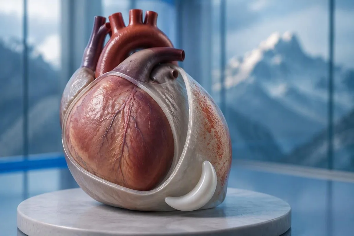
Da – bolečina v prsih pri perikarditisu se pogosto poslabša v ležečem položaju in se izboljša v sedečem položaju ali ob nagnjenju naprej. Vendar pa ta vzorec ne more izključiti srčnega napada; nova nerazložljiva bolečina v prsih potrebuje takojšnjo zdravniško oceno.


- Bolečina glede na položaj se pogosto poslabša v ležečem položaju in se izboljša pri nagnjenju naprej, vendar noben odziv ne potrdi perikarditisa ali izključi srčnega napada.
- Bolečina glede na dihanje se lahko poslabša pri globokem vdihu ali kašlju; pljučna embolija in druge pljučne bolezni lahko povzročijo enak vzorec.
- Nujni simptomi vključujejo hude ali vztrajne bolečine v prsih, težko dihanje, omedlevico, hladno znojenje ali bolečino, ki se širi v roko, čeljust ali hrbet. Pokličite svojo lokalno telefonsko številko za nujne primere brez čakanja na vse simptome.
- Spremembe EKG tipične za akutni perikarditis so bile poročane v do 36 % primerov v smernicah ESC iz leta 2015; normalen EKG ne izključuje bolezni.
- Ehokardiografija oceni tekočino okoli srca in njen vpliv na polnjenje. Izliv, večji od 20 mm, je dejavnik visoke tveganosti, ne pa prag, pod katerim je vsa tekočina neškodljiva.
- Troponin nad 99. percentilom, specifičnim za test, kaže na poškodbo srčne mišice; sam rezultat ne more razlikovati srčnega napada od miokarditisa.
- Preiskave vnetja kot sta CRP in ESR, podpirata oceno in spremljanje, vendar normalni rezultati sami po sebi ne morejo izključiti perikarditisa.
- Nadaljnje spremljanje običajno se pojavi približno v 1 tednu pri ustrezno izbranih bolnikih na domu; poslabšanje simptomov zahteva zgodnejšo ponovno oceno.
Zakaj se bolečina v prsih pri perikarditisu poslabša v ležečem položaju?
Bolečine v prsih pri perikarditisu se pogosto poslabšajo leže in se zmanjšajo sede ali nagnjene naprej. Spremembe položaja spremenijo mehansko razmerje med razdraženim osrčnikom in bližnjimi strukturami, vendar olajšanje v enem položaju je diagnostični namig – ne dokaz, da je srčni napad nemogoč.
The osrčnik je dvoslojni ovoj, ki obdaja srce, njegova zunanja ovojnica pa ima bolečinsko občutljive povezave, ki jih srčna mišica sama nima na enak način. Znana razlaga, da se plasti preprosto “drsijo druga ob drugo”, je nepopolna: gibanje, draženje okoliškega tkiva in posamezni čutni odzivi vplivajo tudi na bolečino.
Hipotetični 42-letnik, ki po respiratorni bolezni občuti ostro bolečino in ne more udobno spati na ravnem, ima vzorec, ki je skladen s perikarditisom. Še vedno bi povprašal o natančnem začetku, naporu, zasoplosti in srčno-žilni anamnezi, preden bi postavil diagnozo; starost in nedavna prehlad ne izginejo koronarne bolezni ali pljučne embolije.
Kantesti je AI analizator krvi ki lahko pomaga razložiti poročane rezultate CRP in troponina po klinični oceni; ne more odločiti, ali je bolečina v prsih, povezana s položajem, varna. Kot Thomas Klein to razliko predstavim jasno: 1 pomirjujoč simptom nikoli ne sme previti nepojasnjene akutne bolečine v prsih; informacije o naši organizaciji pojasnjuje našo izobraževalno vlogo.
Se bolečina pri perikarditisu poslabša pri globokem vdihu?
Bolečine pri perikarditisu se običajno poslabšajo pri globokem vdihu, kašlju ali včasih pri požiranju. Kliniki imenujejo bolečino, povezano z dihanjem, pleuritično, vendar ta opis opredeljuje vzorec bolečine, ne pa 1 specifične bolezni; pljuča, prsni koš in osrčnik lahko vsi povzročijo to bolečino.
Globok vdih premakne diafragmo in spremeni razmerja v prsnem košu, kar lahko poslabša razdraženo ovojnico osrčnika. Povezave, ki vključujejo frenične živce, ki izvirajo predvsem iz C3–C5, pomagajo pojasniti, zakaj nekateri bolniki čutijo preneseno nelagodje v bližini rame ali grebena trapeziusa namesto samo za prsnico.
Bolečina, ki se ob vsakem vdihu ustavi, potrebuje širšo oceno, ko se pojavi nenadoma, še posebej ob zasoplosti ali otekli nogi. Pljučna embolija lahko povzroči isto pritožbo brez klasičnega občutka pritiska; celo nasičenost s kisikom 98% je ne more neodvisno izključiti.
Občutljivost, ko zdravnik pritisne na 1 del prsnega koša, naredi vir iz prsnega koša bolj verjeten, vendar ne zagotavlja absolutnega izključevanja koronarnih arterij. Naša razlaga preiskav za nepojasnjene bolečine ločuje podporne laboratorijske namige od stanj, ki zahtevajo pregled in slikanje.
Kateri drugi simptomi perikarditisa so pomembni?
Simptomi perikarditisa lahko vključujejo vročino, utrujenost, zasoplost in hitrejši srčni utrip ter bolečine v prsih. Noben ni obvezen in njihova kombinacija je pomembnejša od katerega koli 1 simptoma; izrazita oslabelost, omotica ali poslabšanje dihanja spremeni nujnost ocene.
Temperatura nad 38 °C is a higher-risk feature in the 2015 ESC pericardial guideline, particularly when the cause is uncertain. A patient with 37.4°C and considerable pain may still need urgent assessment, however; temperature thresholds help clinicians organize risk rather than define a safe home-care boundary.
A fast pulse can reflect pain, fever, dehydration, an arrhythmia, or impaired cardiac filling. I would interpret a resting pulse of 110 beats/min differently in someone comfortable and well hydrated than in someone pale and faint; our vodilo za testiranje palpitacij explains why laboratory results are only part of that assessment.
Breathlessness when lying flat is not automatically the same thing as chest pain when lying flat. Someone who needs 3 pillows because they cannot breathe comfortably may have congestion, an effusion, or another cardiopulmonary problem; the distinction is covered in our rezultati za zadihanost vodijo, but active breathing difficulty should not wait for online interpretation.
Kdaj bolečina v prsih, povezana s položajem, potrebuje nujno oskrbo?
Call emergency services for severe, persistent, or rapidly worsening chest pain, especially with breathlessness, fainting, cold sweating, or pain spreading to the arm, jaw, or back. In the UK call 999 or 112; elsewhere use your local emergency number, and do not drive yourself.
Sudden maximal pain, collapse, new confusion, or coughing up blood also warrants emergency assessment because the differential includes pulmonary embolism and aortic emergencies. Do not wait 5 or 15 minutes to see whether leaning forward fixes the problem; concerning accompanying symptoms make a waiting experiment unsafe.
New unexplained chest pain that is not severe still deserves prompt, usually same-day, medical assessment rather than a routine appointment several weeks away. The 2021 AHA/ACC chest pain guideline emphasizes structured assessment and discourages using supposedly “atypical” symptoms to dismiss acute coronary syndrome (Gulati et al., 2021).
Stop exertion while arranging help, and follow the emergency dispatcher’s instructions about any medication rather than starting treatment for presumed pericarditis. Our nujnih krvnih preiskav za bolečine v prsih article explains why 1 laboratory result cannot replace an emergency ECG, examination, and appropriately timed testing.
Kako zdravniki ocenjujejo sum na perikarditis?
Clinicians assess suspected pericarditis by combining the history, examination, ECG, echocardiography, and selected blood tests. The widely taught 2015 ESC framework requires at least 2 of 4 findings: typical pain, a pericardial rub, characteristic ECG changes, or a new or worsening pericardial effusion (Adler et al., 2015).
Sharp pain, pain worse lying down, and relief leaning forward are not 3 independent diagnostic criteria; they are different features of the same symptom. This distinction prevents a persuasive story from being mistaken for confirmation, especially when the ECG and imaging have not yet been assessed.
A pericardial friction rub may be transient, so a clinician may listen again after an initial examination rather than treating 1 quiet examination as decisive. Vital signs, signs of poor circulation, heart failure, and the history of recent procedures or systemic illness can matter more urgently than whether the rub is audible.
The 2025 ESC myocarditis and pericarditis guideline gives contemporary imaging and overlapping myocardial involvement a central place in assessment, so the older checklist is not a complete care pathway (Schulz-Menger et al., 2025). Our clinical standards information should be read as documentation about laboratory interpretation—not evidence that software can reproduce this bedside evaluation.
Kaj pokaže EKG pri perikarditisu?
An ECG may show widespread ST-segment elevation and PR-segment depression in acute pericarditis, but these changes are not always present. The 2015 ESC guideline reports characteristic ECG changes in up to 60% of cases, so a normal tracing does not exclude pericarditis (Adler et al., 2015).
A 12-kanalni EKG records electrical activity from several perspectives; clinicians assess which leads are affected, reciprocal changes, rhythm, and comparison with previous tracings. “The ST segment looks elevated” is not enough to separate pericarditis, early repolarization, myocardial infarction, and other causes without the clinical setting.
A heart attack more often produces changes corresponding to a coronary territory, whereas pericarditis may produce a broader pattern—but neither rule is absolute. ECG abnormalities can evolve over urah, and repeating the tracing during recurrent or changing symptoms may reveal information absent from the first recording.
Electrolyte disturbances can add another layer of confusion, particularly when kidney disease or medication effects are present. A potassium concentration around kalij nad can be clinically urgent depending on context, and our potassium and ECG guide explains why ECG interpretation should never be detached from the rest of the assessment.
Zakaj se ehokardiografija uporablja pri perikarditisu?
Echocardiography checks for fluid around the heart, ventricular function, and signs that fluid is impairing cardiac filling. A normal echocardiogram does not exclude uncomplicated pericarditis; an effusion greater than 20 mm is a recognized high-risk feature, not the definition of every dangerous effusion.
The 2015 ESC guideline describes effusions as small below 10 mm, moderate at 10–20 mm, and large above 20 mm, using the echo-free space on imaging. These measurements help communicate size, but the distribution of fluid and its hemodynamic effects still require interpretation rather than a number-only decision.
Cardiac tamponade occurs when pericardial pressure compromises filling; how quickly fluid accumulates can matter more than its absolute volume. A collection that develops over urah may be less well tolerated than a larger, slowly developing collection, and the classic low-pressure, raised-neck-vein, quiet-heart-sound triad is not reliably present in every patient.
The 2025 ESC guideline supports integrating echocardiography with other imaging when the diagnosis or extent of involvement remains uncertain (Schulz-Menger et al., 2025). An elevated NT-proBNP may add information about cardiac strain, but 1 value cannot diagnose tamponade; our NT-proBNP interpretation guide explains several alternative reasons for elevation.
Ali lahko troponin razlikuje perikarditis od srčnega napada?
Troponin identifies heart muscle injury, but an elevated result alone cannot distinguish a heart attack from myocarditis or overlapping myopericarditis. The relevant threshold is the assay-specific zgornjo referenčno mejo 99. percentila, interpreted alongside symptoms, ECG findings, serial measurements, and sometimes imaging.
There is no universal normal troponin number for every laboratory: troponina I in troponina T use different assays, and some laboratories apply sex-specific reference limits. If a hypothetical report lists an upper limit of 14 ng/L, a result of 20 ng/L exceeds that particular limit; it does not independently identify the cause, as our troponin I versus T explanation details.
An early normal troponin may precede a detectable rise, which is why emergency departments use validated serial pathways, often with repeat sampling at 1 or 2 hours for suitable high-sensitivity assays. Selected patients can meet validated single-sample rule-out criteria, but that decision depends on assay, symptom timing, ECG, and risk—not a home interpretation of “normal.”
Pericardial symptoms plus raised troponin prompt assessment for myocardial involvement, especially with arrhythmia, breathlessness, or reduced ventricular function. A 30-year-old with a recent viral illness still needs a proper differential; our myocarditis troponin guide explains why neither youth nor the size of the rise settles the diagnosis.
Kaj dodajo CRP, ESR in krvne preiskave?
CRP, ESR, and a full blood count can support evidence of inflammation, but they do not confirm pericarditis or exclude a heart attack. Many laboratories use a standard CRP upper reference limit around 5 mg/L, while others use different limits; the printed laboratory range takes precedence.
CRP v vrednosti 45 mg/L indicates a substantial inflammatory response in a laboratory with an upper limit of 5 mg/L, but it cannot say whether the source is pericardial, pulmonary, autoimmune, or elsewhere. High-sensitivity CRP measures the same protein at low concentrations; acute illness values should not be interpreted using stable cardiovascular-risk categories.
Kantesti je platforma za razlaga krvne slike z AI that can explain a CRP result against the supplied reference range and help organize its trend alongside other reported results. If CRP changes from 45 to 12 mg/L, the direction may support improvement, but persistent symptoms still matter; our WBC and CRP comparison discusses why these markers can disagree.
ESR often changes more slowly than CRP and is influenced by age, anemia, pregnancy, and protein concentrations, so comparing 2 different markers as though they were interchangeable can mislead follow-up. The 2015 ESC guideline treats inflammatory tests as supportive evidence rather than standalone diagnostic criteria (Adler et al., 2015); our causes of elevated ESR adds context for an unexpectedly persistent elevation.
Katere vzroki in dejavniki tveganja spremenijo načrt oskrbe?
Pericarditis may follow a viral illness, occur after cardiac injury, or accompany autoimmune disease, severe kidney disease, tuberculosis, or other conditions. Vročina nad 38 °C, a large effusion, tamponade, or failure to improve with initial treatment are among the features that can justify hospital assessment and a broader search for causes.
The 2015 ESC guideline lists lack of response to anti-inflammatory treatment within 7 dni as a major adverse prognostic feature, alongside subacute onset and the imaging findings above (Adler et al., 2015). This is a clinician’s reassessment trigger—not permission to wait a week through worsening pain, fever, or breathlessness.
Autoimmune testing is most useful when the history supplies a reason: recurrent episodes, joint symptoms, rashes, mouth ulcers, or known systemic disease. 1 positive antibody does not prove autoimmune pericarditis; our vodnik po rezultatih anti-dsDNA explains why antibody findings need compatible clinical evidence.
Severe kidney dysfunction can change both the cause of pericarditis and medication safety; an eGFR below 15 mL/min/1.73 m² identifies kidney failure-range function but does not itself diagnose uremic pericarditis. Our urgent low-eGFR symptoms guide is relevant to that background, while chest pain in this setting requires direct medical assessment.
Ali izboljšanje z zdravili potrdi perikarditis?
Improvement with ibuprofen, aspirin, or a change in posture does not confirm pericarditis. For appropriately diagnosed uncomplicated cases, clinicians commonly use an anti-inflammatory medicine with colchicine, often for approximately 3 mesece after a first episode, but the regimen must account for kidney function, interactions, and the underlying cause.
A reduction in pain from 8 out of 10 to 3 out of 10 is useful clinical information, but several chest-pain causes can respond to analgesia. Do not use medication response as a reason to cancel assessment, and do not start high-dose anti-inflammatory treatment for undiagnosed chest pain without professional advice.
Colchicine requires particular care with kidney or liver impairment and certain interacting medicines, including some antibiotics; taking 2 medicines together can alter exposure even when each prescription is familiar. Corticosteroids are not routine first-line treatment for every uncomplicated case, and selected bacterial or systemic causes need a different strategy rather than simply more pain relief.
Kantesti can help organize laboratory follow-up for a clinician through our vodnik po trendih varnosti zdravil, but it should not select the treatment or tapering schedule. A plan that includes 1 named prescriber, clear interaction checks, and instructions for worsening symptoms is more useful than a generic dosing chart.
Kdaj je vadba varna po izboljšanju simptomov perikarditisa?
Exercise should be restricted during active pericarditis, and return should follow clinical reassessment rather than pain relief alone. The decision changes substantially if troponin, rhythm testing, or imaging suggests myocardial involvement; 1 symptom-free day is not evidence that strenuous activity is safe.
The older 2015 ESC guideline used a minimum 3-mesečnem restriction for athletes with isolated pericarditis, while contemporary guidance places greater emphasis on individualized remission and risk assessment. Patients should not borrow an athlete’s timetable—or a myocarditis timetable—without knowing which diagnosis their clinician has established.
For a hypothetical 28-year-old runner, the difference between isolated pericarditis and myocardial involvement can change follow-up testing and return-to-training decisions. I would ask whether symptoms, inflammatory markers, ventricular function, and any relevant rhythm concerns have resolved; a normal resting pulse of 65 beats/min is only one small part of that picture.
Daily activity, physical work, and competitive sport are 3 different workloads, so a written return plan should specify what the patient can actually do. Our vodnik po krvnih biomarkerjih can explain reported measurements, but no biomarker page, wearable reading, or AI result constitutes exercise clearance.
Kaj če se bolečina v prsih vrne ali izvidi testov kažejo normalne vrednosti?
Persistent or recurrent chest pain needs reassessment even when an earlier ECG, echocardiogram, or CRP result was normal. Appropriately selected low-risk outpatients are commonly reviewed within about 1 teden, but recurrence with faintness, breathlessness, or worsening pain warrants earlier—and sometimes emergency—assessment.
Residual discomfort, recurrent inflammation, chest-wall pain, and a new unrelated problem can feel similar to the patient, so 2 episodes should not automatically receive the same explanation. Cardiac MRI can sometimes identify pericardial or myocardial involvement when first-line tests leave uncertainty; it is selected for a clinical question, not ordered for every ache.
A follow-up note should record symptom timing, temperature, activity tolerance, medication changes, and the actual laboratory reference ranges. CRP values of 8 and 6 mg/L from different laboratories may not represent a meaningful improvement without knowing the methods and context; our post-discharge result changes guide explains why small numerical changes deserve restraint.
Kantesti je Orodje za analizo krvnih testov z umetno inteligenco that can help organize reported results for discussion, but it cannot verify the cause of renewed chest pain from a PDF. My advice here, as Thomas Klein, is to agree on 3 things before leaving follow-up: the next review date, permitted activity, and the symptoms that require immediate help.
Klinične meje in povezani raziskovalni objavi
Symptoms and laboratory interpretation cannot safely replace an acute chest-pain assessment. Od 3. oktober 2026, the clinical references below support the distinction between pericardial clues, myocardial injury, and emergency risk; the 2 Zenodo resources are background laboratory guides, not trials validating a pericarditis diagnosis.
Kantesti-jev medical advisory information describes the professional expertise behind our educational work, but readers should not equate a medical title with an examination of their own chest pain. A personal care plan still needs 1 accountable treating clinician or team who can examine the patient, review the ECG, and act on imaging or troponin findings.
The Normalno območje aPTT: D-dimer, protein C, vodnik za strjevanje krvi is available as a Zenodo educational publication with DOI 10.5281/zenodo.18262555. Its associated clotting-test interpretation guide is relevant when clinicians consider coagulation questions, but a normal aPTT cannot exclude pulmonary embolism and D-dimer must be used within an appropriate clinical probability pathway.
The Vodnik za serumske beljakovine: krvni test za globuline, albumine in razmerje A/G is a second Zenodo educational publication with DOI 10.5281/zenodo.18316300. The associated serum protein interpretation guide supplies background on systemic laboratory findings; neither albumin nor the A/G ratio independently diagnoses pericarditis, and the discovery links below are searches rather than verified copies of these publications.
Pogosto zastavljena vprašanja
Ali se bolečina v prsih pri perikarditisu poslabša v ležečem položaju?
Perikarditisna bolečina v prsih se pogosto poslabša leže na hrbtu in se izboljša med sedenjem ali predklonom. Ta vzorec je sumljiv, vendar predstavlja le eno komponento klinične ocene in ne more izključiti srčnega napada. Klinični zdravniki združijo anamnezo s pregledom, EKG-jem, ehokardiografijo in izbranimi krvnimi preiskavami. Nova nepojasnjena bolečina v prsih zahteva takojšnjo oceno, z nujno pomočjo pri močni bolečini, kratki sapi, omedlevici ali hladnem znojenju.
Ali bolečina v prsih, ki se ublaži s predklonom, izključuje srčni napad?
Bolečina v prsih, ki se olajša pri nagnjenju naprej, ne izključuje srčnega infarkta. Smernice AHA/ACC iz leta 2021 za bolečine v prsih podpirajo strukturirano oceno, namesto da bi simptome zavrnili, ker zvenijo neklasično. Morda bodo potrebni EKG in ustrezno časovno testiranje visoko občutljivega troponina, zgodnji normalen rezultat pa ni samodejno dokončen. Pokličite reševalno službo ob močni ali vztrajni bolečini ali ob pridruženi sapo, omedlevici ali hladnem znojenju.
Ali je lahko perikarditis z normalnim EKG?
Da, perikarditis se lahko pojavi pri normalnem EKG-ju. Smernice ESC iz leta 2015 za perikarditis so poročale o značilnih spremembah EKG-ja pri do 60 % akutnih primerov, kar pomeni, da znatnemu delu ni ustrezal klasični vzorec. Izsledki EKG-ja se lahko tudi spreminjajo, zato lahko klinični zdravniki ponovijo 12-kanalni zapis, ko se simptomi spremenijo. Sam normalni EKG ne more izključiti perikarditisa ali vsakega srčnega napada.
Bo ultrazvok srca vedno pokazal perikarditis?
Ehokardiogram ne vedno prikaže nekompliciranega perikarditisa, saj lahko do vnetja pride brez vidne zbiralne tekočine. Ehokardiografija je še posebej uporabna za merjenje perikardialne tekočine, ocenjevanje ventrikularne funkcije in iskanje okvarjenega polnjenja. Izliv, večji od 20 mm, je po klasifikaciji ESC iz leta 2015 velik, vendar so lahko manjše, hitro nastajajoče zbirke še vedno nevarne. Dodatno slikanje se lahko upošteva, kadar klinično vprašanje ostane nerešeno.
Ali normalen troponin pomeni, da bolečina v prsih ni resna?
Normalen rezultat troponina ne pomeni samodejno, da bolečina v prsih ni nevarna. Časovni razpored, 99. percentilni limit specifičen za test, izsledki EKG in celovita ocena tveganja določajo, kaj lahko izključimo z rezultatom. Veljavni poti za visoko občutljivi troponin pogosto uporabljajo ponovljene meritve po 1 ali 2 urah, čeprav se lahko izbrani bolniki uvrstijo v pot izključitve z enim samim vzorcem. Troponin tudi samostojno ne izključuje pljučne embolije, srčnih akutnih stanj ali perikarditisa.
Kako dolgo trajajo simptomi perikarditisa?
Simptomi perikarditisa se lahko z ustrezno obravnavo izboljšajo v dneh do tednih, vendar se časovni potek razlikuje glede na vzrok, ponovitev in vpletenost miokarda. Kolhicin se običajno predpisuje približno 3 mesece po prvem nekompliciranem epizodi, kar ne pomeni, da bi bolečina morala trajati celotno obdobje. Pomanjkanje izboljšanja v 7 dneh zdravljenja z protivnetnimi zdravili je priznan sprožilec ponovne ocene v smernicah ESC iz leta 2015. Poslabšanje bolečine, zasoplost ali omedlevanje potrebujejo zgodnejšo oceno, namesto da bi čakali na načrtovani pregled.
Danes pridobite analizo krvnih preiskav z umetno inteligenco
Pridružite se več kot 2 milijonoma uporabnikov po vsem svetu, ki zaupajo Kantesti za takojšnjo, natančno analizo laboratorijskih preiskav. Naložite svoje rezultate krvnih preiskav in v nekaj sekundah prejmite celovito razlago biomarkerjev 15,000+.
📚 Citirane raziskovalne publikacije
Klein, T., Mitchell, S., & Weber, H. (2026). Normalno območje aPTT: D-dimer, protein C, vodnik za strjevanje krvi. Kantesti AI Medical Research.
Klein, T., Mitchell, S., & Weber, H. (2026). Vodnik za serumske beljakovine: krvni test za globuline, albumine in razmerje A/G. Kantesti AI Medical Research.
📖 Zunanje medicinske reference
Gulati M et al. (2021). Smernice 2021 AHA/ACC/ASE/CHEST/SAEM/SCCT/SCMR za oceno in diagnozo bolečine v prsih. Circulation.
Schulz-Menger J et al. (2025). 2025 ESC Guidelines for the management of myocarditis and pericarditis.European Heart Journal.
⚕️ Medicinska izjava o omejitvi odgovornosti
Ta članek je namenjen izobraževalnim namenom in ne predstavlja medicinskega nasveta. Za odločitve o diagnozi in zdravljenju se vedno posvetujte z usposobljenim izvajalcem zdravstvenih storitev.
E-E-A-T zaupanja vredni signali
Izkušnje
Zdravniški klinični pregled delovnih postopkov za interpretacijo laboratorijskih izvidov.
Strokovno znanje
Laboratorijska medicina s poudarkom na tem, kako se biomarkerji obnašajo v kliničnem kontekstu.
Avtoritativnost
Napisal dr. Thomas Klein, pregledala dr. Sarah Mitchell in prof. dr. Hans Weber.
Zanesljivost
Interpretacija na podlagi dokazov z jasnimi nadaljnjimi potmi za zmanjšanje alarmiranja.