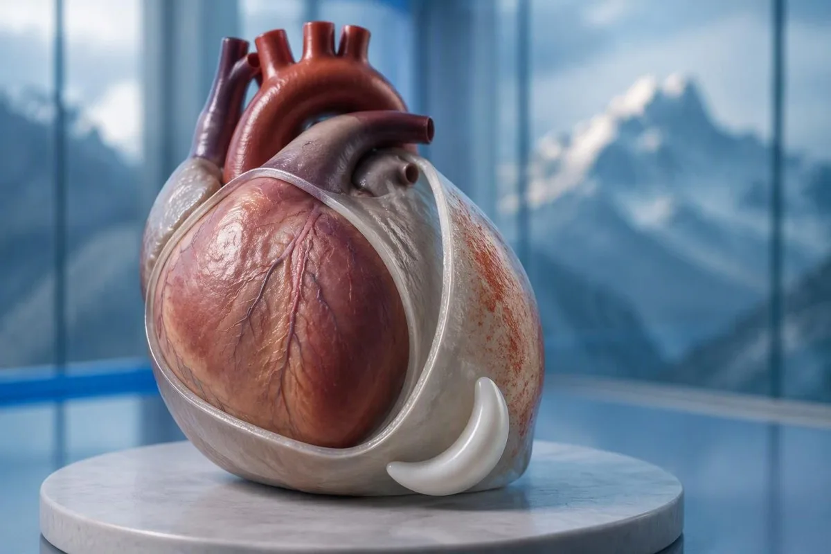
હા—પેરીકાર્ડિટિસની છાતીમાં દુખાવો ઘણીવાર સપાટ સૂવાથી વધી જાય છે અને બેસી રહેવાથી અથવા આગળ ઝૂકવાથી સુધરે છે. પરંતુ તે પેટર્ન હાર્ટ એટેકને નકારી શકતી નથી; નવી અસ્પષ્ટ છાતીમાં દુખાવો માટે તાત્કાલિક તબીબી મૂલ્યાંકનની જરૂર છે.


- સ્થિતિજન્ય દુખાવો ઘણીવાર સપાટ સૂવાથી વધે છે અને આગળ ઝૂકવાથી સુધરે છે, પરંતુ બંનેમાંથી કોઈ પણ પ્રતિભાવ પેરીકાર્ડિટિસની પુષ્ટિ કરતું નથી અથવા હાર્ટ એટેકને નકારતું નથી.
- શ્વાસ સંબંધિત દુખાવો ઊંડા શ્વાસ લેવાથી અથવા ખાંસીથી વધી શકે છે; પલ્મોનરી એમબોલિઝમ અને અન્ય ફેફસાના રોગો સમાન પેટર્ન ઉત્પન્ન કરી શકે છે.
- ઇમરજન્સી લક્ષણો ગંભીર અથવા સતત છાતીમાં દુખાવો, શ્વાસ લેવામાં તકલીફ, બેહોશ થવું, ઠંડો પરસેવો થવો, અથવા હાથ, જડબા અથવા પીઠ સુધી ફેલાતો દુખાવો શામેલ છે. દરેક લક્ષણની રાહ જોયા વિના તમારા સ્થાનિક ઇમરજન્સી નંબર પર કૉલ કરો.
- ECG ફેરફારો 2015 ESC માર્ગદર્શિકામાં 60% જેટલા કેસોમાં તીવ્ર પેરીકાર્ડિટિસ માટે લાક્ષણિક હોવાનું નોંધાયું હતું; સામાન્ય ECG સ્થિતિને નકારી કાઢતું નથી.
- ઇકોકાર્ડિયોગ્રાફી હૃદયની આસપાસના પ્રવાહીનું મૂલ્યાંકન કરે છે અને તેના ભરણ પર અસર કરે છે. 20 mm કરતાં વધુ એફ્યુઝન એ ઉચ્ચ-જોખમ લક્ષણ છે, એવી કોઈ મર્યાદા નથી કે જેનાથી નીચેનું તમામ પ્રવાહી હાનિકારક હોય.
- ટ્રોપોનિન એસે-સ્પેસિફિક 99મી પર્સન્ટાઇલ કરતાં ઉપર હૃદયના સ્નાયુઓને ઈજા સૂચવે છે; પરિણામ એકલું હાર્ટ એટેકને માયોકાર્ડિટિસથી અલગ કરી શકતું નથી.
- दाहક પરીક્ષણો CRP અને ESR જેવા પરીક્ષણો મૂલ્યાંકન અને ફોલો-અપમાં મદદ કરે છે, પરંતુ સામાન્ય પરિણામો એકલા પેરીકાર્ડાઇટિસને નકારી શકતા નથી.
- અનુસરણ યોગ્ય રીતે પસંદ કરાયેલા આઉટપેશન્ટ્સ માટે લગભગ 1 અઠવાડિયામાં સામાન્ય રીતે થાય છે; લક્ષણો વણસવાથી વહેલા ફરીથી મૂલ્યાંકનની જરૂર પડે છે.
પેરીકાર્ડિટિસની છાતીમાં દુખાવો સૂવાથી કેમ વધી શકે છે?
પેરીકાર્ડાઇટિસમાં છાતીમાં દુખાવો ઘણીવાર સૂઈ જવાથી વધી જાય છે અને બેસવાથી અથવા આગળ ઝૂકવાથી ઓછો તીવ્ર બને છે. મુદ્રામાં ફેરફાર બળતરાવાળા પેરીકાર્ડિયમ અને નજીકની રચનાઓ વચ્ચેના યાંત્રિક સંબંધને અસર કરે છે, પરંતુ 1 સ્થિતિમાં રાહત એ નિદાન સંકેત છે—એ પુરાવો નથી કે હાર્ટ એટેક અશક્ય છે.
આ પેરીકાર્ડિયમ હૃદયની આસપાસ બે-સ્તરની કોથળી છે, અને તેની બાહ્ય અસ્તરમાં પીડાદાયક સંવેદનાઓ હોય છે જે હૃદયના સ્નાયુ પોતે તે જ રીતે શેર કરતું નથી. સામાન્ય સમજૂતી કે સ્તરો ફક્ત “એકબીજા પર ઘસાય છે” તે અધૂરી છે: હલનચલન, આસપાસના પેશીઓની બળતરા, અને વ્યક્તિગત સંવેદનાત્મક પ્રતિભાવો પણ પીડાને પ્રભાવિત કરે છે.
એક કાલ્પનિક 42-વર્ષીય વ્યક્તિ જેને શ્વસન બિમારી પછી તીવ્ર દુખાવો થાય છે અને તે આરામથી સપાટ સૂઈ શકતો નથી, તેમાં પેરીકાર્ડાઇટિસને અનુરૂપ પેટર્ન છે. નિદાન જોડતા પહેલા હું હજુ પણ ચોક્કસ શરૂઆત, શ્રમ, શ્વાસની તકલીફ અને કાર્ડિયોવાસ્ક્યુલર ઇતિહાસ વિશે પૂછીશ; ઉંમર અને તાજેતરનો શરદી કોરોનરી રોગ અથવા પલ્મોનરી એમબોલિઝમને દૂર કરતા નથી.
કાન્ટેસ્ટી એક છે AI રક્ત પરીક્ષણ વિશ્લેષક જે ક્લિનિકલ મૂલ્યાંકન પછી CRP અને ટ્રોપોનિનના પરિણામો સમજાવવામાં મદદ કરી શકે છે; તે નક્કી કરી શકતું નથી કે સ્થિતિજન્ય છાતીમાં દુખાવો સલામત છે કે નહીં. થોમસ ક્લાઈન તરીકે, હું આ ભેદને સ્પષ્ટપણે રજૂ કરું છું: 1 આશ્વાસન આપતું લક્ષણ ક્યારેય અસ્પષ્ટ તીવ્ર છાતીની ફરિયાદ કરતાં વધુ મહત્વનું ન હોવું જોઈએ; માહિતી વિશે અમારી સંસ્થા અમારી શૈક્ષણિક ભૂમિકા સમજાવે છે.
શું ઊંડા શ્વાસ લેવાથી પેરીકાર્ડિટિસનો દુખાવો વધે છે?
પેરીકાર્ડાઇટિસનો દુખાવો સામાન્ય રીતે ઊંડો શ્વાસ લેવાથી, ખાંસી ખાવાથી, અથવા ક્યારેક ગળવાથી વધી જાય છે. ચિકિત્સકો શ્વાસ-સંબંધિત પીડાને પ્લ્યુરિટીક કહે છે, પરંતુ તે વર્ણન 1 ચોક્કસ રોગને બદલે પીડા પેટર્નને ઓળખે છે; ફેફસાં, છાતીની દીવાલ અને પેરીકાર્ડિયમ તે બધા ઉત્પન્ન કરી શકે છે.
ઊંડો શ્વાસ ડાયાફ્રેમને ખસેડે છે અને છાતીની અંદરના સંબંધોને બદલે છે, જે બળતરાવાળા પેરીકાર્ડિયલ અસ્તરને વધારી શકે છે. ફ્રેનિક ચેતાઓમાં સંડોવાયેલા જોડાણો, મુખ્યત્વે C3–C5, થી ઉદ્ભવતા, સમજાવવામાં મદદ કરે છે કે શા માટે કેટલાક દર્દીઓને સ્તનની ડીંટડીના હાડકાની પાછળને બદલે ખભા અથવા ટ્રેપેઝિયસ રિજની નજીક સંદર્ભિત અસ્વસ્થતા લાગે છે.
દર શ્વાસ સાથે પકડાઈ જતો દુખાવો જ્યારે તે અચાનક શરૂ થાય છે, ખાસ કરીને શ્વાસની તકલીફ અથવા સોજો પગની સાથે, ત્યારે વધુ વ્યાપક મૂલ્યાંકનની જરૂર પડે છે. પલ્મોનરી એમબોલિઝમ ક્લાસિક દબાણ સંવેદના વિના સમાન ફરિયાદ પેદા કરી શકે છે; ઓક્સિજન સંતૃપ્તિ પણ 98% તેને એકલા નકારી શકતું નથી.
જ્યારે કોઈ ચિકિત્સક છાતીના 1 ભાગ પર દબાણ કરે છે ત્યારે કોમળતા છાતી-દીવાલના સ્ત્રોતને વધુ સંભવિત બનાવે છે, પરંતુ તે સંપૂર્ણ કોરોનરી નિયમ-આઉટ પ્રદાન કરતું નથી. અમારી સમજૂતી અસ્પષ્ટ પીડા માટે પરીક્ષણો સહાયક પ્રયોગશાળા સંકેતોને એવી પરિસ્થિતિઓથી અલગ પાડે છે જેમાં પરીક્ષા અને ઇમેજિંગની જરૂર પડે છે.
પેરીકાર્ડિટિસના કયા અન્ય લક્ષણો મહત્વપૂર્ણ છે?
પેરીકાર્ડાઇટિસના લક્ષણોમાં તાવ, થાક, શ્વાસની તકલીફ અને છાતીમાં દુખાવા ઉપરાંત ઝડપી ધબકારાનો સમાવેશ થઈ શકે છે. કોઈ પણ ફરજિયાત નથી, અને તેમના સંયોજનનું મહત્વ કોઈ 1 લક્ષણ કરતાં વધુ છે; નોંધપાત્ર નબળાઇ, બેહોશી, અથવા શ્વાસમાં બગડતા ફેરફારો મૂલ્યાંકનની તાકીદ બદલી નાખે છે.
એક તાપમાન જે ઉપર હોય 38°C 2015 ESC pericardial guideline માં એક ઉચ્ચ-જોખમી લક્ષણ છે, ખાસ કરીને જ્યારે કારણ અનિશ્ચિત હોય. 37.4°C અને નોંધપાત્ર પીડા ધરાવતા દર્દીને તાત્કાલિક મૂલ્યાંકનની જરૂર પડી શકે છે; જોકે, તાપમાનની મર્યાદા ક્લિનિશિયનોને જોખમ ગોઠવવામાં મદદ કરે છે, ઘરની સંભાળ માટે સુરક્ષિત સીમા વ્યાખ્યાયિત કરવાને બદલે.
ઝડપી પલ્સ પીડા, તાવ, નિર્જલીકરણ, અનિયમિત ધબકારા, અથવા ક્ષતિગ્રસ્ત હૃદય ભરવાનું પ્રતિબિંબિત કરી શકે છે. હું આરામની પલ્સનું અર્થઘટન કરીશ 110 beats/min આરામદાયક અને સારી રીતે હાઇડ્રેટેડ વ્યક્તિમાં નિસ્તેજ અને બેહોશ વ્યક્તિ કરતાં અલગ; અમારું પલ્પિટેશન્સ ટેસ્ટિંગ ગાઇડ સમજાવે છે કે લેબોરેટરી પરિણામો એ મૂલ્યાંકનનો માત્ર એક ભાગ શા માટે છે.
સપાટ સૂતી વખતે શ્વાસ લેવામાં તકલીફ એ સપાટ સૂતી વખતે છાતીમાં દુખાવો જેવી જ વસ્તુ નથી. કોઈ વ્યક્તિ જેને 3 pillows ની જરૂર છે કારણ કે તેઓ આરામથી શ્વાસ લઈ શકતા નથી તેમને કન્જેશન, ઇફ્યુઝન, અથવા અન્ય કાર્ડિયોપલ્મોનરી સમસ્યા હોઈ શકે છે; આ તફાવત અમારા શ્વાસ લેવામાં તકલીફના પરિણામો માર્ગદર્શન આપે છે, માં આવરી લેવામાં આવ્યો છે, પરંતુ સક્રિય શ્વાસની તકલીફ ઓનલાઈન અર્થઘટનની રાહ ન જોવી જોઈએ.
સ્થિતિજન્ય છાતીમાં દુખાવો ક્યારે કટોકટીની સંભાળની જરૂર છે?
ગંભીર, સતત, અથવા ઝડપથી વધી રહેલી છાતીમાં દુખાવો, ખાસ કરીને શ્વાસ લેવામાં તકલીફ, બેહોશી, ઠંડો પરસેવો, અથવા હાથ, જડબા, અથવા પીઠ સુધી ફેલાતા દુખાવા માટે તાત્કાલિક સેવાઓ પર કૉલ કરો. યુકેમાં કૉલ કરો 999 or 112; અન્યત્ર તમારા સ્થાનિક ઇમરજન્સી નંબરનો ઉપયોગ કરો, અને જાતે ડ્રાઇવ કરશો નહીં.
અચાનક મહત્તમ પીડા, પતન, નવી મૂંઝવણ, અથવા લોહી ખાંસી થવી પણ ઇમરજન્સી મૂલ્યાંકનની બાંયધરી આપે છે કારણ કે ડિફરન્શિયલ પલ્મોનરી એમબોલિઝમ અને એઓર્ટિક ઇમરજન્સીનો સમાવેશ કરે છે. રાહ ન જુઓ 5 or 15 minutes જોવા માટે કે આગળ ઝૂકવાથી સમસ્યા હલ થાય છે કે કેમ; ચિંતાજનક સાથેના લક્ષણો રાહ જોવાનો પ્રયોગ અસુરક્ષિત બનાવે છે.
નવી અસ્પષ્ટ છાતીમાં દુખાવો જે ગંભીર નથી તે હજુ પણ થોડા અઠવાડિયા દૂરના નિયમિત એપોઇન્ટમેન્ટને બદલે તાત્કાલિક, સામાન્ય રીતે તે જ દિવસે, તબીબી મૂલ્યાંકનને યોગ્ય છે. 2021 AHA/ACC chest pain guideline સંરચિત મૂલ્યાંકન પર ભાર મૂકે છે અને તીવ્ર કોરોનરી સિન્ડ્રોમને નકારવા માટે કથિત “અસામાન્ય” લક્ષણોનો ઉપયોગ કરવાનું નિરુત્સાહિત કરે છે (Gulati et al., 2021).
મદદ ગોઠવતી વખતે શ્રમ બંધ કરો, અને કોઈપણ દવા વિશે ઇમરજન્સી ડિસ્પેચરની સૂચનાઓનું પાલન કરો, ધારણા પેરીકાર્ડિટિસ માટે સારવાર શરૂ કરવાને બદલે. અમારો તાત્કાલિક છાતી-દુખાવો બ્લડ ટેસ્ટ લેખ સમજાવે છે કે શા માટે 1 લેબોરેટરી પરિણામ ઇમરજન્સી ECG, પરીક્ષા, અને યોગ્ય રીતે સમયસર પરીક્ષણનું સ્થાન લઈ શકતું નથી.
ચિકિત્સકો શંકાસ્પદ પેરીકાર્ડિટિસનું મૂલ્યાંકન કેવી રીતે કરે છે?
ક્લિનિશિયનો ઇતિહાસ, પરીક્ષા, ECG, ઇકોકાર્ડિયોગ્રાફી, અને પસંદ કરેલા રક્ત પરીક્ષણોને સંયોજિત કરીને શંકાસ્પદ પેરીકાર્ડિટિસનું મૂલ્યાંકન કરે છે. વ્યાપકપણે શીખવાયેલ 2015 ESC ફ્રેમવર્કને ઓછામાં ઓછું જરૂરી છે 2 of 4 તારણો: લાક્ષણિક પીડા, પેરીકાર્ડિયલ રબ, લાક્ષણિક ECG ફેરફારો, અથવા નવો અથવા બગડતો પેરીકાર્ડિયલ એફ્યુઝન (Adler et al., 2015).
તીવ્ર પીડા, સૂઈ જવાથી પીડા વધવી, અને આગળ ઝૂકવાથી રાહત એ 3 સ્વતંત્ર નિદાન માપદંડ નથી; તે એક જ લક્ષણની જુદી જુદી વિશેષતાઓ છે. આ ભેદભેદ પુષ્ટિ માટે ખોટી રીતે સમજાવતી વાર્તાને અટકાવે છે, ખાસ કરીને જ્યારે ECG અને ઇમેજિંગનું મૂલ્યાંકન હજુ સુધી થયું નથી.
A પેરીકાર્ડિયલ ફ્રિક્શન રબ અસ્થાયી હોઈ શકે છે, તેથી ચિકિત્સક પ્રારંભિક પરીક્ષા પછી ફરીથી સાંભળી શકે છે, તેના બદલે 1 શાંત પરીક્ષાને નિર્ણાયક માની લે છે. વાઇટલ સંકેતો, નબળા પરિભ્રમણના સંકેતો, હૃદયની નિષ્ફળતા, અને તાજેતરની પ્રક્રિયાઓ અથવા પ્રણાલીગત બીમારીનો ઇતિહાસ રબ સંભળાતો હોય કે ન હોય તેના કરતાં વધુ તાકીદે મહત્વનો હોઈ શકે છે.
આ 2025 ESC માયોકાર્ડિટિસ અને પેરીકાર્ડિટિસ માર્ગદર્શિકા મૂલ્યાંકનમાં સમકાલીન ઇમેજિંગ અને ઓવરલેપિંગ માયોકાર્ડિયલ સંડોવણીને કેન્દ્રીય સ્થાન આપે છે, તેથી જૂની ચેકલિસ્ટ સંપૂર્ણ સંભાળ માર્ગ નથી (Schulz-Menger et al., 2025). અમારી ક્લિનિકલ ધોરણો માહિતી લેબોરેટરી અર્થઘટન વિશે દસ્તાવેજીકરણ તરીકે વાંચવી જોઈએ - કોઈ પુરાવા નથી કે સોફ્ટવેર આ બેડસાઇડ મૂલ્યાંકનનું પુનરાવર્તન કરી શકે છે.
પેરીકાર્ડિટિસમાં ECG શું દર્શાવે છે?
એક ECG તીવ્ર પેરીકાર્ડિટિસમાં વ્યાપક ST-સેગમેન્ટ એલિવેશન અને PR-સેગમેન્ટ ડિપ્રેશન દર્શાવી શકે છે, પરંતુ આ ફેરફારો હંમેશા હાજર નથી. 2015 ESC માર્ગદર્શિકા Up to 60% કેસોમાં લાક્ષણિક ECG ફેરફારો દર્શાવે છે, તેથી સામાન્ય ટ્રેસિંગ પેરીકાર્ડિટિસને બાકાત રાખતું નથી (Adler et al., 2015).
A 12-લીડ ECG અનેક પરિપ્રેક્ષ્યોથી ઇલેક્ટ્રિકલ પ્રવૃત્તિ રેકોર્ડ કરે છે; ચિકિત્સકો મૂલ્યાંકન કરે છે કે કઈ લીડ્સ અસરગ્રસ્ત છે, પરસ્પર વિરોધી ફેરફારો, લય, અને અગાઉના ટ્રેસિંગ સાથે સરખામણી. “ST સેગમેન્ટ elevated દેખાય છે” ક્લિનિકલ સંદર્ભ વિના પેરીકાર્ડિટિસ, પ્રારંભિક રિપોલરાઇઝેશન, માયોકાર્ડિયલ ઇન્ફાર્ક્શન અને અન્ય કારણોને અલગ કરવા માટે પૂરતું નથી.
હાર્ટ એટેક વધુ વખત કોરોનરી વિસ્તારને અનુરૂપ ફેરફારો ઉત્પન્ન કરે છે, જ્યારે પેરીકાર્ડિટિસ વ્યાપક પેટર્ન ઉત્પન્ન કરી શકે છે - પરંતુ કોઈ પણ નિયમ સંપૂર્ણ નથી. ECG અસાધારણતા વિકસિત થઈ શકે છે કલાકોની અંદર, અને પ્રથમ રેકોર્ડિંગમાંથી ગેરહાજર માહિતી જાહેર કરવા માટે પુનરાવર્તિત અથવા બદલાતા લક્ષણો દરમિયાન ટ્રેસિંગને પુનરાવર્તિત કરવું.
ઇલેક્ટ્રોલાઇટ ડિસ્ટર્બન્સ ગૂંચવણનો બીજો સ્તર ઉમેરી શકે છે, ખાસ કરીને જ્યારે કિડની રોગ અથવા દવાઓની અસરો હાજર હોય. આસપાસ પોટેશિયમ સાંદ્રતા 6.0 mmol/L થી ઉપર સંદર્ભ પર આધાર રાખીને ક્લિનિકલી તાકીદે હોઈ શકે છે, અને અમારું પોટેશિયમ અને ECG માર્ગદર્શિકા સમજાવે છે કે ECG અર્થઘટન ક્યારેય મૂલ્યાંકનના બાકીના ભાગથી અલગ ન હોવું જોઈએ.
પેરીકાર્ડિટિસ માટે ઇકોકાર્ડિયોગ્રાફીનો ઉપયોગ શા માટે થાય છે?
ઇકોકાર્ડિયોગ્રાફી હૃદયની આસપાસ પ્રવાહી, વેન્ટ્રિક્યુલર ફંક્શન અને પ્રવાહી કાર્ડિયાક ફિલિંગને નુકસાન પહોંચાડી રહ્યું છે કે કેમ તેના સંકેતો તપાસે છે. સામાન્ય ઇકોકાર્ડિયોગ્રામ અનકમ્પ્લિકેટેડ પેરીકાર્ડિટિસને બાકાત રાખતું નથી; તેના કરતાં વધુ એફ્યુઝન 20 mm is a recognized high-risk feature, not the definition of every dangerous effusion.
The 2015 ESC guideline describes effusions as small below 10 mm, moderate at 10–20 mm, and large above 20 mm, using the echo-free space on imaging. These measurements help communicate size, but the distribution of fluid and its hemodynamic effects still require interpretation rather than a number-only decision.
Cardiac tamponade occurs when pericardial pressure compromises filling; how quickly fluid accumulates can matter more than its absolute volume. A collection that develops over કલાકોની અંદર may be less well tolerated than a larger, slowly developing collection, and the classic low-pressure, raised-neck-vein, quiet-heart-sound triad is not reliably present in every patient.
આ 2025 ESC guideline supports integrating echocardiography with other imaging when the diagnosis or extent of involvement remains uncertain (Schulz-Menger et al., 2025). An elevated NT-proBNP may add information about cardiac strain, but 1 value cannot diagnose tamponade; our NT-proBNP interpretation guide explains several alternative reasons for elevation.
શું ટ્રોપોનિન પેરીકાર્ડિટિસને હાર્ટ એટેકથી અલગ કરી શકે છે?
Troponin identifies heart muscle injury, but an elevated result alone cannot distinguish a heart attack from myocarditis or overlapping myopericarditis. The relevant threshold is the assay-specific 99મા પર્સેન્ટાઇલની ઉપરની રેફરન્સ મર્યાદા કરતાં ઉપરનું કોઈપણ મૂલ્ય અસામાન્ય છે, પરંતુ નિદાન લક્ષણો અને વધે-ઘટે એવા પેટર્ન પર આધાર રાખે છે, એક જ અલગ માપ પર નહીં; એટલે જ AI ક્યારેય છાતીના દુખાવાને ક્લિયર ન કરવું જોઈએ. એક, interpreted alongside symptoms, ECG findings, serial measurements, and sometimes imaging.
There is no universal normal troponin number for every laboratory: ટ્રોપોનિન I અને ટ્રોપોનિન T use different assays, and some laboratories apply sex-specific reference limits. If a hypothetical report lists an upper limit of 14 ng/L, a result of 20 ng/L exceeds that particular limit; it does not independently identify the cause, as our troponin I versus T explanation details.
An early normal troponin may precede a detectable rise, which is why emergency departments use validated serial pathways, often with repeat sampling at 1 or 2 hours for suitable high-sensitivity assays. Selected patients can meet validated single-sample rule-out criteria, but that decision depends on assay, symptom timing, ECG, and risk—not a home interpretation of “normal.”
Pericardial symptoms plus raised troponin prompt assessment for myocardial involvement, especially with arrhythmia, breathlessness, or reduced ventricular function. A 30-year-old with a recent viral illness still needs a proper differential; our myocarditis troponin guide explains why neither youth nor the size of the rise settles the diagnosis.
CRP, ESR, અને બ્લડ કાઉન્ટ્સ શું ઉમેરે છે?
CRP, ESR, and a full blood count can support evidence of inflammation, but they do not confirm pericarditis or exclude a heart attack. Many laboratories use a standard CRP upper reference limit around 5 mg/L, while others use different limits; the printed laboratory range takes precedence.
CRP નું મૂલ્ય 45 mg/L indicates a substantial inflammatory response in a laboratory with an upper limit of 5 mg/L, but it cannot say whether the source is pericardial, pulmonary, autoimmune, or elsewhere. High-sensitivity CRP measures the same protein at low concentrations; acute illness values should not be interpreted using stable cardiovascular-risk categories.
કાન્ટેસ્ટી એક છે AI બ્લડ ટેસ્ટ રિપોર્ટ સમજો પ્લેટફોર્મ that can explain a CRP result against the supplied reference range and help organize its trend alongside other reported results. If CRP changes from 45 to 12 mg/L, the direction may support improvement, but persistent symptoms still matter; our WBC and CRP comparison discusses why these markers can disagree.
ESR often changes more slowly than CRP and is influenced by age, anemia, pregnancy, and protein concentrations, so comparing 2 different markers as though they were interchangeable can mislead follow-up. The 2015 ESC guideline treats inflammatory tests as supportive evidence rather than standalone diagnostic criteria (Adler et al., 2015); our causes of elevated ESR adds context for an unexpectedly persistent elevation.
કયા કારણો અને જોખમી લક્ષણો સંભાળ યોજના બદલે છે?
Pericarditis may follow a viral illness, occur after cardiac injury, or accompany autoimmune disease, severe kidney disease, tuberculosis, or other conditions. 38°Cથી ઉપર તાવ 38°C, a large effusion, tamponade, or failure to improve with initial treatment are among the features that can justify hospital assessment and a broader search for causes.
આ 2015 ESC guideline lists lack of response to anti-inflammatory treatment within 7 દિવસ as a major adverse prognostic feature, alongside subacute onset and the imaging findings above (Adler et al., 2015). This is a clinician’s reassessment trigger—not permission to wait a week through worsening pain, fever, or breathlessness.
Autoimmune testing is most useful when the history supplies a reason: recurrent episodes, joint symptoms, rashes, mouth ulcers, or known systemic disease. 1 positive antibody does not prove autoimmune pericarditis; our anti-dsDNA પરિણામ માર્ગદર્શિકા explains why antibody findings need compatible clinical evidence.
Severe kidney dysfunction can change both the cause of pericarditis and medication safety; an eGFR below 15 mL/min/1.73 m² identifies kidney failure-range function but does not itself diagnose uremic pericarditis. Our urgent low-eGFR symptoms guide is relevant to that background, while chest pain in this setting requires direct medical assessment.
શું દવાઓ સાથે સુધારો પેરીકાર્ડિટિસની પુષ્ટિ કરે છે?
Improvement with ibuprofen, aspirin, or a change in posture does not confirm pericarditis. For appropriately diagnosed uncomplicated cases, clinicians commonly use an anti-inflammatory medicine with colchicine, often for approximately 3 મહિના કરતાં વધુ after a first episode, but the regimen must account for kidney function, interactions, and the underlying cause.
A reduction in pain from 8 out of 10 to 3 out of 10 is useful clinical information, but several chest-pain causes can respond to analgesia. Do not use medication response as a reason to cancel assessment, and do not start high-dose anti-inflammatory treatment for undiagnosed chest pain without professional advice.
Colchicine requires particular care with kidney or liver impairment and certain interacting medicines, including some antibiotics; taking 2 medicines together can alter exposure even when each prescription is familiar. Corticosteroids are not routine first-line treatment for every uncomplicated case, and selected bacterial or systemic causes need a different strategy rather than simply more pain relief.
Kantesti can help organize laboratory follow-up for a clinician through our દવા-સુરક્ષા ટ્રેન્ડ માર્ગદર્શિકા, but it should not select the treatment or tapering schedule. A plan that includes 1 named prescriber, clear interaction checks, and instructions for worsening symptoms is more useful than a generic dosing chart.
પેરીકાર્ડિટિસના લક્ષણો સુધર્યા પછી કસરત ક્યારે સુરક્ષિત છે?
Exercise should be restricted during active pericarditis, and return should follow clinical reassessment rather than pain relief alone. The decision changes substantially if troponin, rhythm testing, or imaging suggests myocardial involvement; 1 symptom-free day is not evidence that strenuous activity is safe.
The older 2015 ESC guideline used a minimum 3-મહિના restriction for athletes with isolated pericarditis, while contemporary guidance places greater emphasis on individualized remission and risk assessment. Patients should not borrow an athlete’s timetable—or a myocarditis timetable—without knowing which diagnosis their clinician has established.
For a hypothetical 28-year-old runner, the difference between isolated pericarditis and myocardial involvement can change follow-up testing and return-to-training decisions. I would ask whether symptoms, inflammatory markers, ventricular function, and any relevant rhythm concerns have resolved; a normal resting pulse of 65 beats/min is only one small part of that picture.
Daily activity, physical work, and competitive sport are 3 different workloads, so a written return plan should specify what the patient can actually do. Our બ્લડ બાયોમાર્કર માર્ગદર્શિકા can explain reported measurements, but no biomarker page, wearable reading, or AI result constitutes exercise clearance.
જો છાતીમાં દુખાવો પાછો ફરે અથવા પરીક્ષણો સામાન્ય દેખાય તો શું?
Persistent or recurrent chest pain needs reassessment even when an earlier ECG, echocardiogram, or CRP result was normal. Appropriately selected low-risk outpatients are commonly reviewed within about 1 અઠવાડિયું, but recurrence with faintness, breathlessness, or worsening pain warrants earlier—and sometimes emergency—assessment.
Residual discomfort, recurrent inflammation, chest-wall pain, and a new unrelated problem can feel similar to the patient, so 2 episodes should not automatically receive the same explanation. Cardiac MRI can sometimes identify pericardial or myocardial involvement when first-line tests leave uncertainty; it is selected for a clinical question, not ordered for every ache.
A follow-up note should record symptom timing, temperature, activity tolerance, medication changes, and the actual laboratory reference ranges. CRP values of 8 and 6 mg/L from different laboratories may not represent a meaningful improvement without knowing the methods and context; our post-discharge result changes guide explains why small numerical changes deserve restraint.
કાન્ટેસ્ટી એક છે AI-સંચાલિત બ્લડ ટેસ્ટ એનાલિસિસ ટૂલ that can help organize reported results for discussion, but it cannot verify the cause of renewed chest pain from a PDF. My advice here, as Thomas Klein, is to agree on 3 things before leaving follow-up: the next review date, permitted activity, and the symptoms that require immediate help.
ક્લિનિકલ સીમાઓ અને સંબંધિત સંશોધન પ્રકાશનો
Symptoms and laboratory interpretation cannot safely replace an acute chest-pain assessment. મુજબ ઓક્ટોબર 3, 2026, the clinical references below support the distinction between pericardial clues, myocardial injury, and emergency risk; the 2 Zenodo resources are background laboratory guides, not trials validating a pericarditis diagnosis.
Kantesti’s medical advisory information describes the professional expertise behind our educational work, but readers should not equate a medical title with an examination of their own chest pain. A personal care plan still needs 1 accountable treating clinician or team who can examine the patient, review the ECG, and act on imaging or troponin findings.
આ aPTT નોર્મલ રેન્જ: D-Dimer, પ્રોટીન C બ્લડ ક્લોટિંગ માર્ગદર્શિકા is available as a Zenodo educational publication with DOI ૧૦.૫૨૮૧/ઝેનોડો.૧૮૨૬૨૫૫૫. Its associated clotting-test interpretation guide is relevant when clinicians consider coagulation questions, but a normal aPTT cannot exclude pulmonary embolism and D-dimer must be used within an appropriate clinical probability pathway.
આ સીરમ પ્રોટીન માર્ગદર્શિકા: ગ્લોબ્યુલિન, આલ્બ્યુમિન અને એ/જી રેશિયો બ્લડ ટેસ્ટ is a second Zenodo educational publication with DOI ૧૦.૫૨૮૧/ઝેનોડો.૧૮૩૧૬૩૦૦. The associated serum protein interpretation guide supplies background on systemic laboratory findings; neither albumin nor the A/G ratio independently diagnoses pericarditis, and the discovery links below are searches rather than verified copies of these publications.
વારંવાર પૂછાતા પ્રશ્નો
શું પેરીકાર્ડાઇટિસમાં છાતીમાં દુખાવો નીચે સૂવાથી વધી જાય છે?
પેરિકાર્ડાઇટિસની છાતીમાં દુખાવો ઘણીવાર સપાટ સૂવાથી વધી જાય છે અને બેસવાથી અથવા આગળ ઝૂકવાથી સુધરે છે. તે પેટર્ન સૂચક છે, પરંતુ તે ક્લિનિકલ આકારણીનો માત્ર 1 ઘટક રજૂ કરે છે અને હાર્ટ એટેકને નકારી શકે નહીં. ચિકિત્સકો ઇતિહાસને તપાસ, ECG, ઇકોકાર્ડિયોગ્રાફી અને પસંદ કરેલા રક્ત પરીક્ષણો સાથે જોડે છે. છાતીમાં નવો અસ્પષ્ટ દુખાવો તાત્કાલિક આકારણીની જરૂર છે, જેમાં ગંભીર દુખાવો, શ્વાસ લેવામાં તકલીફ, બેહોશી અથવા ઠંડો પરસેવો માટે તાત્કાલિક મદદ લેવી.
આગળ નમવાથી છાતીમાં દુખાવો હાર્ટ એટેકને નકારી કાઢે છે?
આગળ ઝૂકવાથી છાતીમાં દુખાવો ઓછો થાય છે, તેનો અર્થ એ નથી કે હાર્ટ એટેક નથી. 2021 AHA/ACC છાતીમાં દુખાવાની માર્ગદર્શિકા લક્ષણોને બિન-પરંપરાગત ગણીને અવગણવાને બદલે વ્યવસ્થિત મૂલ્યાંકનને સમર્થન આપે છે. ECG અને યોગ્ય સમયે હાઇ-સેન્સિટિવિટી ટ્રોપોનિન પરીક્ષણ જરૂરી હોઈ શકે છે, અને પ્રારંભિક સામાન્ય પરિણામ આપોઆપ નિર્ણાયક નથી. ગંભીર અથવા સતત દુખાવો અથવા શ્વાસ લેવામાં તકલીફ, બેહોશી, અથવા ઠંડો પરસેવો જેવા લક્ષણો જણાય તો તાત્કાલિક સેવાઓનો સંપર્ક કરો.
શું સામાન્ય ECG સાથે પેરીકાર્ડાઇટિસ થઈ શકે છે?
હા, પેરીકાર્ડાઇટિસ સામાન્ય ECG સાથે થઈ શકે છે. 2015 ESC પેરીકાર્ડિયલ માર્ગદર્શિકાએ તીવ્ર કિસ્સાઓમાં 60% સુધી લાક્ષણિક ECG ફેરફારો નોંધ્યા હતા, જેનો અર્થ છે કે નોંધપાત્ર પ્રમાણમાં ક્લાસિક પેટર્ન દર્શાવવામાં આવી ન હતી. ECG તારણો પણ વિકસિત થઈ શકે છે, તેથી ક્લિનિશિયન લક્ષણો બદલાય ત્યારે 12-લીડ ટ્રેસિંગનું પુનરાવર્તન કરી શકે છે. ફક્ત સામાન્ય ECG પેરીકાર્ડાઇટિસ અથવા દરેક હાર્ટ એટેકને નકારી શકતું નથી.
શું ઇકોકાર્ડિયોગ્રામ હંમેશા પેરીકાર્ડાઇટિસ બતાવશે?
એક ઇકોકાર્ડિયોગ્રામ હંમેશા સરળ પેરીકાર્ડિટિસને હંમેશા બતાવતું નથી કારણ કે દૃશ્યમાન પ્રવાહી સંગ્રહ વિના સોજો આવી શકે છે. ઇકોકાર્ડિયોગ્રાફી ખાસ કરીને પેરીકાર્ડિયલ પ્રવાહી માપવા, વેન્ટ્રિક્યુલર કાર્યનું મૂલ્યાંકન કરવા અને ક્ષતિગ્રસ્ત ભરવાની શોધવા માટે ઉપયોગી છે. 2015 ESC વર્ગીકરણમાં 20 mm કરતાં વધુનો સ્ત્રાવ મોટો ગણવામાં આવે છે, પરંતુ નાના ઝડપથી એકઠા થતા સંગ્રહો હજુ પણ જોખમી હોઈ શકે છે. જ્યારે ક્લિનિકલ પ્રશ્ન વણઉકેલાયેલો રહે ત્યારે વધુ ઇમેજિંગનો વિચાર કરી શકાય છે.
શું સામાન્ય ટ્રોપોનિનનો અર્થ એ છે કે છાતીમાં દુખાવો ગંભીર નથી?
સામાન્ય ટ્રોપોનિન પરિણામનો અર્થ એ નથી કે છાતીમાં દુખાવો હાનિકારક છે. ટાઇમિંગ, એસે-સ્પેસિફિક 99મો પર્સેન્ટાઇલ લિમિટ, ECG તારણો અને એકંદર જોખમ મૂલ્યાંકન નક્કી કરે છે કે પરિણામ શું બાકાત રાખી શકે છે. માન્ય હાઇ-સેન્સિટિવિટી ટ્રોપોનિન પાથવે ઘણીવાર 1 અથવા 2 કલાકોમાં પુનરાવર્તિત માપનનો ઉપયોગ કરે છે, જોકે પસંદગીના દર્દીઓ સિંગલ-સેમ્પલ રૂલ-આઉટ પાથવે માટે લાયક ઠરી શકે છે. ટ્રોપોનિન પલ્મોનરી એમબોલિઝમ, એઓર્ટિક ઇમરજન્સી અથવા પેરીકાર્ડાઇટિસને સ્વતંત્ર રીતે બાકાત રાખતું નથી.
પેરીકાર્ડાઇટિસના લક્ષણો કેટલો સમય ચાલે છે?
યોગ્ય સારવાર સાથે પેરીકાર્ડિટિસના લક્ષણો દિવસોથી અઠવાડિયા સુધીમાં સુધરી શકે છે, પરંતુ કારણ, પુનરાવર્તન અને મ્યોકાર્ડિયલ સંડોવણી સાથે સમય બદલાય છે. પ્રથમ બિન-જટિલ એપિસોડ પછી લગભગ 3 મહિના માટે કોલચિસિન સામાન્ય રીતે સૂચવવામાં આવે છે, જેનો અર્થ એ નથી કે તે સમયગાળા દરમિયાન દુખાવો ચાલુ રહેવો જોઈએ. 2015 ESC માર્ગદર્શિકામાં બળતરા વિરોધી સારવારના 7 દિવસની અંદર સુધારણાનો અભાવ એ પુનઃમૂલ્યાંકન ટ્રિગર તરીકે ઓળખાય છે. વણસી રહેલો દુખાવો, શ્વાસ લેવામાં તકલીફ અથવા બેહોશી થવી એ સુનિશ્ચિત સમીક્ષાની રાહ જોવાને બદલે વહેલા મૂલ્યાંકનની જરૂર છે.
આજે જ AI-સંચાલિત બ્લડ ટેસ્ટ વિશ્લેષણ મેળવો
વિશ્વભરના 2 મિલિયનથી વધુ વપરાશકર્તાઓ જોડાઓ જેઓ તાત્કાલિક, ચોક્કસ લેબ ટેસ્ટ વિશ્લેષણ માટે Kantesti પર વિશ્વાસ કરે છે. તમારાં બ્લડ ટેસ્ટ રિપોર્ટ અપલોડ કરો અને સેકન્ડોમાં 15,000+ બાયોમાર્કર્સની વ્યાપક સમજૂતી મેળવો.
📚 સંદર્ભિત સંશોધન પ્રકાશનો
ક્લાઇન, ટી., મિચેલ, એસ., અને વેબર, એચ. (2026). aPTT નોર્મલ રેન્જ: D-Dimer, પ્રોટીન C બ્લડ ક્લોટિંગ માર્ગદર્શિકા. Kantesti AI Medical Research.
ક્લાઇન, ટી., મિચેલ, એસ., અને વેબર, એચ. (2026). સીરમ પ્રોટીન માર્ગદર્શિકા: ગ્લોબ્યુલિન, આલ્બ્યુમિન અને એ/જી રેશિયો બ્લડ ટેસ્ટ. Kantesti AI Medical Research.
📖 બાહ્ય તબીબી સંદર્ભો
ગુલાટી એમ. વગેરે. (2021). 2021 AHA/ACC/ASE/CHEST/SAEM/SCCT/SCMR છાતીના દુખાવાની મૂલ્યાંકન અને નિદાન માટેનું માર્ગદર્શિકા. Circulation.
Schulz-Menger J et al. (2025). 2025 ESC Guidelines for the management of myocarditis and pericarditis. European Heart Journal.
⚕️ તબીબી અસ્વીકરણ
આ લેખ માત્ર શૈક્ષણિક હેતુઓ માટે છે અને તે તબીબી સલાહનું સ્વરૂપ નથી. નિદાન અને સારવાર સંબંધિત નિર્ણય માટે હંમેશા લાયક આરોગ્યસેવા પ્રદાતા સાથે પરામર્શ કરો.
E-E-A-T વિશ્વાસ સંકેતો
અનુભવ
લેબ રિપોર્ટની વ્યાખ્યાયન વર્કફ્લોઝનું ડૉક્ટર-આધારિત ક્લિનિકલ સમીક્ષણ.
કુશળતા
લેબોરેટરી મેડિસિનનો ફોકસ કે બાયોમાર્કર્સ ક્લિનિકલ સંદર્ભમાં કેવી રીતે વર્તે છે તેના પર.
સત્તાવાદ
ડૉ. થોમસ ક્લાઇન દ્વારા લખાયેલ અને ડૉ. સારાહ મિચેલ તથા પ્રો. ડૉ. હાન્સ વેબર દ્વારા સમીક્ષિત.
વિશ્વસનીયતા
પુરાવા આધારિત વ્યાખ્યાયન સાથે સ્પષ્ટ અનુસરણ માર્ગો, જેથી ચિંતા/અલાર્મ ઓછું થાય.