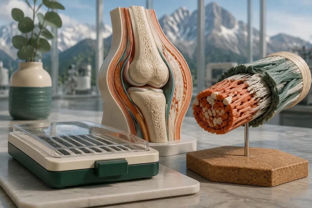
基因检测可确认分子层面已明确的 EDS 亚型,包括经典型和血管型 EDS,但通常无法确认高活动型 EDS。阴性基因面板和意义未明变异回答的是不同问题。

要点
- 可检测的类型: 2017 年 EDS 分类描述了 13 种亚型;其中 12 种已有明确的分子病因,可通过适当的基因检测进行评估。
- 高活动型 EDS: 目前没有普遍认可的分子检测可以确认或排除 hEDS;诊断需要进行临床评估。
- 经典型 EDS: COL5A1 和 COL5A2 是主要相关基因;特定的 COL1A1 变异可能导致与经典型相重叠的表型。
- 血管型 EDS: 结合临床表现解读时,致病性或可能致病的 COL3A1 变异可确认 vEDS。
- 变异分类: 临床实验室使用 5 个类别,从致病到良性;VUS 不属于阳性诊断。
- 可能致病: ACMG/AMP 框架将该分类的确定性设定为高于 90%,而非对症状严重程度作出 90% 的预测。
- 隐性遗传型 EDS: 确诊通常需要相关基因的两份拷贝分别存在一个致病变异。
- 家族风险: 许多显性遗传型 EDS 亚型每次妊娠的遗传概率为 50%;隐性遗传风险则取决于父母双方的检测结果。
基因检测可以确认哪些 EDS 类型?
Ehlers-Danlos 遗传检测可确诊分子定义的亚型,但通常不能确诊高活动性 EDS。 2017 年分类确定了 13 种 EDS 亚型,其中 12 种有已确立的遗传病因;确诊需要存在分类适当的变异、符合正确的遗传模式,并有相符的临床表现。
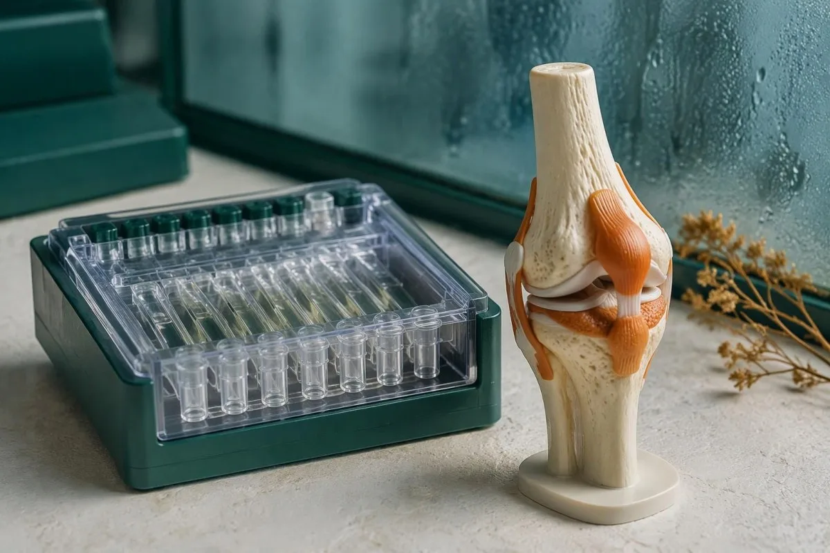
Ehlers-Danlos 综合征的基因检测检查的是 DNA,而不是血液中循环的胶原蛋白含量。经典型、血管型、脊柱后侧凸型以及一些较罕见的 EDS 类型都可以通过这种方式进行检查,但包含 20 或 40 个基因的检测 panel 并不会自动覆盖所有相关变异类型;实验室采用的方法与其基因列表同样重要。
国际分类将可识别的临床表现模式与其分子病因区分开来(Malfait et al., 2017)。其 12 种分子定义类别并不意味着普遍达到 100% 的检出率:实验室可能未纳入某个基因、无法检测技术上较难分析的区域,或发现目前尚无法可靠分类的变异;我们对 遗传检测结果类别 的说明在另一种遗传性疾病中探讨了同样的区别。
Kantesti 是一款 AI 验血分析器,可帮助解读常规实验室检查结果,但不能替代 EDS 分子诊断。作为一个组织, Kantesti Ltd 的临床范围 区分了科普解读与专科评估:无论是一份正常的血液检查报告,还是 AI 生成的解读,都不能确定某人是否患有遗传性结缔组织疾病。
哪些基因可确认经典型和类经典型 EDS?
经典型 EDS 通常与 COL5A1 或 COL5A2 有关,而 TNXB 相关经典样 EDS 通常需要存在两个致病变异。 名称相似并不意味着遗传方式、实验室方法或临床表现相同;特定的 COL1A1 变异和 AEBP1 相关疾病也存在进一步的区别。
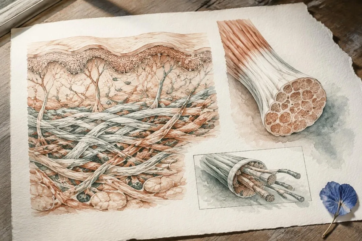
经典型 EDS 的特征包括关节过度活动,以及典型的皮肤过度伸展性和萎缩性瘢痕,但家族内部的表现程度各异。COL5A1 和 COL5A2 编码 V 型胶原的组成成分;杂合致病或可能致病变异可支持确诊;但任意一种 COL1A1 变异都不足以支持确诊,因为该基因中的不同变异会导致截然不同的结缔组织表型。
TNXB 相关经典样 EDS 常导致皮肤过度伸展和容易瘀青,但通常没有经典型 EDS 的典型萎缩性瘢痕。TNXB 位于一个高度相似的假基因 TNXA 旁边,因此检测报告应说明检测方法如何区分两者,以及如何检出缺失或重排;有些变异还涉及邻近的 CYP21A2,可能使结缔组织表现与一种独立的内分泌疾病相关联。
AEBP1 相关经典样 EDS 有时称为经典样 EDS 2 型,是在 2017 年分类发布后才得到描述的,通常为隐性遗传。这就是为什么对于宣称涵盖所有 EDS 类型的检测 panel,需要核查其实际基因列表;我们的 基因特异性家族风险指南 explains why a condition name alone cannot determine inheritance or the meaning of a negative result.
基因检测可以确认血管型 EDS 吗?
Vascular EDS is usually confirmed by a pathogenic or likely pathogenic COL3A1 variant. COL3A1 encodes type III collagen, and the combination of molecular evidence with a personal or family history of arterial or organ complications carries more diagnostic weight than a high joint-flexibility score.
Generalized joint hypermobility is not required for vascular EDS, so a Beighton score of 2/9 does not rule it out. Features prompting specialist consideration include arterial dissection or rupture, unexplained bowel perforation, spontaneous pneumothorax, and a compatible family history; isolated bruising or flexibility is much less specific and should not be treated as equivalent evidence.
A confirmed COL3A1 result can change vascular surveillance, procedural planning, pregnancy counseling, and testing for relatives. Different variant mechanisms can influence average patterns of disease, but they do not provide a reliable countdown to an individual's first complication; for a heterozygous dominant variant, the usual transmission probability is 50% per pregnancy, even when an affected parent has relatively few symptoms.
Sudden severe chest, abdominal, back, or head pain, collapse, or new neurological symptoms require emergency assessment—not waiting several weeks for a panel result. Call the local emergency number, such as 999 or 112 where applicable, and report known or suspected vascular EDS; our 中风急症警示征象 explain why new speech, vision, balance, or limb changes need immediate action.
哪些较罕见的 EDS 亚型已有明确的遗传病因?
The 9 other molecularly defined subtypes in the 2017 classification have distinct gene and inheritance requirements. A broad EDS panel can investigate them, but confirmation depends on variant mechanism—not simply finding any change in a gene associated with connective tissue.
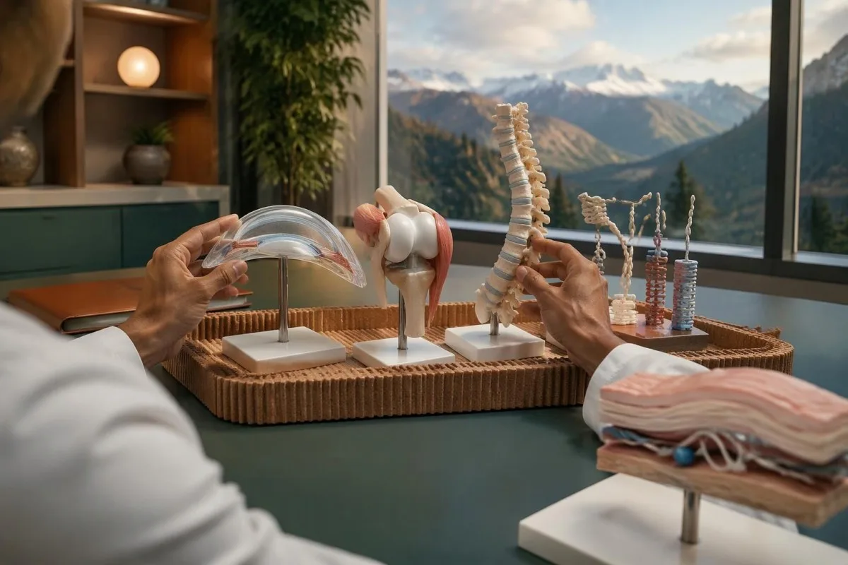
Arthrochalasia EDS involves specific COL1A1 or COL1A2 changes affecting processing around exon 6, rather than every variant in either gene. Dermatosparaxis EDS involves biallelic ADAMTS2 variants, while cardiac-valvular EDS involves biallelic COL1A2 variants with an appropriate loss-of-function mechanism; these 3 diagnoses illustrate why gene name, variant location, and biological effect must be interpreted together.
Kyphoscoliotic EDS can involve 2 disease-causing variants in PLOD1 or FKBP14, with congenital hypotonia and early progressive spinal curvature providing useful clinical clues. Brittle cornea syndrome involves ZNF469 or PRDM5 and warrants careful ophthalmic assessment because corneal thinning changes management; a connective-tissue panel therefore needs to be matched to eye, skeletal, hearing, and muscle findings, not flexibility alone.
Spondylodysplastic EDS involves B4GALT7, B3GALT6, or SLC39A13, whereas musculocontractural EDS involves CHST14 or DSE; both groups usually follow recessive inheritance. Short stature, characteristic skeletal changes, or congenital contractures can direct testing more efficiently than adding dozens of unrelated genes, and 1 pathogenic variant in a recessive gene is ordinarily an incomplete explanation until the second allele is investigated.
Myopathic EDS involves COL12A1 and can follow dominant or recessive inheritance, while periodontal EDS involves heterozygous pathogenic variants in C1R or C1S. Severe early periodontal disease and lack of attached gingiva are particularly informative for the latter; unlike some metabolic conditions described in our enzyme and DNA results, most EDS diagnoses do not have a routine enzyme-screening shortcut.
为什么阴性基因面板不能排除高活动型 EDS?
A negative EDS panel does not exclude hypermobile EDS because hEDS lacks a routinely accepted diagnostic molecular test. The result means the laboratory did not identify reportable explanatory variants within its tested genes and methods—not that all inherited connective-tissue causes have been eliminated.
Most clinical EDS panels primarily investigate alternatives to hEDS, such as classical or vascular EDS and overlapping connective-tissue disorders. A hypothetical person with a Beighton score of 7/9, recurrent instability, and qualifying systemic findings could receive a negative 30-gene panel and still meet clinical hEDS criteria; the panel and the examination are answering different questions.
Research into hEDS genetics is active, but a candidate gene association is not the same as a validated test applicable to everyone with the condition. Before paying for a test described as hypermobile EDS genetic testing, ask for 3 things: the gene-disease evidence, the diagnostic validation, and the criteria used to decide whether a result confirms hEDS.
Neither orthostatic symptoms nor fatigue independently establishes hEDS, and neither symptom needs a positive genetic panel to deserve treatment. A person may need a separate standing or tilt-table assessment for suspected POTS; distinguishing 2 coexisting conditions avoids using one diagnosis as an explanation for every symptom or mistaking an association for proof of causation.
无法通过基因检测确认时,如何诊断 hEDS?
Hypermobile EDS is diagnosed through clinical criteria that assess generalized hypermobility, associated features, and exclusion of alternative explanations. Under the established 2017 adult criteria, the Beighton threshold is generally at least 5/9 through age 50 and at least 4/9 above age 50.
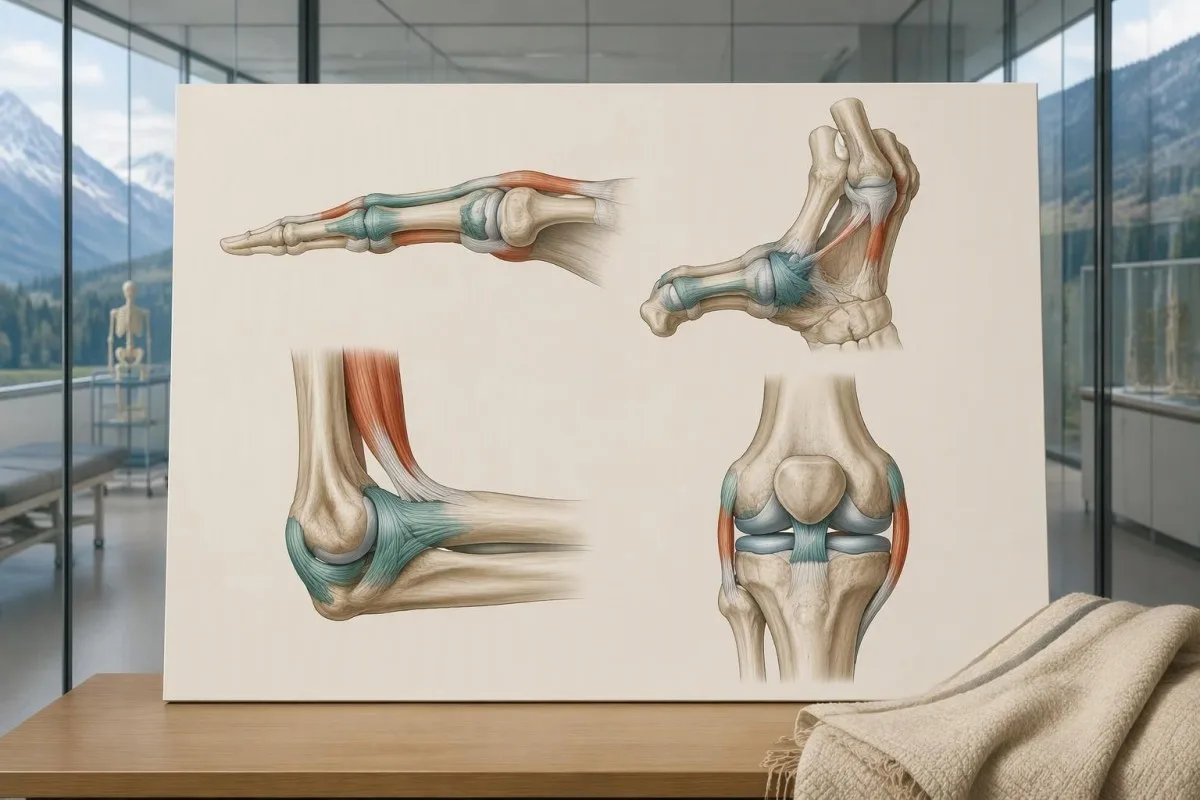
The 2017 criteria require all 3 major criteria, not just a high Beighton score. The second criterion requires at least 2 of 3 feature groups: systemic manifestations, a qualifying first-degree family history, or specified musculoskeletal complications; within the systemic group, at least 5 of 12 listed findings are required, so unrelated symptoms cannot simply be added together to reach a diagnosis.
Age and prior injury can alter measured flexibility, and historical hypermobility becomes relevant when someone is 1 point below the age-specific cutoff. The pediatric framework recommends a Beighton threshold of at least 6/9 for generalized joint hypermobility and reserves hEDS assessment for biological maturity (Tofts et al., 2023); childhood flexibility should not automatically become a lifelong adult diagnostic label.
Alternative explanations include other inherited connective-tissue disorders, neuromuscular conditions, and acquired rheumatologic disease; the exclusions depend on the person's findings rather than a mandatory universal blood-test package. An anti-RNP result, for example, belongs to an autoimmune connective-tissue assessment, not EDS DNA testing; clinicians may also diagnose a hypermobility spectrum disorder when symptoms are significant but all 3 hEDS criteria are not met.
哪些人需要进行 EDS 基因面板检测,而不是靶向检测?
An EDS panel is most useful when clinical findings suggest a molecularly defined subtype or several overlapping inherited disorders. When a family already has 1 known pathogenic variant, targeted testing for that exact variant is usually more informative than starting with a new broad panel.

Marked skin fragility, characteristic scars, congenital hip dislocation, progressive early scoliosis, unusual corneal findings, or arterial complications can justify clinical genetics assessment. A genetics service may select a broader heritable connective-tissue or aortopathy panel rather than an EDS-only panel when 2 or more diagnostic possibilities overlap; the aim is to investigate the phenotype, not to maximize the number of genes purchased.
Testing the most clearly affected relative first often makes later family testing easier to interpret. A negative targeted result for a known familial variant is fundamentally different from an unexplained negative panel in the first person tested; this distinction also appears in our explanation of informative negative genetic results, although the condition-specific screening recommendations are different.
Routine panel testing is not always helpful for isolated hypermobility without features of a molecularly defined disorder, because additional genes can generate additional VUS findings without clarifying the diagnosis. Some laboratories need specialized methods for particular variant classes, as illustrated by repeat-expansion testing methods; before ordering 1 panel, ask which plausible diagnoses its technical design can actually address.
EDS 基因面板检测在技术上可能漏检什么?
An EDS panel can miss variants outside its coverage, certain deletions or rearrangements, and low-level mosaicism. Sequencing and deletion/duplication analysis are complementary methods, so a report stating that 99% of covered bases were read does not mean 99% of every possible EDS cause was excluded.
Small sequence changes are not the same as changes in gene copy number. Ask whether the assay includes deletion/duplication analysis, how poorly covered regions are completed, and whether complex loci such as TNXB receive validated additional testing; our copy-number interpretation guide shows why a reassuring number of copies can still leave unresolved questions in a different genetic setting.
Exome sequencing focuses mainly on protein-coding regions, while genome sequencing examines a broader DNA space; neither approach guarantees an explanation after 1 negative panel. Deep intronic variants, complex structural changes, mosaicism, and uncertain gene-disease relationships may require specialist analysis, and selected cases may benefit from RNA studies that investigate whether a suspected variant changes how a gene transcript is processed.
Kantesti is an AI blood test interpretation platform whose routine laboratory explanations do not validate an EDS panel's sequencing sensitivity. Our 技术和临床标准 should not be interpreted as 100% accuracy for germline diagnosis; the molecular laboratory must provide its own assay validation, limitations, accredited reporting process, and variant-specific interpretation.
致病变异与意义未明变异有何不同?
A pathogenic or likely pathogenic variant has enough evidence to support disease causation; a variant of uncertain significance does not. The ACMG/AMP framework uses 5 categories: pathogenic, likely pathogenic, uncertain significance, likely benign, and benign, with classification based on evidence rather than how unusual a DNA change looks.
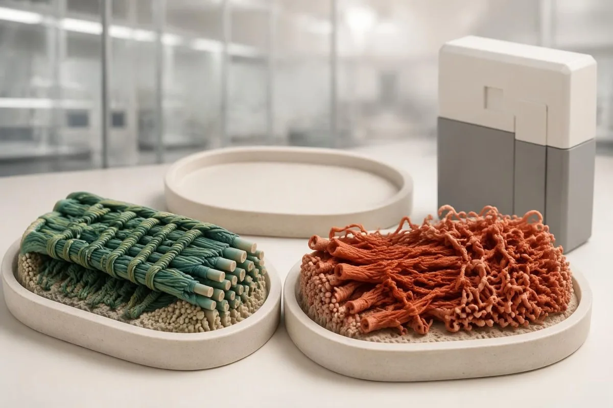
Evidence can include population frequency, observations in affected families, segregation, well-validated functional studies, and the expected disease mechanism. A variant present in 1 person with hypermobility is not automatically causal, especially when that feature is common; Richards et al. (2015) specifically state that a VUS should not be used in clinical decision-making, preventing uncertain evidence from becoming a definitive diagnosis.
The ACMG/AMP definition of likely pathogenic corresponds to greater than 90% certainty that a variant is disease-causing. That percentage describes confidence in classification—not a 90% chance of arterial rupture, severe disability, or transmission—and even an established pathogenic variant must fit the gene, inheritance pattern, and clinical question before it can explain an individual's presentation.
A pathogenic classification also does not make every carrier's prognosis identical: penetrance and severity can vary with the gene, variant mechanism, and family context. Our explanation of variant effects and family risk illustrates this separation in another inherited disorder; for EDS, request the exact DNA and protein notation, laboratory classification, and report date rather than relying on a shortened portal label.
对意义未明的 EDS 变异应该怎么做?
An EDS VUS should trigger clarification, not an automatic EDS diagnosis or gene-specific treatment. The next steps are to review phenotype fit, check inheritance and assay limitations, and ask whether carefully selected family testing could provide evidence; testing 10 relatives without a clear question may add little.

Consider a clearly hypothetical adult whose panel finds 1 COL3A1 VUS and no pathogenic variants. The person does not have genetically confirmed vascular EDS on that basis, but a history of arterial dissection would still justify specialist evaluation; management follows the real clinical risk while the molecular uncertainty is investigated, rather than either ignoring the history or treating the VUS as proof.
For a recessive subtype, 2 variants must usually affect opposite gene copies, called being in trans, to explain the disorder. If both changes sit on the same copy, called being in cis, the other copy may remain functional; our discussion of recessive carrier result interpretation explains why parental testing can sometimes answer a specific phasing question rather than simply produce more results.
A VUS may later become benign, likely benign, likely pathogenic, or pathogenic as evidence changes, but there is no universal 6- or 12-month reclassification deadline. Ask who initiates reinterpretation, whether the laboratory contacts the ordering clinician, and how to update family findings; a new clinical event or informative relative's result can be more useful than repeatedly ordering the same unchanged panel.
EDS 基因面板检测的阳性和阴性结果意味着什么?
A positive EDS panel result can confirm a subtype only when classification, inheritance, and phenotype align. A negative result can lower the likelihood of adequately tested molecular diagnoses, but it neither confirms hEDS nor excludes it; 1 result must be interpreted against the reason testing was ordered.
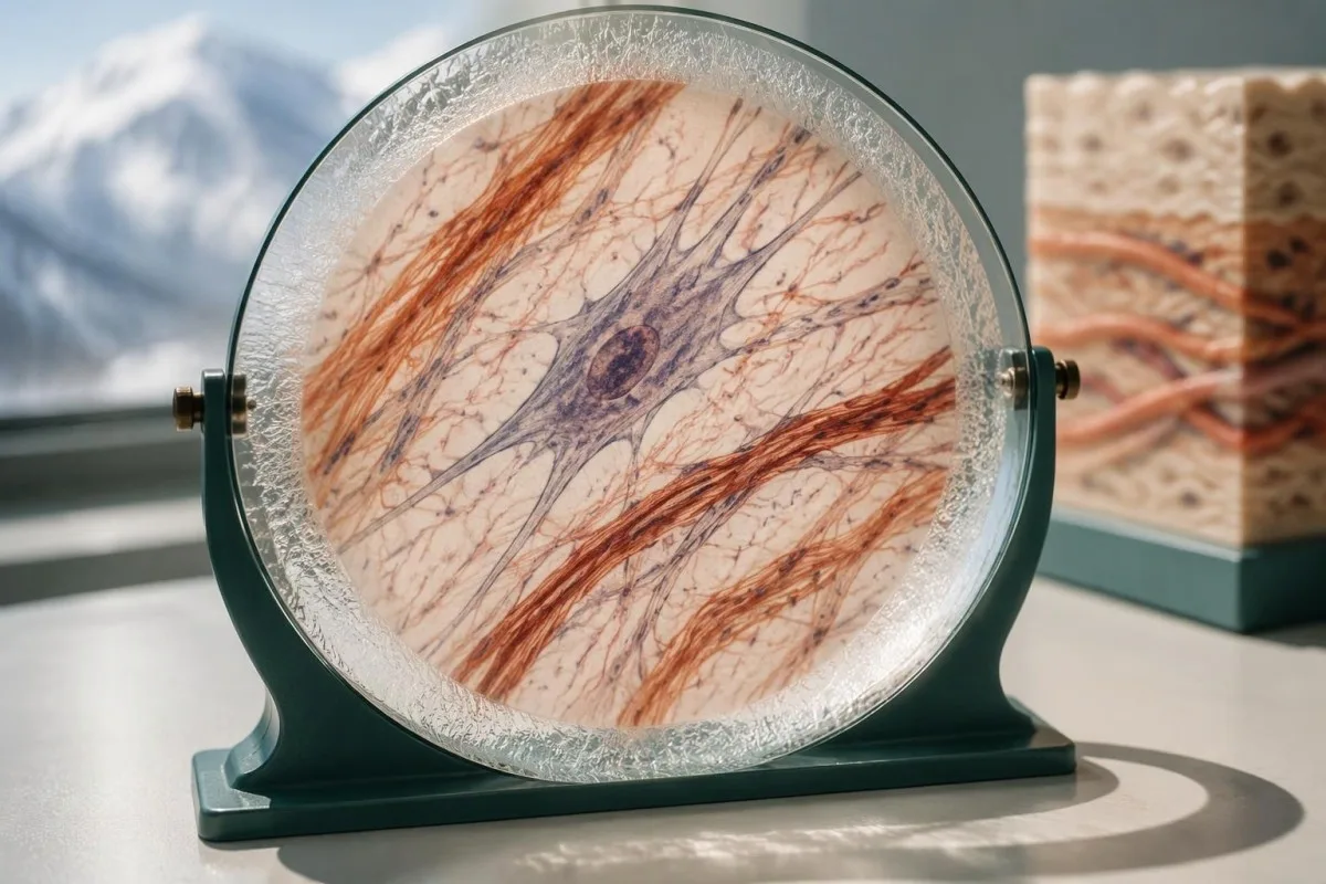
A hypothetical heterozygous pathogenic COL5A1 finding in someone with characteristic scars and hyperextensible skin can support classical EDS. By contrast, 1 pathogenic ADAMTS2 variant in a person without a second explanatory allele usually indicates carrier status rather than confirmed dermatosparaxis EDS; a report can therefore contain a genuine pathogenic finding without that finding explaining the person's symptoms.
The meaning of a negative result changes with pretest probability. When the examination strongly suggests a rare molecular subtype, the next question is whether the assay covered the relevant gene and variant mechanisms; when findings are nonspecific, expanding from 30 to 300 genes may increase uncertain or incidental findings without making the answer more useful or making hEDS genetically testable.
Clinical genetics may recommend a targeted technical follow-up, a different diagnostic hypothesis, or later reanalysis rather than immediately repeating the same panel. The relationship between phenotype and molecular findings also matters in gene and biochemical confirmation, though EDS does not share that condition's biochemical tests; ask for a written explanation of what remains plausible after 1 negative result.
EDS 检测结果对儿童及其他亲属有何影响?
Family testing should follow the confirmed gene and inheritance pattern, not the broad EDS label. For many dominant subtypes, a heterozygous affected parent has a 50% transmission probability per pregnancy; when both parents carry a pathogenic variant for the same recessive subtype, the affected-child probability is usually 25%.
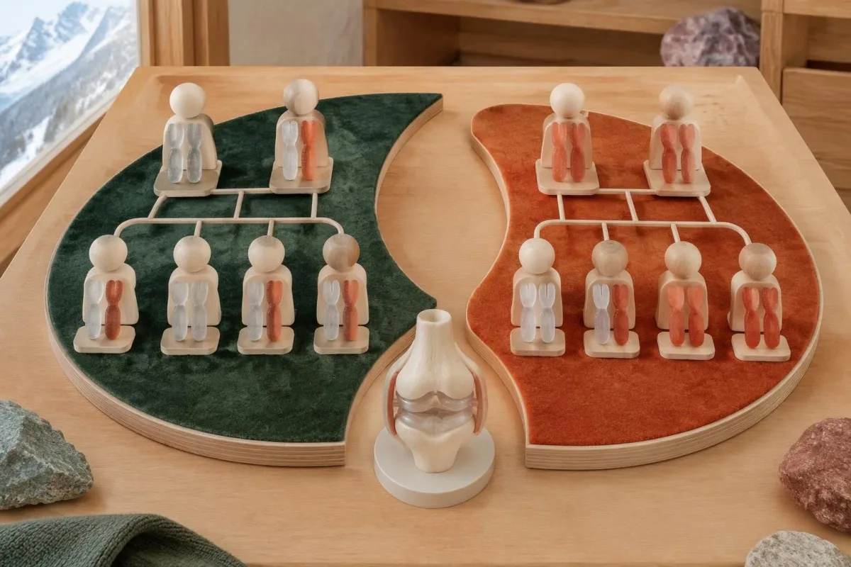
The standard recessive model gives a 25% chance of an affected child, a 50% chance of a carrier child, and a 25% chance of a child inheriting neither familial variant when both parents are carriers. Each pregnancy is a new event, not a running quota; partner testing must investigate the relevant gene sufficiently rather than checking only the variant carried by the first parent.
A negative family history does not exclude dominant EDS because a variant may arise de novo, relatives may have subtle findings, or family information may be incomplete. Testing children can be appropriate when a confirmed condition changes childhood care, particularly vascular EDS; this differs from some adult-onset predictive testing decisions, where timing and the child's future autonomy carry a different balance.
Hypermobile EDS can cluster in families, but no routine molecular result currently provides the same variant-specific cascade-testing pathway. Relatives should consent separately to testing and data sharing, and a family's 3 generations of history should distinguish documented diagnoses from suspected flexibility; our family consent and tracking guide explains why shared health information does not remove individual privacy or the need for personal assessment.
常规血液检查或 AI 解读能诊断 EDS 吗?
Routine blood tests and AI blood-test interpretation cannot diagnose EDS or classify a germline variant independently. A CBC, ferritin, thyroid panel, or coagulation screen may investigate symptoms and alternative explanations, but there is no routine collagen blood concentration with a diagnostic cutoff for the 13 EDS subtypes.
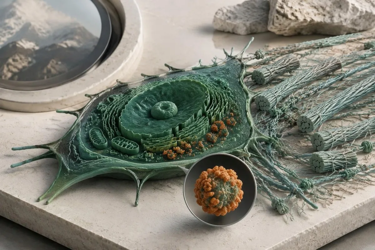
Kantesti is an AI-powered blood test analysis tool that can explain routine biomarker results alongside reported symptoms. For example, a hypothetical adult with ferritin of 9 ng/mL and hemoglobin of 13.0 g/dL may have depleted iron stores despite hemoglobin within their laboratory's reference interval; our guide to 贫血前缺铁 explains why that finding deserves attention without becoming evidence for or against EDS.
Normal platelet counts, PT, and aPTT do not exclude connective-tissue fragility, and bruising can have more than 1 cause. Conversely, iron deficiency, thyroid dysfunction, or a bleeding disorder should not be dismissed simply because someone already has hEDS; our complete iron study interpretation addresses a treatable contributor to fatigue, not an inherited collagen diagnosis or a supplement-based cure.
An EDS genetic report contains sensitive information about both the individual and potentially their relatives, so it should not be treated as an ordinary screenshot for unrestricted sharing. Before sending even 1 report to an online service, check purpose, retention, deletion, and whether genetic-report analysis is supported; Kantesti AI's privacy information describes our data-handling approach, not authorization to replace a genetic counselor or molecular laboratory.
去遗传学门诊时应该带什么资料?哪些来源有帮助?
Bring the complete genetic report, relevant medical records, and a structured family history to your genetics appointment. The 3 most useful questions are whether the result explains your phenotype, what the assay could have missed, and whether the finding changes your own or your relatives' care.

Record major complications with ages, documented diagnoses, and the exact relationship of affected relatives; a 3-generation pedigree can be more informative than an unverified statement that everyone is flexible. Include ophthalmology findings, relevant imaging, characteristic scarring, congenital orthopedic problems, and the full laboratory methods page, because a portal summary may omit gene coverage, deletion testing, or the distinction between a VUS and a pathogenic result.
Before leaving, ask who coordinates 1 next step and who will receive any amended report: the genetics service, the ordering clinician, or the molecular laboratory. Kantesti's 医疗内容标准 support clear separation of established evidence, hypothetical examples, and unresolved questions; this article's EDS recommendations draw on the international classification, ACMG/AMP interpretation guidance, and pediatric hypermobility framework cited below.
The 2 Zenodo publications listed in our research references cover urinalysis and iron studies, not EDS genetic-test validation. Our 尿液分析解读指南 may help explain a separate urine report, but neither urine urobilinogen nor iron-binding capacity confirms an EDS subtype; these publications should not be read as evidence that our platform performs molecular EDS diagnosis.
常见问题解答
基因检测能诊断埃勒斯-当洛综合征吗?
A genetic test can confirm molecularly defined Ehlers-Danlos subtypes when it identifies an appropriately classified variant with the correct inheritance pattern and compatible clinical findings. The 2017 classification includes 13 subtypes, and 12 have established molecular causes that appropriate testing can investigate. Hypermobile EDS does not have a routinely accepted diagnostic molecular test. A panel's gene list, technical coverage, and variant interpretation determine what its result can establish.
EDS基因检测面板结果为阴性,能否排除关节过度活动型 EDS?
A negative EDS panel does not rule out hypermobile EDS because hEDS is diagnosed clinically rather than by a validated routine molecular test. A hypothetical negative 30-gene panel means no explanatory reportable variant was found within that assay's scope. The result can help evaluate other inherited disorders, but it cannot prove or disprove hEDS. The next step is a clinical assessment that considers hypermobility, associated features, and alternative diagnoses.
COL3A1变异是否意味着我患有血管型 EDS?
只有当 COL3A1 变异被判定为致病或可能致病,并结合临床表现进行解读时,才能支持对血管型 EDS 的确诊。临床实验室采用 5 种变异分类;意义未明的 COL3A1 变异并不能确诊血管型 EDS。即使检测结果不确定,严重且原因不明的动脉或器官并发症仍需由专科医生评估。突发剧烈疼痛、昏厥或神经系统症状应立即接受急诊救治,不应等待变异重新分类。
EDS基因检测面板中的 VUS 是什么意思?
意义未明变异(VUS)是指现有证据不足以可靠判定该 DNA 变异是否会导致疾病。ACMG/AMP 框架将 VUS 与可能致病变异区分开来;后者表示导致疾病的确定性超过 90%。VUS 不应单独作为 EDS 诊断、预测性检测或基因特异性治疗的依据。临床遗传学专家可能会建议开展针对性的家系研究或日后重新解读,同时根据症状和风险本身的临床意义进行管理。
为什么在一个 EDS 基因中发现了两个变异?
两种变异可能具有重要意义,因为多种 EDS 亚型遵循隐性遗传模式,通常需要基因的两个拷贝都发生致病性改变。实验室必须确定这两种改变是否为致病或可能致病变异,以及它们是否位于反式,即分别影响两个拷贝。位于顺式、影响同一拷贝的两种改变,可能无法解释一种隐性遗传病。发现一个致病变异和一个意义未明变异(VUS),也不能自动确认分子诊断。
如果我患有 EDS,我的孩子是否应该接受检测?
Children's testing depends on the confirmed subtype, whether a familial variant is known, and whether the result changes care during childhood. Many dominant EDS subtypes have a 50% transmission probability per pregnancy, making targeted testing useful when appropriate. When both parents carry variants for the same recessive subtype, the usual affected-child probability is 25% per pregnancy. Hypermobile EDS currently requires clinical evaluation rather than routine variant-specific family testing.
正常的血液检查能排除埃勒斯-当洛斯综合征吗?
常规血液检查无法排除埃勒斯-当洛斯综合征,因为标准检查项目无法测量导致 EDS 的遗传性结构变化。CBC、PT 或 aPTT 结果正常,并不能排除结缔组织脆弱性,也没有常规胶原蛋白血液检测阈值可以诊断已分类的 13 种亚型。血液检查可以发现导致症状的其他因素,例如缺铁或甲状腺功能异常。分子遗传学定义的 EDS 需要进行适当的遗传学解读,而高活动型 EDS 则需要临床评估。
获取血液检查结果解读
上传您的实验室报告,Kantesti 的 AI 会在几秒内用通俗语言解释每项生物标志物。全球已有超过 200 万人使用。.
参考
- Klein, T., Mitchell, S., & Weber, H. (2026). 尿胆原在尿液测试:完整尿液分析指南 2026。. Kantesti AI 医学研究。. DOI
- Klein, T., Mitchell, S., & Weber, H. (2026). 铁代谢检测指南:总铁结合力、铁饱和度和结合能力。. Kantesti AI 医学研究。. DOI
- Malfait F et al. (2017). The 2017 international classification of the Ehlers-Danlos syndromes. American Journal of Medical Genetics Part C: Seminars in Medical Genetics. DOI
- Richards S 等(2015)。. 序列变异解读标准与指南:美国医学遗传学与基因组学学会和分子病理学协会联合共识建议。. 《医学遗传学》。. DOI PubMed
- Tofts LJ et al. (2023). Pediatric joint hypermobility: a diagnostic framework and narrative review. 《Orphanet罕见病杂志》。. DOI
- 200万+分析的检测项目
- 127+国家
- 100+语言
关于医疗团队


E-E-A-T信任信号
经验
由医生主导的临床审阅:实验室解读工作流程。.
专业知识
实验室医学重点:生物标志物在临床情境中的表现。.
权威
经 Thomas Klein 医生医学审核,并由 Sarah Mitchell 医生和 Hans Weber 教授参与撰写。.
可信度
基于循证的解读,并提供清晰的后续路径以减少警报。.

医疗免责声明
本文仅用于教育目的,不构成医疗建议。诊断和治疗决策请始终咨询合格的医疗专业人员。.
发表 · 联系编辑团队
由 Kantesti Ltd 发布,在英格兰和威尔士注册,注册公司编号:. 17090423, ,英国伦敦。.
