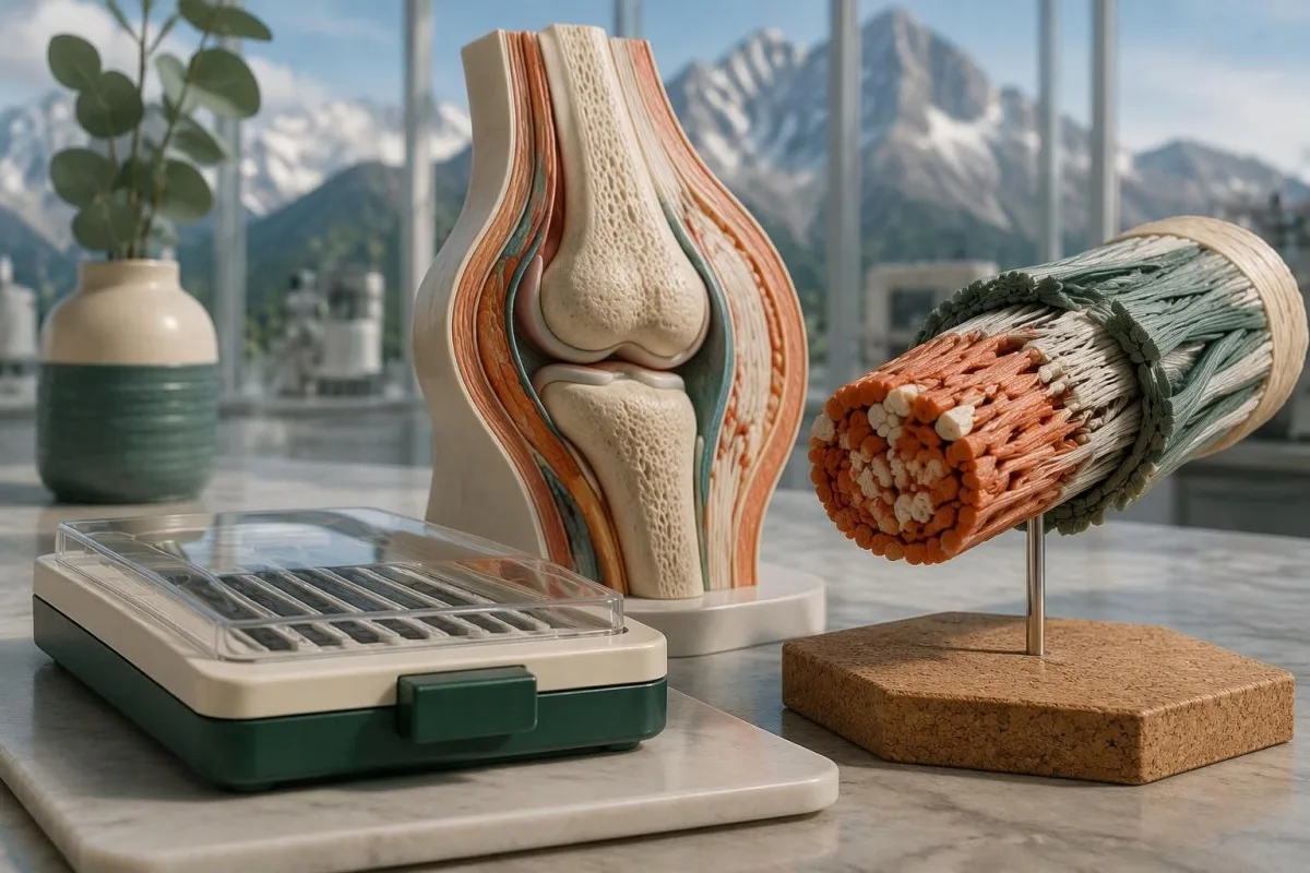
Testarea genetică poate confirma subtipurile EDS definite molecular, inclusiv EDS clasic și vascular, dar nu poate confirma în mod obișnuit EDS hipermobil. Un panel negativ și o variantă cu semnificație incertă răspund la întrebări diferite.

Idei principale
- Tipuri detectabile: Clasificarea EDS din 2017 descrie 13 subtipuri; 12 au cauze moleculare stabilite, care pot fi investigate prin testare genetică adecvată.
- EDS hipermobil: Nu există niciun test molecular acceptat în mod obișnuit care să poată confirma sau exclude hEDS; diagnosticul necesită o evaluare clinică.
- EDS clasic: COL5A1 și COL5A2 sunt genele principale; anumite variante COL1A1 pot produce un fenotip clasic suprapus.
- EDS vascular: O variantă patogenă sau probabil patogenă a COL3A1 poate confirma vEDS atunci când este interpretată împreună cu constatările clinice.
- Clasificarea variantelor: Laboratoarele clinice folosesc 5 categorii, de la patogen la benign; o VUS nu reprezintă un diagnostic pozitiv.
- Probabil patogen: Cadrul ACMG/AMP folosește o certitudine de peste 90% pentru această clasificare, nu o predicție de 90% a severității simptomelor.
- EDS recesiv: Confirmarea necesită de obicei 2 variante cauzatoare de boală care afectează copii opuse ale genei relevante.
- Riscul familial: Multe subtipuri dominante de EDS au o probabilitate de transmitere de 50% la fiecare sarcină; riscurile recesive depind de rezultatele ambilor părinți.
Ce tipuri de EDS pot fi confirmate printr-un test genetic?
Testarea genetică pentru sindromul Ehlers-Danlos poate confirma subtipurile definite molecular, dar nu și, de regulă, EDS hipermobil. Clasificarea din 2017 identifică 13 subtipuri de EDS, cu cauze genetice stabilite pentru 12; confirmarea necesită o variantă clasificată corespunzător, tiparul corect de moștenire și constatări clinice compatibile.
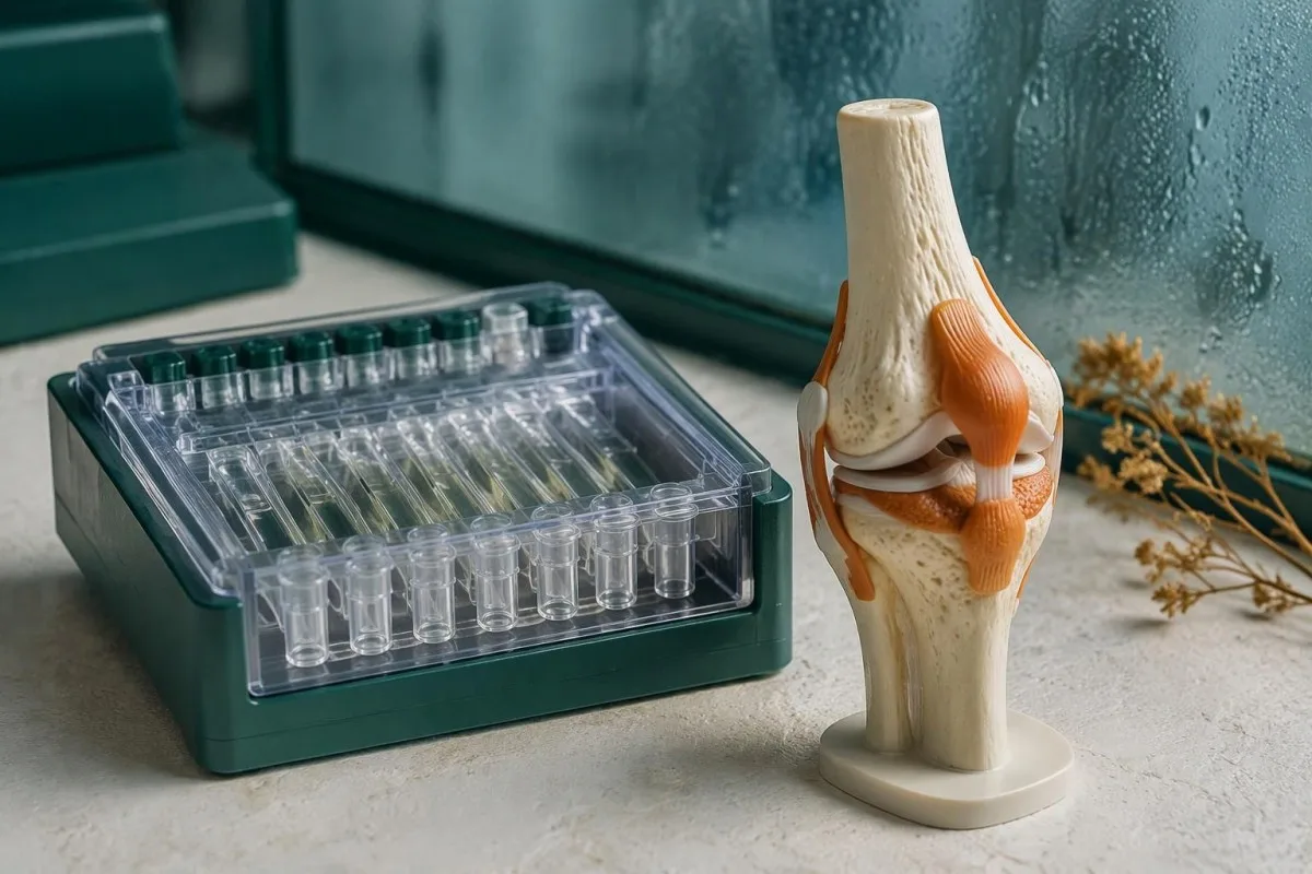
Un test genetic pentru sindromul Ehlers-Danlos analizează ADN-ul, nu cantitatea de colagen care circulă în sânge. Formele clasice, vasculare, cifoscoliotice și câteva forme mai rare de EDS pot fi investigate astfel, dar un panel care conține 20 sau 40 de gene nu acoperă automat toate tipurile relevante de variante; metodele laboratorului contează la fel de mult ca lista de gene.
Clasificarea internațională separă tiparele clinice recognoscibile de cauzele lor moleculare (Malfait et al., 2017). Cele 12 categorii definite molecular nu implică o rată universală de detecție de 100%: un laborator poate omite o genă, poate să nu detecteze o regiune dificilă din punct de vedere tehnic sau poate identifica o modificare care nu poate fi încă clasificată cu certitudine; explicația noastră despre categoriile rezultatelor genetice explorează aceeași distincție în cazul unei alte afecțiuni ereditare.
Kantesti este un analizator de analize de sânge bazat pe inteligență artificială, care ajută la explicarea rezultatelor de laborator de rutină, nu un substitut pentru diagnosticul molecular al EDS. Ca organizație, domeniul clinic al Kantesti Ltd face distincția între interpretarea educațională și evaluarea de specialitate: nici un panel de sânge normal și nici o explicație generată de inteligența artificială nu pot stabili dacă o persoană are o afecțiune ereditară a țesutului conjunctiv.
Ce gene confirmă EDS clasic și EDS cu aspect clasic?
EDS clasic implică de obicei COL5A1 sau COL5A2, în timp ce EDS de tip clasic-like asociat cu TNXB necesită de regulă 2 variante cauzatoare de boală. Denumirile similare nu înseamnă moștenire, metode de laborator sau constatări clinice identice; anumite variante COL1A1 și boala asociată cu AEBP1 adaugă și alte distincții.
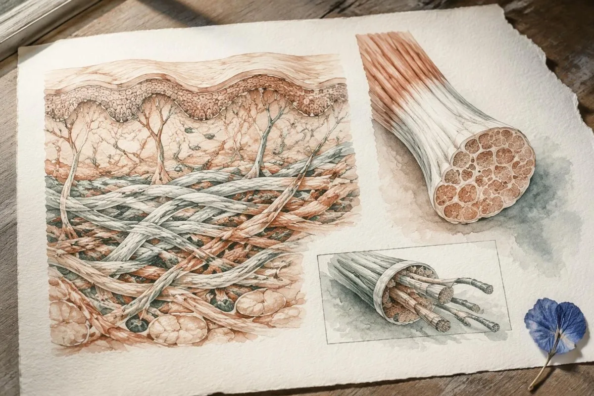
EDS clasic combină hipermobilitatea articulară cu hiperextensibilitatea caracteristică a pielii și cicatrizarea atrofică, deși manifestările variază în cadrul familiilor. COL5A1 și COL5A2 codifică componente ale colagenului de tip V, iar o variantă heterozigotă patogenă sau probabil patogenă poate susține confirmarea; o constatare arbitrară în COL1A1 nu poate, deoarece variante diferite în acea singură genă cauzează fenotipuri ale țesutului conjunctiv substanțial diferite.
EDS de tip clasic-like asociat cu TNXB provoacă adesea hiperextensibilitate și apariția ușoară a vânătăilor, fără cicatricile atrofice tipice EDS-ului clasic. TNXB este situată lângă o pseudogenă foarte asemănătoare, TNXA, astfel că raportul ar trebui să explice cum le diferențiază testul și cum detectează delețiile sau rearanjările; unele modificări implică și CYP21A2, aflată în vecinătate, putând asocia constatările privind țesutul conjunctiv cu o afecțiune endocrină distinctă.
EDS de tip clasic-like asociat cu AEBP1, numit uneori EDS de tip clasic-like 2, a fost caracterizat după clasificarea din 2017 și este, în general, recesiv. De aceea, trebuie verificată lista efectivă de gene a unui panel prezentat ca acoperind toate tipurile de EDS; ghidul nostru despre riscul familial specific genei explains why a condition name alone cannot determine inheritance or the meaning of a negative result.
Poate testarea genetică să confirme EDS vascular?
Vascular EDS is usually confirmed by a pathogenic or likely pathogenic COL3A1 variant. COL3A1 encodes type III collagen, and the combination of molecular evidence with a personal or family history of arterial or organ complications carries more diagnostic weight than a high joint-flexibility score.
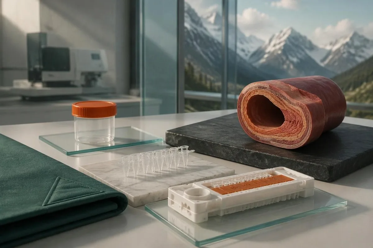
Generalized joint hypermobility is not required for vascular EDS, so a Beighton score of 2/9 does not rule it out. Features prompting specialist consideration include arterial dissection or rupture, unexplained bowel perforation, spontaneous pneumothorax, and a compatible family history; isolated bruising or flexibility is much less specific and should not be treated as equivalent evidence.
A confirmed COL3A1 result can change vascular surveillance, procedural planning, pregnancy counseling, and testing for relatives. Different variant mechanisms can influence average patterns of disease, but they do not provide a reliable countdown to an individual's first complication; for a heterozygous dominant variant, the usual transmission probability is 50% per pregnancy, even when an affected parent has relatively few symptoms.
Sudden severe chest, abdominal, back, or head pain, collapse, or new neurological symptoms require emergency assessment—not waiting several weeks for a panel result. Call the local emergency number, such as 999 or 112 where applicable, and report known or suspected vascular EDS; our semnele de alarmă ale unei urgențe de tip accident vascular cerebral explain why new speech, vision, balance, or limb changes need immediate action.
Ce subtipuri mai rare de EDS au cauze genetice stabilite?
The 9 other molecularly defined subtypes in the 2017 classification have distinct gene and inheritance requirements. A broad EDS panel can investigate them, but confirmation depends on variant mechanism—not simply finding any change in a gene associated with connective tissue.
Arthrochalasia EDS involves specific COL1A1 or COL1A2 changes affecting processing around exon 6, rather than every variant in either gene. Dermatosparaxis EDS involves biallelic ADAMTS2 variants, while cardiac-valvular EDS involves biallelic COL1A2 variants with an appropriate loss-of-function mechanism; these 3 diagnoses illustrate why gene name, variant location, and biological effect must be interpreted together.
Kyphoscoliotic EDS can involve 2 disease-causing variants in PLOD1 or FKBP14, with congenital hypotonia and early progressive spinal curvature providing useful clinical clues. Brittle cornea syndrome involves ZNF469 or PRDM5 and warrants careful ophthalmic assessment because corneal thinning changes management; a connective-tissue panel therefore needs to be matched to eye, skeletal, hearing, and muscle findings, not flexibility alone.
Spondylodysplastic EDS involves B4GALT7, B3GALT6, or SLC39A13, whereas musculocontractural EDS involves CHST14 or DSE; both groups usually follow recessive inheritance. Short stature, characteristic skeletal changes, or congenital contractures can direct testing more efficiently than adding dozens of unrelated genes, and 1 pathogenic variant in a recessive gene is ordinarily an incomplete explanation until the second allele is investigated.
Myopathic EDS involves COL12A1 and can follow dominant or recessive inheritance, while periodontal EDS involves heterozygous pathogenic variants in C1R or C1S. Severe early periodontal disease and lack of attached gingiva are particularly informative for the latter; unlike some metabolic conditions described in our enzyme and DNA results, most EDS diagnoses do not have a routine enzyme-screening shortcut.
De ce un panel negativ nu exclude EDS hipermobil?
A negative EDS panel does not exclude hypermobile EDS because hEDS lacks a routinely accepted diagnostic molecular test. The result means the laboratory did not identify reportable explanatory variants within its tested genes and methods—not that all inherited connective-tissue causes have been eliminated.
Most clinical EDS panels primarily investigate alternatives to hEDS, such as classical or vascular EDS and overlapping connective-tissue disorders. A hypothetical person with a Beighton score of 7/9, recurrent instability, and qualifying systemic findings could receive a negative 30-gene panel and still meet clinical hEDS criteria; the panel and the examination are answering different questions.
Research into hEDS genetics is active, but a candidate gene association is not the same as a validated test applicable to everyone with the condition. Before paying for a test described as hypermobile EDS genetic testing, ask for 3 things: the gene-disease evidence, the diagnostic validation, and the criteria used to decide whether a result confirms hEDS.
Neither orthostatic symptoms nor fatigue independently establishes hEDS, and neither symptom needs a positive genetic panel to deserve treatment. A person may need a separate standing or tilt-table assessment for suspected POTS; distinguishing 2 coexisting conditions avoids using one diagnosis as an explanation for every symptom or mistaking an association for proof of causation.
Cum este diagnosticat hEDS când testarea genetică nu îl poate confirma?
Hypermobile EDS is diagnosed through clinical criteria that assess generalized hypermobility, associated features, and exclusion of alternative explanations. Under the established 2017 adult criteria, the Beighton threshold is generally at least 5/9 through age 50 and at least 4/9 above age 50.
The 2017 criteria require all 3 major criteria, not just a high Beighton score. The second criterion requires at least 2 of 3 feature groups: systemic manifestations, a qualifying first-degree family history, or specified musculoskeletal complications; within the systemic group, at least 5 of 12 listed findings are required, so unrelated symptoms cannot simply be added together to reach a diagnosis.
Age and prior injury can alter measured flexibility, and historical hypermobility becomes relevant when someone is 1 point below the age-specific cutoff. The pediatric framework recommends a Beighton threshold of at least 6/9 for generalized joint hypermobility and reserves hEDS assessment for biological maturity (Tofts et al., 2023); childhood flexibility should not automatically become a lifelong adult diagnostic label.
Alternative explanations include other inherited connective-tissue disorders, neuromuscular conditions, and acquired rheumatologic disease; the exclusions depend on the person's findings rather than a mandatory universal blood-test package. An anti-RNP result, for example, belongs to an autoimmune connective-tissue assessment, not EDS DNA testing; clinicians may also diagnose a hypermobility spectrum disorder when symptoms are significant but all 3 hEDS criteria are not met.
Cine are nevoie de un panel EDS în locul unui test țintit?
An EDS panel is most useful when clinical findings suggest a molecularly defined subtype or several overlapping inherited disorders. When a family already has 1 known pathogenic variant, targeted testing for that exact variant is usually more informative than starting with a new broad panel.

Marked skin fragility, characteristic scars, congenital hip dislocation, progressive early scoliosis, unusual corneal findings, or arterial complications can justify clinical genetics assessment. A genetics service may select a broader heritable connective-tissue or aortopathy panel rather than an EDS-only panel when 2 or more diagnostic possibilities overlap; the aim is to investigate the phenotype, not to maximize the number of genes purchased.
Testing the most clearly affected relative first often makes later family testing easier to interpret. A negative targeted result for a known familial variant is fundamentally different from an unexplained negative panel in the first person tested; this distinction also appears in our explanation of informative negative genetic results, although the condition-specific screening recommendations are different.
Routine panel testing is not always helpful for isolated hypermobility without features of a molecularly defined disorder, because additional genes can generate additional VUS findings without clarifying the diagnosis. Some laboratories need specialized methods for particular variant classes, as illustrated by repeat-expansion testing methods; before ordering 1 panel, ask which plausible diagnoses its technical design can actually address.
Ce poate omite din punct de vedere tehnic un panel EDS?
An EDS panel can miss variants outside its coverage, certain deletions or rearrangements, and low-level mosaicism. Sequencing and deletion/duplication analysis are complementary methods, so a report stating that 99% of covered bases were read does not mean 99% of every possible EDS cause was excluded.
Small sequence changes are not the same as changes in gene copy number. Ask whether the assay includes deletion/duplication analysis, how poorly covered regions are completed, and whether complex loci such as TNXB receive validated additional testing; our copy-number interpretation guide shows why a reassuring number of copies can still leave unresolved questions in a different genetic setting.
Exome sequencing focuses mainly on protein-coding regions, while genome sequencing examines a broader DNA space; neither approach guarantees an explanation after 1 negative panel. Deep intronic variants, complex structural changes, mosaicism, and uncertain gene-disease relationships may require specialist analysis, and selected cases may benefit from RNA studies that investigate whether a suspected variant changes how a gene transcript is processed.
Kantesti is an AI blood test interpretation platform whose routine laboratory explanations do not validate an EDS panel's sequencing sensitivity. Our standarde tehnice și clinice should not be interpreted as 100% accuracy for germline diagnosis; the molecular laboratory must provide its own assay validation, limitations, accredited reporting process, and variant-specific interpretation.
Prin ce se deosebesc variantele patogene de un VUS?
A pathogenic or likely pathogenic variant has enough evidence to support disease causation; a variant of uncertain significance does not. The ACMG/AMP framework uses 5 categories: pathogenic, likely pathogenic, uncertain significance, likely benign, and benign, with classification based on evidence rather than how unusual a DNA change looks.
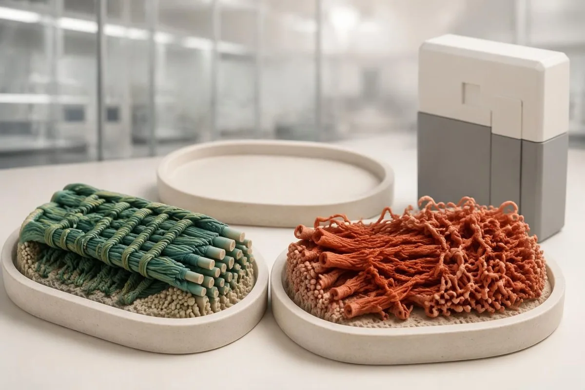
Evidence can include population frequency, observations in affected families, segregation, well-validated functional studies, and the expected disease mechanism. A variant present in 1 person with hypermobility is not automatically causal, especially when that feature is common; Richards et al. (2015) specifically state that a VUS should not be used in clinical decision-making, preventing uncertain evidence from becoming a definitive diagnosis.
The ACMG/AMP definition of likely pathogenic corresponds to greater than 90% certainty that a variant is disease-causing. That percentage describes confidence in classification—not a 90% chance of arterial rupture, severe disability, or transmission—and even an established pathogenic variant must fit the gene, inheritance pattern, and clinical question before it can explain an individual's presentation.
A pathogenic classification also does not make every carrier's prognosis identical: penetrance and severity can vary with the gene, variant mechanism, and family context. Our explanation of variant effects and family risk illustrates this separation in another inherited disorder; for EDS, request the exact DNA and protein notation, laboratory classification, and report date rather than relying on a shortened portal label.
Ce ar trebui să faceți în cazul unei variante EDS cu semnificație incertă?
An EDS VUS should trigger clarification, not an automatic EDS diagnosis or gene-specific treatment. The next steps are to review phenotype fit, check inheritance and assay limitations, and ask whether carefully selected family testing could provide evidence; testing 10 relatives without a clear question may add little.

Consider a clearly hypothetical adult whose panel finds 1 COL3A1 VUS and no pathogenic variants. The person does not have genetically confirmed vascular EDS on that basis, but a history of arterial dissection would still justify specialist evaluation; management follows the real clinical risk while the molecular uncertainty is investigated, rather than either ignoring the history or treating the VUS as proof.
For a recessive subtype, 2 variants must usually affect opposite gene copies, called being in trans, to explain the disorder. If both changes sit on the same copy, called being in cis, the other copy may remain functional; our discussion of recessive carrier result interpretation explains why parental testing can sometimes answer a specific phasing question rather than simply produce more results.
A VUS may later become benign, likely benign, likely pathogenic, or pathogenic as evidence changes, but there is no universal 6- or 12-month reclassification deadline. Ask who initiates reinterpretation, whether the laboratory contacts the ordering clinician, and how to update family findings; a new clinical event or informative relative's result can be more useful than repeatedly ordering the same unchanged panel.
Ce înseamnă rezultatele pozitive și negative ale unui panel EDS?
A positive EDS panel result can confirm a subtype only when classification, inheritance, and phenotype align. A negative result can lower the likelihood of adequately tested molecular diagnoses, but it neither confirms hEDS nor excludes it; 1 result must be interpreted against the reason testing was ordered.
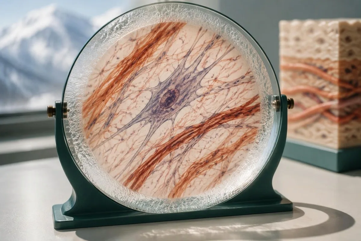
A hypothetical heterozygous pathogenic COL5A1 finding in someone with characteristic scars and hyperextensible skin can support classical EDS. By contrast, 1 pathogenic ADAMTS2 variant in a person without a second explanatory allele usually indicates carrier status rather than confirmed dermatosparaxis EDS; a report can therefore contain a genuine pathogenic finding without that finding explaining the person's symptoms.
The meaning of a negative result changes with pretest probability. When the examination strongly suggests a rare molecular subtype, the next question is whether the assay covered the relevant gene and variant mechanisms; when findings are nonspecific, expanding from 30 to 300 genes may increase uncertain or incidental findings without making the answer more useful or making hEDS genetically testable.
Clinical genetics may recommend a targeted technical follow-up, a different diagnostic hypothesis, or later reanalysis rather than immediately repeating the same panel. The relationship between phenotype and molecular findings also matters in gene and biochemical confirmation, though EDS does not share that condition's biochemical tests; ask for a written explanation of what remains plausible after 1 negative result.
Cum afectează rezultatele EDS copiii și alte rude?
Family testing should follow the confirmed gene and inheritance pattern, not the broad EDS label. For many dominant subtypes, a heterozygous affected parent has a 50% transmission probability per pregnancy; when both parents carry a pathogenic variant for the same recessive subtype, the affected-child probability is usually 25%.
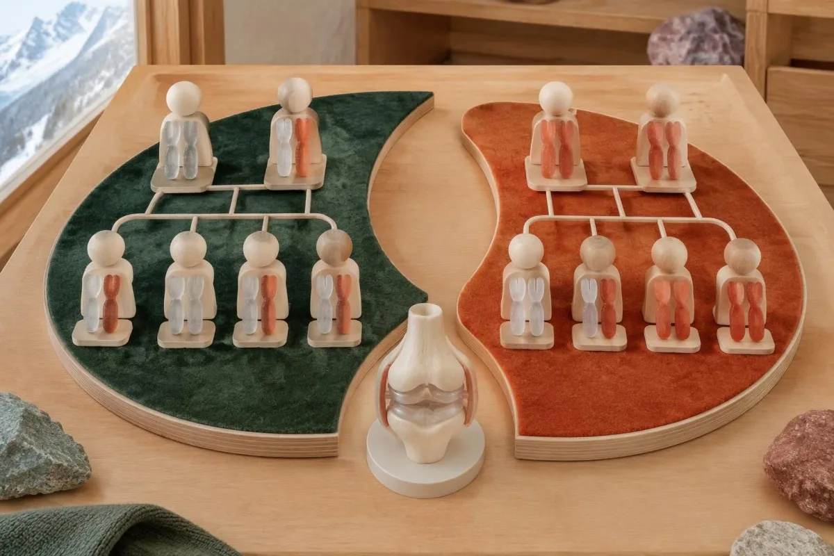
The standard recessive model gives a 25% chance of an affected child, a 50% chance of a carrier child, and a 25% chance of a child inheriting neither familial variant when both parents are carriers. Each pregnancy is a new event, not a running quota; partner testing must investigate the relevant gene sufficiently rather than checking only the variant carried by the first parent.
A negative family history does not exclude dominant EDS because a variant may arise de novo, relatives may have subtle findings, or family information may be incomplete. Testing children can be appropriate when a confirmed condition changes childhood care, particularly vascular EDS; this differs from some adult-onset predictive testing decisions, where timing and the child's future autonomy carry a different balance.
Hypermobile EDS can cluster in families, but no routine molecular result currently provides the same variant-specific cascade-testing pathway. Relatives should consent separately to testing and data sharing, and a family's 3 generations of history should distinguish documented diagnoses from suspected flexibility; our family consent and tracking guide explains why shared health information does not remove individual privacy or the need for personal assessment.
Pot analizele de sânge de rutină sau interpretarea AI să diagnosticheze EDS?
Routine blood tests and AI blood-test interpretation cannot diagnose EDS or classify a germline variant independently. A CBC, ferritin, thyroid panel, or coagulation screen may investigate symptoms and alternative explanations, but there is no routine collagen blood concentration with a diagnostic cutoff for the 13 EDS subtypes.
Kantesti is an AI-powered blood test analysis tool that can explain routine biomarker results alongside reported symptoms. For example, a hypothetical adult with ferritin of 9 ng/mL and hemoglobin of 13.0 g/dL may have depleted iron stores despite hemoglobin within their laboratory's reference interval; our guide to deficitul de fier înainte de anemie explains why that finding deserves attention without becoming evidence for or against EDS.
Normal platelet counts, PT, and aPTT do not exclude connective-tissue fragility, and bruising can have more than 1 cause. Conversely, iron deficiency, thyroid dysfunction, or a bleeding disorder should not be dismissed simply because someone already has hEDS; our complete iron study interpretation addresses a treatable contributor to fatigue, not an inherited collagen diagnosis or a supplement-based cure.
An EDS genetic report contains sensitive information about both the individual and potentially their relatives, so it should not be treated as an ordinary screenshot for unrestricted sharing. Before sending even 1 report to an online service, check purpose, retention, deletion, and whether genetic-report analysis is supported; Kantesti AI's privacy information describes our data-handling approach, not authorization to replace a genetic counselor or molecular laboratory.
Ce ar trebui să aduceți la consultația de genetică și ce surse sunt utile?
Bring the complete genetic report, relevant medical records, and a structured family history to your genetics appointment. The 3 most useful questions are whether the result explains your phenotype, what the assay could have missed, and whether the finding changes your own or your relatives' care.
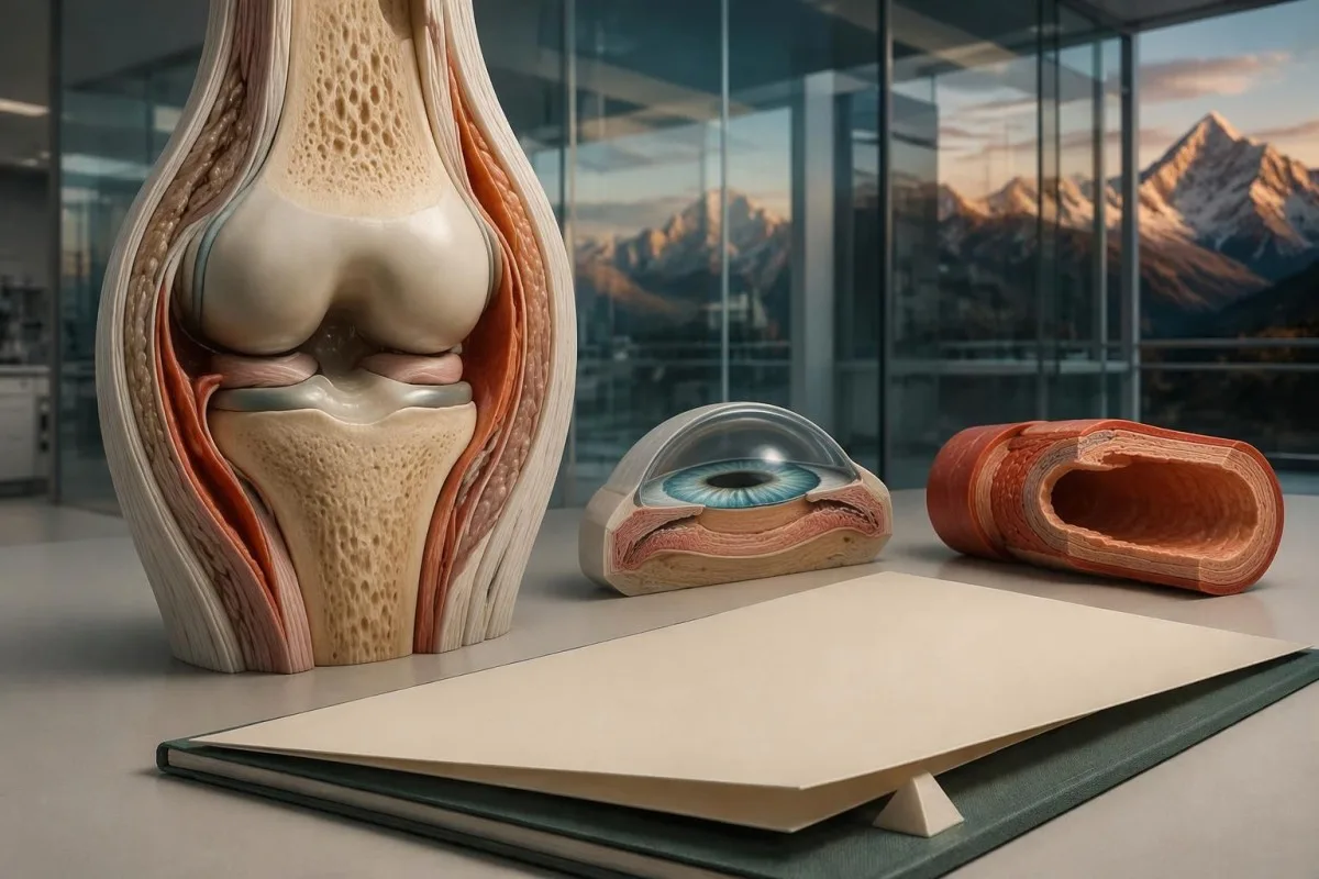
Record major complications with ages, documented diagnoses, and the exact relationship of affected relatives; a 3-generation pedigree can be more informative than an unverified statement that everyone is flexible. Include ophthalmology findings, relevant imaging, characteristic scarring, congenital orthopedic problems, and the full laboratory methods page, because a portal summary may omit gene coverage, deletion testing, or the distinction between a VUS and a pathogenic result.
Before leaving, ask who coordinates 1 next step and who will receive any amended report: the genetics service, the ordering clinician, or the molecular laboratory. Kantesti's standarde de conținut medical support clear separation of established evidence, hypothetical examples, and unresolved questions; this article's EDS recommendations draw on the international classification, ACMG/AMP interpretation guidance, and pediatric hypermobility framework cited below.
The 2 Zenodo publications listed in our research references cover urinalysis and iron studies, not EDS genetic-test validation. Our ghid de interpretare a sumarului de urină may help explain a separate urine report, but neither urine urobilinogen nor iron-binding capacity confirms an EDS subtype; these publications should not be read as evidence that our platform performs molecular EDS diagnosis.
Întrebări frecvente
Poate un test genetic să diagnosticheze sindromul Ehlers-Danlos?
A genetic test can confirm molecularly defined Ehlers-Danlos subtypes when it identifies an appropriately classified variant with the correct inheritance pattern and compatible clinical findings. The 2017 classification includes 13 subtypes, and 12 have established molecular causes that appropriate testing can investigate. Hypermobile EDS does not have a routinely accepted diagnostic molecular test. A panel's gene list, technical coverage, and variant interpretation determine what its result can establish.
Un rezultat negativ al panelului EDS exclude EDS hipermobil?
A negative EDS panel does not rule out hypermobile EDS because hEDS is diagnosed clinically rather than by a validated routine molecular test. A hypothetical negative 30-gene panel means no explanatory reportable variant was found within that assay's scope. The result can help evaluate other inherited disorders, but it cannot prove or disprove hEDS. The next step is a clinical assessment that considers hypermobility, associated features, and alternative diagnoses.
O variantă COL3A1 înseamnă că am sindrom Ehlers-Danlos vascular?
O variantă COL3A1 susține confirmarea sindromului Ehlers-Danlos vascular doar dacă este patogenă sau probabil patogenă și este interpretată în contextul constatărilor clinice. Laboratoarele clinice folosesc 5 categorii de variante, iar o variantă COL3A1 cu semnificație incertă nu confirmă diagnosticul de sindrom Ehlers-Danlos vascular. Complicațiile arteriale sau de organ severe, neexplicate, justifică în continuare evaluarea de către un specialist chiar și atunci când testarea nu este concludentă. Durerea bruscă și severă, colapsul sau simptomele neurologice necesită îngrijiri de urgență, nu așteptarea reclasificării variantei.
Ce înseamnă o VUS pe un panel genetic pentru EDS?
O variantă cu semnificație incertă înseamnă că dovezile disponibile nu permit stabilirea cu certitudine a faptului că acea modificare a ADN-ului cauzează boala. Cadrul ACMG/AMP distinge VUS de „probabil patogenă”, care corespunde unei certitudini de peste 90% că modificarea cauzează boala. Un VUS nu ar trebui să determine, de unul singur, diagnosticul de EDS, testarea predictivă sau tratamentul specific genei. Genetica clinică poate recomanda testarea țintită a membrilor familiei sau reinterpretarea ulterioară, în timp ce simptomele și riscurile sunt gestionate pe baza meritelor lor clinice.
De ce au fost găsite două variante într-o genă EDS?
Două variante pot fi importante, deoarece mai multe subtipuri de EDS urmează un tipar de moștenire recesivă și necesită de obicei modificări cauzatoare de boală în ambele copii ale genei. Laboratorul trebuie să stabilească dacă cele 2 modificări sunt patogene sau probabil patogene și dacă sunt în trans, afectând copii opuse. Două modificări în cis, care afectează aceeași copie, pot să nu explice o afecțiune recesivă. Identificarea unei variante patogene și a unei VUS nu oferă automat nici confirmarea moleculară.
Ar trebui să fie testați copiii mei dacă am EDS?
Children's testing depends on the confirmed subtype, whether a familial variant is known, and whether the result changes care during childhood. Many dominant EDS subtypes have a 50% transmission probability per pregnancy, making targeted testing useful when appropriate. When both parents carry variants for the same recessive subtype, the usual affected-child probability is 25% per pregnancy. Hypermobile EDS currently requires clinical evaluation rather than routine variant-specific family testing.
Analizele de sânge normale pot exclude sindromul Ehlers-Danlos?
Analizele uzuale de sânge normale nu pot exclude sindromul Ehlers-Danlos, deoarece panourile standard nu măsoară modificările structurale ereditare responsabile de SED. Un CBC, un timp de protrombină (PT) sau un timp de tromboplastină parțial activată (aPTT) normal nu exclude fragilitatea țesutului conjunctiv, iar niciun prag uzual al colagenului în sânge nu diagnostichează cele 13 subtipuri clasificate. Analizele de sânge pot identifica factori separați care contribuie la simptome, precum deficitul de fier sau disfuncția tiroidiană. SED definit molecular necesită interpretare genetică adecvată, în timp ce SED hipermobil necesită evaluare clinică.
Aflați cum se citesc analizele de sânge
Încărcați buletinul de analize, iar AI-ul Kantesti explică fiecare biomarker în limbaj simplu, în câteva secunde. Folosit de peste 2 milioane de persoane din întreaga lume.
Referințe
- Klein, T., Mitchell, S., & Weber, H. (2026). Testul de urobilinogen în urină: Ghid complet de analiză de urină 2026. Kantesti AI Medical Research. DOI
- Klein, T., Mitchell, S., & Weber, H. (2026). Ghid privind studiile de fier: TIBC, saturația fierului și capacitatea de legare. Kantesti AI Medical Research. DOI
- Malfait F et al. (2017). The 2017 international classification of the Ehlers-Danlos syndromes. American Journal of Medical Genetics Part C: Seminars in Medical Genetics. DOI
- Richards S et al. (2015). Standarde și ghiduri pentru interpretarea variantelor de secvență: o recomandare de consens comună a American College of Medical Genetics and Genomics și a Association for Molecular Pathology. Genetics in Medicine. DOI PubMed
- Tofts LJ et al. (2023). Pediatric joint hypermobility: a diagnostic framework and narrative review. Orphanet Journal of Rare Diseases. DOI
- 2M+Teste analizate
- 127+Țări
- 100+Limbi
Despre echipa medicală


Semnale de încredere E-E-A-T
Experienţă
Revizuire clinică condusă de medici a fluxurilor de lucru pentru interpretarea analizelor.
Expertiză
Focalizare pe medicina de laborator asupra modului în care biomarkerii se comportă în context clinic.
Autoritate
Revizuit medical de Dr. Thomas Klein, cu contribuții din partea Dr. Sarah Mitchell și Prof. Dr. Hans Weber.
Încredere
Interpretare bazată pe dovezi, cu căi clare de urmărire pentru a reduce alarmele.
Declinare a răspunderii medicale
Acest articol are doar scop educațional și nu constituie sfat medical. Consultă întotdeauna un furnizor calificat de servicii medicale pentru decizii privind diagnosticarea și tratamentul.
Publicat · Contactați echipa editorială
Publicat de Kantesti Ltd, înregistrată în Anglia și Țara Galilor, numărul companiei. 17090423, Londra, Regatul Unit.
