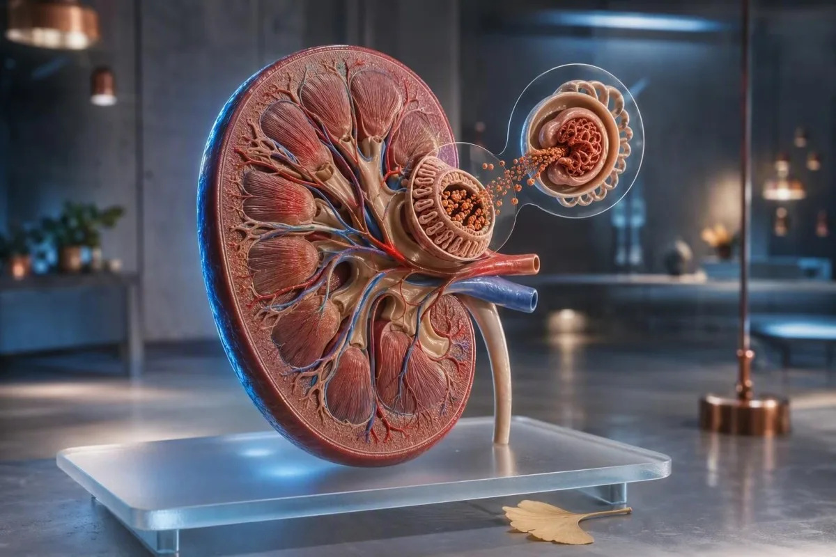
Кілька бульбашок після швидкого сечовипускання зазвичай є фізичним явищем. Піна, яка багаторазово покриває унітаз, залишається після змивання або супроводжується набряками, потребує тесту на альбумін у сечі, а не здогадок.


- Короткочасні бульбашки які зникають за кілька секунд, зазвичай відображають швидкість сечовипускання, залишки засобу для чищення унітазу або концентровану сечу, а не хворобу нирок.
- Стійка пінаста сеча викликає більше занепокоєння, коли щільний білий шар залишається протягом 1-2 хвилин при багаторазових випадках.
- ACR сечі нижче 3 мг/ммоль (30 мг/г) є нормою або легким підвищенням екскреції альбуміну у більшості дорослих.
- Альбумінурія від 3 до 30 мг/ммоль (30-300 мг/г) слід підтвердити повторними ранковими зразками протягом щонайменше 3 місяців.
- ACR вище 30 мг/ммоль (300 мг/г) є вираженою альбумінурією і потребує термінової клінічної оцінки.
- Сигнали тривоги включають набряк обличчя або ніг, видиму кров, задишку, зменшення виділення сечі, лихоманку або вагітність з головним болем і високим кров'яним тиском.
- Збір має значення: уникайте інтенсивних фізичних навантажень протягом 24 годин, використовуйте чисту середню порцію ранкової сечі та повідомте лікаря про менструацію або інфекцію сечовивідних шляхів (ІСШ).
- Аналізи крові допомагають: креатинін, eGFR, сироватковий альбумін, глюкоза та HbA1c, щоб уточнити, чи відображає білок у сечі захворювання нирок, діабет або системне захворювання.
Як відрізнити безпечні бульбашки від стійкої пінистої сечі
Причини пінистої сечі варіюються від швидкого струменя, що б'є у воду, до альбуміну, що просочується крізь нирковий фільтр. Нешкідливі бульбашки великі, прозорі та швидко зникають; піна, що викликає занепокоєння, дрібніша, біла, повторювана і тримається приблизно 1-2 хвилини, незважаючи на чисту ємність.
Поверхневий натяг пояснює більшість одноразових бульбашок. Сильний перший ранковий струмінь, зневоднення, концентровані розчинені речовини або залишки миючого засобу можуть затримувати повітря на поверхні води без втрати білка; це особливо поширене після тривалого перельоту, лихоманки або важкого тренування.
Патерн має більше значення, ніж одноразове спостереження. За 15 років моєї клінічної практики я прошу пацієнтів помічати піну протягом трьох окремих днів, використовуючи чисту, невибілену ємність; повторюваний шар дрібної піни є кращою причиною для тестування, ніж драматичний вигляд після фізичних навантажень.
Сама по собі піна не може діагностувати протеїнурію, оскільки фосфати, жовчні солі, сперма та запалення сечовивідних шляхів також можуть змінювати поверхневий натяг. Якщо сеча каламутна, а не піниста, диференціальний діагноз зміщується в бік кристалів, клітин або інфекції; наш посібник з каламутної сечі допомагає розрізнити ці патерни.
Просте правило домашнього спостереження
Не фотографуйте унітаз і не намагайтеся робити домашні “струшувальні тести”. Натомість відзначте, чи з'являється піна в чистій ємності щонайменше 3 з 7 днів, чи тримається вона довше 1 хвилини, і чи присутні набряки або зміни артеріального тиску. Ці деталі роблять лабораторний запит набагато кориснішим.
Чому білок у сечі може створювати стабільну піну
Піниста сеча білок найчастіше є альбуміном, білком з негативним зарядом, який зазвичай залишається в кровотоці. Коли нирковий фільтр стає більш проникним, альбумін знижує поверхневий натяг і може утворювати дрібні, стійкі бульбашки.
Здорова доросла людина зазвичай виділяє менше 30 мг альбуміну на день, хоча рутинний тест-смужка може не виявити низького рівня втрати альбуміну. Більш чутливе співвідношення альбумін-креатинін у сечі, або ACR, коригує альбумін відповідно до концентрації сечі і є кращим для початкового скринінгу.
Альбумін – не єдиний білок, який має значення. Легкі ланцюги з розладів плазматичних клітин, тубулярні білки після певних ліків та транзиторні білки під час лихоманки можуть виходити за межі тест-смужки, призначеної головним чином для альбуміну; ось чому негативна смужка не вирішує всіх випадків стійкої піни.
Доктор Томас Кляйн аналізує результати аналізу сечі разом із сироватковим креатиніном, eGFR, альбуміном та глюкозою, а не розглядає візуальний симптом як діагноз. Наш довідник із сироваткового білка пояснює, чому низький рівень альбуміну в крові та підвищений ACR у сечі разом мають більшу вагу, ніж кожен показник окремо.
Поширені причини пінистої сечі, не пов'язані з нирками
Більшість тимчасової пінистої сечі має не ниркову причину: концентрована сеча, швидке сечовипускання, нещодавня еякуляція, залишки мила, лихоманка або інтенсивні фізичні навантаження. Ці причини зазвичай минають протягом 24-48 годин після нормалізації гідратації та відновлення.
Зневоднення робить сечу темнішою та більш концентрованою, тому бульбашки можуть здаватися щільнішими, навіть коли виділення альбуміну в нормі. Прагніть до блідо-солом'яної сечі протягом наступного дня, а не до надмірного споживання рідини; випивання кількох літрів швидко може саме по собі знизити рівень натрію у схильних людей.
Фізичні навантаження можуть тимчасово підвищити рівень альбуміну в сечі. 10-кілометровий забіг, інтенсивна силова тренування або тривалий вплив тепла можуть призвести до появи білка в зразку, зібраному безпосередньо після цього, тому лабораторії зазвичай рекомендують уникати інтенсивних фізичних навантажень за 24 години до тестування ACR.
Сперма в уретрі після еякуляції може викликати тимчасові бульбашки та незначне позитивне тестування на білок. Видима рожева або чайного кольору сеча повністю змінює питання і повинна бути оцінена за допомогою цього посібника з кров у сечі, особливо після 45 років або у будь-кого, хто піддавався впливу куріння.
Коли стійка піна свідчить про клінічно значущу втрату білка
Стійка піна вимагає тестування сечі на білок, коли вона виникає повторно, особливо у поєднанні з діабетом, високим кров'яним тиском, набряками або зниженим eGFR. Альбумінурія може бути раннім попередженням про стан нирок, навіть коли креатинін залишається в межах лабораторного діапазону.
KDIGO визначає ACR менше 3 мг/ммоль, або 30 мг/г, як A1; 3-30 мг/ммоль як A2; і вище 30 мг/ммоль як A3 альбумінурію. Категорії найточніше прогнозують ризик захворювань нирок та серцево-судинних захворювань у поєднанні з eGFR, а не використовуються ізольовано (KDIGO, 2024).
Діабет та гіпертензія є найпоширенішими хронічними причинами альбумінурії у світі, але вони не єдині. Гломерулонефрит, ожиріння-асоційована гломерулопатія, вовчак, прееклампсія, апное уві сні та деякі протизапальні препарати також можуть сприяти цьому; стійкі результати вимагають від лікаря з'ясування механізму.
Кантесті – це Аналізатор крові зі штучним інтелектом що ставить креатинін, eGFR, сироватковий альбумін, глюкозу та HbA1c в один поздовжній контекст. Він не може виміряти ACR сечі з кров'яної панелі, але може допомогти визначити метаболічний або нирковий патерн, який робить результат аналізу сечі більш терміновим.
Які тести на білок у сечі найкраще відповідають на це питання
Дослідження ACR випадкової порції ранкової сечі є найкращим первинним тестом для підозри на втрату альбуміну у більшості дорослих. Співвідношення білка до креатиніну, або PCR, є корисним, коли підозрюється неальбуміновий білок або коли потрібно кількісно визначити загальний білок.
ACR повідомляється як мг/ммоль у Великобританії та багатьох інших країнах, або мг/г у Сполучених Штатах. ACR 3 мг/ммоль дорівнює приблизно 30 мг/г; це перерахунок важливий, оскільки пацієнти іноді плутають різні одиниці з великою зміною результату.
Результат тесту на білок на тест-смужці — сліди або 1+ — є ознакою скринінгу, а не діагнозом. Лужна сеча, концентрована сеча та забруднення можуть спричинити хибнопозитивні результати, тоді як розведена сеча та неальбумінові білки можуть дати помилкове заспокоєння; кількісне тестування усуває цю невизначеність.
24-годинний збір тепер зарезервований для окремих випадків, таких як дуже висока втрата білка, оцінка вагітності або суперечливі зразки випадкової порції. Його легко недозібрати, тому прочитайте наш практичний посібник зі збору сечі за 24 години перед його початком.
Як точно зібрати сечу для тестування на білок
Точне тестування сечі на білок починається з чистого зразка середньої порції ранкової сечі, зібраного через 24 години без інтенсивних фізичних навантажень. Перевага надається першій сечі після пробудження, оскільки положення тіла, гідратація та денна активність менше впливають на виведення альбуміну.
Вимийте руки, використайте наданий стерильний контейнер, почніть мочитися в унітаз, потім зберіть середню порцію, не торкаючись внутрішньої сторони контейнера або кришки. Доставте зразок негайно; якщо затримка неминуча, дотримуйтесь інструкцій лабораторії щодо охолодження, а не залишайте його в теплому автомобілі.
Відкладіть нетермінові дослідження під час симптоматичної інфекції сечовивідних шляхів, лихоманки, діареї, менструації або протягом 24 годин після інтенсивних фізичних навантажень. Ці стани можуть тимчасово підвищити виведення альбуміну, тоді як менструальне забруднення також може призвести до появи крові та білка; результатів лейкоцитарної естерази може прояснити можливий інфекційний патерн.
Ортостатична протеїнурія заслуговує на згадку, особливо у підлітків та молодих людей. Білок, що з'являється в денній сечі, але відсутній у зразку першої ранкової сечі, часто є доброякісним, хоча лікар повинен підтвердити цей патерн, а не припускати його.
Як інтерпретувати результати ACR та співвідношення білок/креатинін
ACR нижче 3 мг/ммоль зазвичай заспокоює, тоді як підтверджений ACR 3 мг/ммоль або вище вимагає подальшого спостереження на основі ризику. Один підвищений зразок не встановлює хронічного захворювання нирок, оскільки фізичні навантаження, інфекція та сплески глюкози можуть спричинити короткочасну альбумінурію.
Для дорослих без діабету нещодавно підвищений ACR зазвичай повторюють за допомогою ранкового зразка протягом тижнів. Хронічне захворювання нирок вимагає наявності відхилень протягом щонайменше 3 місяців, якщо немає чіткого гострого ураження нирок або помітно підвищеного білка, що потребує швидшого перегляду (KDIGO, 2024).
ACR вище 70 мг/ммоль, приблизно 700 мг/г, зазвичай вимагає термінового обговорення зі спеціалістом, навіть якщо eGFR збережений, особливо при наявності крові в сечі. Нефротичний діапазон протеїнурії, як правило, визначається PCR вище 300 мг/ммоль або виведенням білка вище 3,5 г/добу, часто супроводжується набряками та низьким рівнем сироваткового альбуміну.
Кантесті – це сервісі інтерпретації тестів AI що може організувати ниркові маркери крові за датами та позначити тенденцію до зниження eGFR. Інтерпретація тенденцій підтримує, але ніколи не замінює, клінічну мікроскопію сечі, підтвердження ACR та огляд медикаментів.
Аналізи крові, які роблять результати тесту на пінусту сечу значущими
Креатинін, eGFR, сироватковий альбумін, глюкоза, HbA1c, електроліти та ліпіди допомагають пояснити, чи відображає білок у сечі пошкодження ниркової фільтрації чи ширший метаболічний розлад. Нормальний креатинін не виключає ранньої альбумінурії.
eGFR розраховується на основі креатиніну та знижується пізніше, ніж незначне витоком альбуміну, у багатьох людей з діабетом. eGFR нижче 60 мл/хв/1,73 м² протягом 3 місяців або довше відповідає одному критерію хронічного захворювання нирок, але значення повинні інтерпретуватися з урахуванням віку, м'язової маси та нещодавнього захворювання.
Низький рівень альбуміну в сироватці крові нижче приблизно 35 г/л у поєднанні з вираженою протеїнурією та новими набряками гомілок викликає занепокоєння щодо значної втрати білка нирками. Навпаки, високий рівень альбуміну з темною сечею частіше відображає зневоднення; наші керівництво з eGFR після зневоднення пояснює, чому повторне тестування після одужання може бути доцільним.
Американська діабетична асоціація рекомендує щонайменше щорічне тестування UACR та eGFR при цукровому діабеті 2 типу та при цукровому діабеті 1 типу тривалістю 5 років або більше (ADA Professional Practice Committee, 2024). HbA1c показує нам середній рівень глікемії, але різке підвищення глюкози все ще може тимчасово збільшити рівень альбуміну в сечі до того, як зміниться HbA1c.
Ліки, добавки та хвороби, які можуть впливати на білок у сечі
Протизапальні знеболювальні засоби, літій, деякі види протиракової терапії та важкі інфекції можуть впливати на ниркову фільтрацію або обробку білка нирковими канальцями. Не припиняйте прийом призначених ліків лише тому, що сеча виглядає пінистою; попросіть оглянути результати аналізу сечі та крові на функцію нирок.
НПЗП, такі як ібупрофен та напроксен, можуть зменшувати кровотік у нирках, особливо під час зневоднення, серцевої недостатності або наявного ХХН. Ризик зростає, коли НПЗП поєднуються з діуретиком та інгібітором АПФ або БРА, що клініцисти іноді називають “потрійною комбінацією”.”
Препарати з групи SGLT2 можуть спричиняти невелике початкове зниження eGFR, але зазвичай зменшують альбумінурію з часом у належним чином відібраних пацієнтів. Ця очікувана гемодинамічна зміна відрізняється від прогресуючого пошкодження; див. зміни eGFR при лікуванні SGLT2 для практичних відмінностей.
Високі дози вітаміну С можуть перешкоджати деяким тест-смужкам для сечі, тоді як креатин може ускладнити інтерпретацію креатиніну, не обов'язково шкодячи ниркам. Принесіть на прийом повний список рецептурних препаратів, безрецептурних знеболювальних, протеїнових порошків та рослинних продуктів — важливі назви та дозування.
Цукровий діабет та кров'яний тиск: перевірки ризику з найвищим пріоритетом
Діабет та гіпертензія роблять стійку піну більш клінічно значущою, оскільки обидва стани можуть спричиняти альбумінурію за роки до появи симптомів. Артеріальний тиск 140/90 мм рт. ст. або вище та ACR 3 мг/ммоль або вище повинні спонукати до структурованого первинного медичного огляду.
Для багатьох людей з ХХН та альбумінурією клініцисти прагнуть до стандартизованого систолічного артеріального тиску в офісі нижче 120 мм рт. ст., коли це можливо, хоча цільові показники повинні бути індивідуалізовані для віку, ризику падінь, вагітності та методу вимірювання. Домашні вимірювання, проведені неправильно, не повинні бути єдиною підставою для зміни лікування.
Альбумінурія також є маркером судинного ризику. Причина, чому нас турбує підвищення ACR у поєднанні з високим артеріальним тиском, полягає в тому, що разом вони вказують на ендотеліальний та гломерулярний стрес, тоді як будь-яке з цих знахідок один раз під час гострого захворювання може бути тимчасовим; наш посібник з лабораторних досліджень гіпертензії окреслює ширший підхід до обстеження.
Кантесті – це Платформа для інтерпретації біомаркерів AI використовується у 127+ країнах для порівняння рівня глюкози, HbA1c, ниркових маркерів та ліпідних тенденцій. Для стійкої пінистої сечі відповідним наступним тестом залишається лабораторний аналіз ACR сечі, який ідеально переглядається з тією ж клінічною командою, що керує артеріальним тиском або діабетом.
Пінаста сеча у вагітних та дітей потребує різних правил
Піниста сеча під час вагітності потребує оцінки в той же день, якщо вона супроводжується головним болем, змінами зору, болем у верхній частині живота, раптовими набряками або підвищеним артеріальним тиском. Протеїнурія після 20 тижнів може бути частиною прееклампсії, але сам вигляд ніколи не є діагностичним.
При вагітності співвідношення білка до креатиніну 30 мг/ммоль або більше, або 300 мг білка за 24 години, підтверджує значну протеїнурію у відповідному клінічному контексті. Нормальний вигляд зразка сечі не може виключити прееклампсію, якщо артеріальний тиск становить 140/90 мм рт. ст. або вище з тривожними симптомами.
Діти часто мають тимчасову протеїнурію після лихоманки, фізичних навантажень або зневоднення. Стійкий білок у трьох зразках, високий артеріальний тиск, набряки або кров у сечі вимагають педіатричного обстеження; особливо корисним є тестування вранці для виключення постуральної протеїнурії у підлітків.
Вагітність змінює фільтрацію, тому показники креатиніну, які виглядають “нормальними” для невагітних дорослих, можуть не бути заспокійливими. Наш посібник до значення GFR при вагітності надає контекст, але термінові симптоми слід негайно звертатися до приймального відділення пологового будинку, а не до онлайн-інтерпретації.
Тривожні сигнали: коли пінаста сеча потребує невідкладної медичної допомоги
Негайно зверніться за медичною допомогою при пінистій сечі з задишкою, швидким наростанням набряків ніг або обличчя, значним зменшенням кількості сечі, видимої крові, лихоманкою з болем у боці або симптомами попередження вагітності. Ці комбінації можуть вказувати на значне ниркове, серцеве, інфекційне захворювання або захворювання, пов'язане з вагітністю.
Новий набряк обличчя після пробудження, тісне взуття до вечора та збільшення ваги більш ніж на 2 кг за 2-3 дні можуть свідчити про затримку рідини. Коли ці симптоми супроводжуються стійкою піною, лікарі оперативно перевіряють білок у сечі, креатинін, альбумін, кров'яний тиск, а іноді й результати обстеження грудної клітки.
Сеча кольору чаю після інтенсивних фізичних навантажень із сильним болем у м'язах не є типовою доброякісною піною; вона може сигналізувати про вивільнення міоглобіну і вимагає термінового аналізу крові та сечі. Те саме стосується сечі кольору коли зі зменшеним виділенням, що може свідчити про кровотечу з ниркових клубочків або пошкодження м'язів.
Доктор Томас Кляйн радить пацієнтам не чекати результатів домашнього тесту за допомогою смужки, коли присутні тривожні ознаки. Щодо змін кольору, пов'язаних з навантаженням, наша стаття про гематурію у бігунів пояснює доброякісний спектр і ознаки, які не є доброякісними.
Що роблять лікарі після позитивного результату тесту на білок у сечі
Після позитивного результату ACR або PCR лікарі зазвичай підтверджують результат, вимірюють артеріальний тиск і eGFR, досліджують осад сечі та переглядають дані про діабет, ліки, аутоімунні симптоми та сімейний анамнез. Наступний крок залежить від кількості, стійкості та супутньої крові або зниженої фільтрації.
Мікроскопія сечі може виявити еритроцити, лейкоцити та циліндри, які один лише показник білка пояснити не може. Еритроцитарні циліндри або дисморфні еритроцити вказують на клубочкове походження та прискорюють направлення до нефролога; прості гіалінові циліндри можуть з'являтися після зневоднення або фізичних навантажень, як зазначено в нашому посібнику з гіалінових циліндрів.
Якщо альбумінурія підтверджена, лікування зазвичай спрямоване на причину: контроль артеріального тиску, управління рівнем глюкози, підтримка у боротьбі із зайвою вагою та курінням, а також призначення нефропротекторних препаратів, якщо це показано. УЗД нирок, імунологічне тестування, електрофорез сироватки крові або біопсія нирки є вибірковими тестами, а не автоматичними наслідками одного позитивного результату тесту за допомогою смужки 1+.
Клінічна методологія Kantesti перевіряється відповідно до структурованих стандартів безпеки, і читачі можуть ознайомитися з нашим підхід до медичної валідації. Наша роль полягає в тому, щоб полегшити обговорення наявних результатів аналізів крові, а не ставити діагноз гломерулярного розладу за зображенням або одним лабораторним показником.
Практичний семиденний план при стійкій піні
При стійкій пінистій сечі без тривожних ознак призначте первинний аналіз сечі на ACR та загальний аналіз сечі протягом 1-2 тижнів, бажано використовуючи зразок ранкової сечі. Запишіть артеріальний тиск, рівень гідратації, фізичні навантаження, ліки та чи триває піна довше 1 хвилини протягом семи днів.
День 1-3: пийте достатньо води, уникайте інтенсивних фізичних навантажень і спостерігайте тільки в чистій унітазній чаші без додавання миючих засобів. Не намагайтеся “змити” проблему надмірною кількістю води; мета — отримати репрезентативний зразок, а не штучно розведений.
День 4-7: попросіть зробити загальний аналіз сечі та ACR, і запитайте, чи доцільно перевіряти креатинін, eGFR, калій, альбумін сироватки крові, глюкозу та HbA1c у ваших обставинах. Принесіть попередні результати, оскільки зміна ACR з 2 до 8 мг/ммоль має більше значення, ніж будь-яке з цих чисел без контексту; трекінг результатів у динаміці може допомогти організувати дати.
Станом на 4 вересня 2026 року розумне повідомлення залишається заспокійливим, але твердим: більшість поодиноких бульбашок нешкідливі, тоді як повторна дрібна піна заслуговує на кількісний аналіз сечі. Якщо вам потрібна допомога у підготовці запитань для лікаря, наш команду медичного консультування встановлює клінічні стандарти, що лежать в основі нашого освітнього контенту.
Часті запитання
Чи завжди піниста сеча є ознакою наявності білка в сечі?
Ні, піниста сеча не завжди є ознакою втрати білка. Швидкий струмінь, зневоднення, залишки засобу для чищення туалету, лихоманка, фізичні вправи та сперма можуть утворювати бульбашки, які зникають за лічені секунди. Стійка дрібна біла піна, яка повторюється кілька днів, більше свідчить про протеїнурію і повинна бути перевірена за допомогою співвідношення альбумін/креатинін у сечі. Співвідношення ACR 3 мг/ммоль, еквівалентне 30 мг/г, або вище, вважається аномальним і вимагає клінічного контексту та часто повторного тестування.
Як довго піна в сечі повинна тривати, перш ніж варто хвилюватися?
Піна, яка залишається щільним шаром приблизно 1-2 хвилини при повторних випадках, викликає більше занепокоєння, ніж великі бульбашки, що швидко зникають. Тривалість не є діагностичним тестом, оскільки хімічні речовини в туалеті та концентрація сечі можуть впливати на стабільність бульбашок. Якщо така картина повторюється протягом 3 або більше днів, призначте аналіз сечі та дослідження сечі на ACR протягом 1-2 тижнів. Зверніться за невідкладною допомогою раніше, якщо піна з'являється разом з набряком, видимою кров'ю, задишкою, лихоманкою або зменшенням об'єму сечі.
Який тест на білок у сечі мені слід запитати?
Найкращим першим тестом для більшості дорослих є співвідношення альбумін/креатинін у ранковій сечі, яке називається ACR або UACR. ACR нижче 3 мг/ммоль, або 30 мг/г, зазвичай вважається нормальним або незначно підвищеним, тоді як 3-30 мг/ммоль вказує на помірно підвищену альбумінурію. Співвідношення білок/креатинін може бути додано, коли підозрюється втрата загального білка або неальбумінового білка. Рутинний тест-смужка є корисним скринінговим методом, але менш точним, ніж ACR, для виявлення низького рівня втрати альбуміну.
Чи може зневоднення викликати пінисту сечу?
Так, зневоднення може зробити сечу більш концентрованою і спричинити появу більш помітних бульбашок або піни без стійкої втрати білка нирками. Це часто трапляється при темно-жовтому кольорі сечі, відчутті спраги, впливі спеки, блюванні, діареї або тривалому періоді без вживання рідини. Поверніться до звичайного споживання рідини протягом 24-48 годин перед нетерміновим аналізом, але уникайте вживання надмірної кількості води лише для розведення результату. Якщо піна зберігається після гідратації, зверніться за тестуванням сечі ACR.
Чи може фізичне навантаження спричинити появу білка та піни в сечі?
Інтенсивні фізичні навантаження можуть спричинити тимчасову альбумінурію та пінисту сечу, особливо після бігу, силових тренувань, змагань на витривалість або тренувань у спеку. Ефект зазвичай минає протягом 24-48 годин, тому зразок для визначення співвідношення альбумін/креатинін (ACR) ідеально збирати після 24-годинного уникнення інтенсивних фізичних навантажень. Стійка протеїнурія в ранковому зразку після відновлення менш імовірно пояснюється лише фізичними навантаженнями. Темна або забарвлена як "кола" сеча із сильним болем у м'язах потребує термінової медичної оцінки.
Який рівень ACR є небезпечним?
Підвищений рівень ACR понад 30 мг/ммоль або 300 мг/г вказує на виражену альбумінурію та потребує негайного медичного обстеження, особливо якщо eGFR знижений або в сечі присутня кров. Рівень ACR понад 70 мг/ммоль (близько 700 мг/г) зазвичай вимагає термінового обговорення зі спеціалістом, навіть якщо рівень креатиніну в межах норми. Ризик визначається поєднанням ACR, eGFR, артеріального тиску, статусу діабету, симптомів та повторних вимірювань. Одиничне підвищення ACR зазвичай слід підтверджувати, якщо клінічна ситуація не є невідкладною.
Отримайте аналіз крові з підтримкою ШІ вже сьогодні
Приєднуйтесь до понад 2 мільйонів користувачів у всьому світі, які довіряють Kantesti для миттєвого та точного аналізу лабораторних тестів. Завантажте результати аналізу крові та отримайте комплексну інтерпретацію біомаркерів 15,000+ за секунди.
📚 Дослідження з посиланнями на публікації
Klein, T., Mitchell, S., & Weber, H. (2026). Клінічна валідаційна рамка v2.0 (сторінка медичної валідації). Медичні дослідження ШІ Kantesti.
Klein, T., Mitchell, S., & Weber, H. (2026). ШІ аналізатор аналізу крові: проаналізовано 2,5 млн тестів | Глобальний медичний звіт 2026. Медичні дослідження ШІ Kantesti.
📖 Зовнішні медичні посилання
⚕️ Медична відмова від відповідальності
Ця стаття призначена лише для освітніх цілей і не становить медичної поради. Завжди консультуйтеся з кваліфікованим медичним працівником щодо рішень про діагностику та лікування.
Сигнали довіри E-E-A-T
Досвід
Лікарський клінічний огляд робочих процесів інтерпретації лабораторних показників.
Експертиза
Лабораторна медицина з акцентом на те, як біомаркери поводяться в клінічному контексті.
Авторитетність
Написано доктором Томасом Кляйном за редакційного перегляду докторки Сари Мітчелл і професора доктора Ганса Вебера.
Довірливість
Інтерпретація на основі доказів із чіткими шляхами подальших дій, щоб зменшити тривогу.