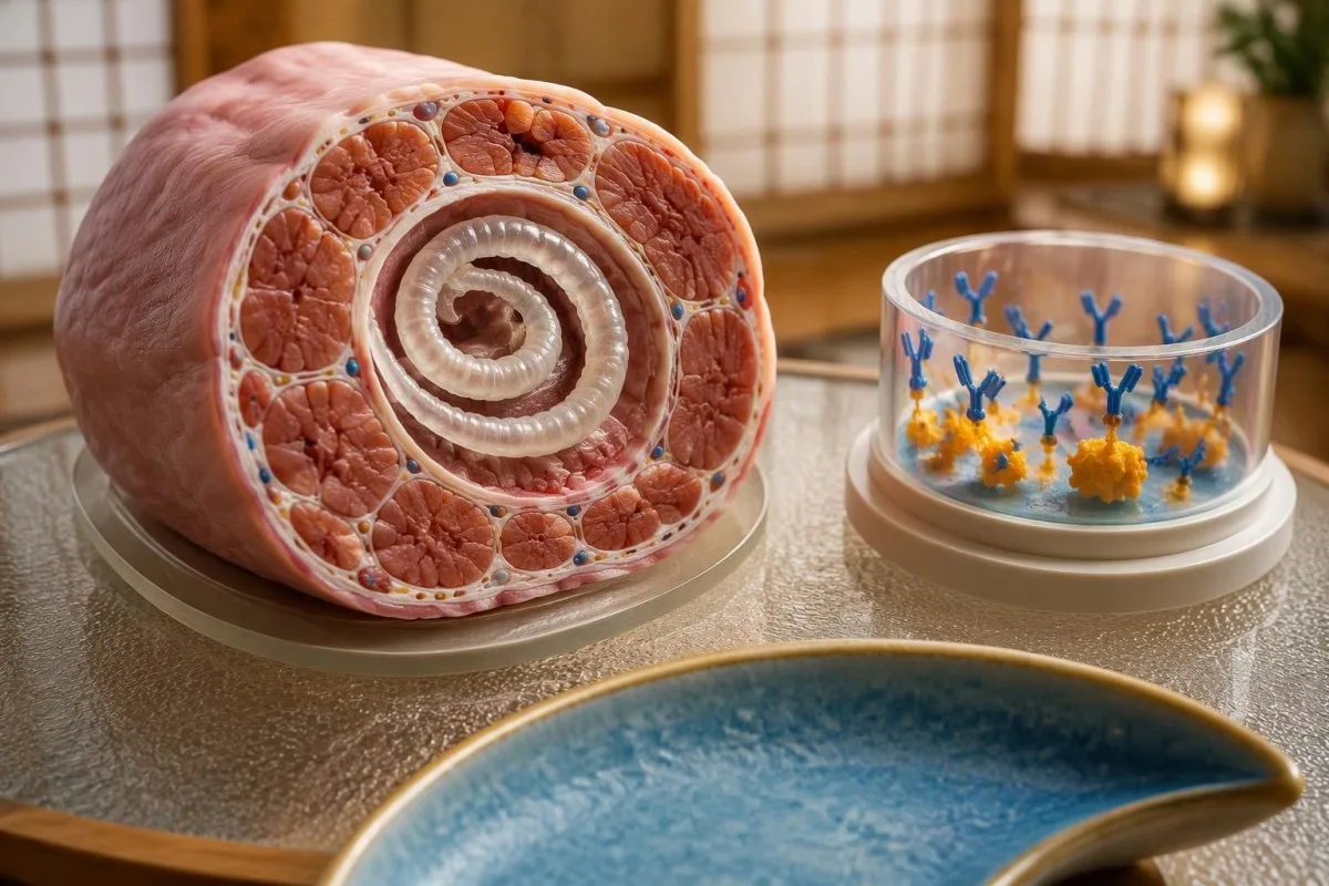
A positive result supports exposure to Trichinella, but does not establish when infection occurred or whether it explains today's symptoms. Early negative results can miss infection; the meal date, symptoms, supporting laboratory findings, and repeat testing determine the next step.


- Positive antibodies support Trichinella exposure, not necessarily recent infection; antibodies can remain detectable for 10 years or longer.
- Testing window varies: Trichinella IgG may become detectable approximately 12–60 days after infection, so an early negative cannot reliably exclude it.
- Repeat testing is commonly arranged 2–3 weeks after an early negative or equivocal result, with additional sampling if the exposure was very recent.
- Symptom timing matters: digestive symptoms can begin within 1–2 days, while fever, muscle soreness, and eyelid swelling usually develop later.
- Eosinophils above 500 cells/µL support a tissue-invasive parasite differential, but allergies, medications, and other conditions can produce the same finding.
- Assay numbers are laboratory-specific; an antibody index of 2.0 is not a worm count, a severity score, or a universal treatment threshold.
- Urgent assessment is needed for chest pain, breathlessness, confusion, or progressive weakness after a plausible exposure—do not wait 2–3 weeks for repeat antibodies.
- Early treatment can matter before antibody confirmation; clinicians may treat a convincing recent exposure and compatible illness while arranging specialist testing.
What does a positive Trichinella antibody result mean?
A positive trichinella antibody test means the assay detected an immune response that recognizes Trichinella antigens. After undercooked pork or wild game, it supports possible trichinellosis, but 1 positive result cannot distinguish recent infection from older exposure or cross-reactivity without the meal date, symptoms, and laboratory context.
Antibodies are evidence of recognition, not a direct parasite count. Trichinella IgG can remain detectable for 10 years or longer, so a positive result months after a hunting trip may not explain a new episode of muscle discomfort; Gottstein, Pozio, and Nöckler describe this persistence in their 2009 clinical review.
Consider a hypothetical 46-year-old who develops fever, sore calves, and eyelid swelling 3 weeks after eating insufficiently cooked bear meat. That combination makes a positive antibody result much more persuasive than the same result in someone without symptoms or a credible exposure; our guide to positive antibody results explains this broader distinction.
Kantesti is an AI blood test analyzer that can organize a Trichinella result alongside eosinophils, muscle enzymes, and sample dates, but 1 report cannot establish a parasitic diagnosis. My editorial emphasis as Thomas Klein, MD, is straightforward: the exposure timeline belongs beside the laboratory result, not in a separate conversation after treatment decisions have already been made.
What does the Trichinella antibody assay actually measure?
Most routine Trichinella serology detects IgG antibodies using an enzyme immunoassay, often with larval excretory-secretory antigens. Results may be reported as positive, negative, equivocal, or an assay index; 1 numerical index cannot be compared with another laboratory's index unless the methods and interpretation rules match.
A laboratory cutoff separates assay signals, not stages of disease. A report showing 1.2, for example, is uninterpretable without that laboratory's negative, equivocal, and positive boundaries; copying a cutoff from an unrelated laboratory can turn an uncertain result into a falsely confident answer.
Ask for the test method, antibody class, reference interval, and collection date—4 details that often disappear when a result is copied into a patient portal message. Our explanation of qualitative versus quantitative tests helps clarify why a numerical antibody signal may still be interpreted primarily as a category rather than a precise biological measurement.
Trichinella IgG does not identify the species or date the meal. An assay built with antigens from T. spiralis may detect responses to other Trichinella species, while its performance depends on the antigen preparation and validation population; there is no universal index threshold such as 2.0 that proves recent infection or dictates treatment.
Why can early Trichinella testing be negative?
An early negative trichinella antibody test can occur because detectable antibodies have not developed yet. CDC's DPDx resource places initial IgG detection at approximately 12–60 days after infection, with variation related to the infecting dose, host response, and test method; testing during the first week is particularly poor for exclusion.
Symptoms and antibody production run on different clocks. A person may have digestive upset on day 2 and muscle symptoms around week 2 while serology is still negative; tissue involvement does not guarantee that an assay has crossed its detection threshold on the same day.
A negative result on day 7 therefore answers a narrow question: detectable antibodies were not found in that sample. It does not answer whether the recent meal contained viable larvae; the same timing-versus-method problem appears in ehrlichiosis antibody testing, although the organisms, preferred early tests, and diagnostic rules are different.
A later negative lowers suspicion more than an early negative, but no single day creates perfect certainty. If symptoms, eosinophilia, and a shared-meal cluster are convincing, a clinician may consult a reference laboratory even after a negative sample at 6–8 weeks; immunosuppression or an uncertain exposure date can complicate interpretation further.
When should you repeat the Trichinella antibody test?
Repeat Trichinella serology is commonly obtained 2–3 weeks after an early negative or equivocal result when exposure and symptoms remain concerning. The interval is measured from the first sample, but interpretation must also consider time since the meal; a second sample can still be too early if the first was collected immediately.
Seroconversion means a previously negative sample becomes positive. In a compatible illness, a negative sample at day 10 followed by a positive sample at day 31 supports a recent immune response more strongly than 1 positive sample collected without an earlier baseline, although cross-reactivity and assay changes still need consideration.
Use the same laboratory and method when feasible, and ask whether paired samples can be compared together. A change from an index of 1.1 to 1.4 may reflect analytical variation rather than a meaningful rise; our blood test timeline guide explains why dates and methods matter when interpreting changing results.
There is no universal fourfold-rise rule for every Trichinella ELISA index. A reference laboratory may use its own validated criteria, and a clinician may arrange another sample around 6–8 weeks after exposure if earlier specimens remain negative; worsening symptoms should trigger reassessment now, not simply another calendar reminder.
Which exposure details and symptoms support trichinosis diagnosis?
Trichinosis diagnosis becomes more likely when a plausible meat exposure is followed by digestive symptoms, then fever, muscle soreness, eyelid swelling, or eosinophilia. Digestive symptoms can begin within 1–2 days, while systemic features often emerge over subsequent weeks; the sequence is more informative than any single symptom.
The animal species and preparation method change the probability of exposure. Ask about bear, wild boar, walrus, and other relevant game, as well as pork, homemade sausage, jerky, curing, smoking, and tasting meat during preparation; a meal 3 weeks earlier may be more relevant than the memorable restaurant meal from yesterday.
Eyelid swelling is a useful clue only in context. Swelling after a shared wild-game meal, fever, and sore muscles suggests a different workup from longstanding morning puffiness without systemic symptoms; our puffy eyes laboratory guide covers thyroid, kidney, and allergy alternatives that 1 Trichinella result cannot eliminate.
Other diners can provide stronger evidence than another nonspecific test. If 3 people who ate the same batch develop similar symptoms, tell the clinician even if their antibody results differ; portion size, uneven cooking, infecting dose, and sample timing can produce different illness and testing patterns after a single shared exposure.
How do eosinophils and muscle enzymes help interpret the result?
Eosinophilia and elevated creatine kinase support, but do not prove, trichinellosis in a compatible exposure. An absolute eosinophil count above 500 cells/µL is generally considered elevated, while CK must be judged against the local upper limit; neither normal eosinophils nor normal CK independently excludes infection.
Use the absolute eosinophil count rather than the percentage alone. A white-cell count of 8,000 cells/µL with 12% eosinophils gives an absolute count of 960 cells/µL; our high eosinophils guide explains why that calculation is more useful than comparing percentages across visits.
CK measures muscle injury from many causes, including strenuous exercise, medications, and inflammatory muscle disease. A CK of 1,100 U/L with a laboratory upper limit of 200 U/L is 5.5 times that limit, but does not measure Trichinella burden; see creatine kinase warning signs for symptom-based reasons to seek prompt assessment.
The combination matters more than an isolated abnormality. New eosinophilia, rising CK, fever, and a credible exposure within the preceding 2–8 weeks form a more coherent pattern than elevated CK alone; AST and LDH may also rise with muscle injury, so an abnormal AST should not automatically be labeled liver disease.
Can a positive Trichinella antibody test be false positive?
Yes—cross-reactive antibodies and assay limitations can produce a positive Trichinella result without recent trichinellosis. Cross-reactions with other helminth infections are described, and performance varies by antigen preparation; a weak positive in someone with no plausible exposure has a different meaning from the same result after a documented outbreak.
Other parasite exposures belong in the differential, not in an automatic testing bundle. Depending on travel and residence history, clinicians may consider conditions such as toxocariasis, filariasis, schistosomiasis, or strongyloidiasis; our Strongyloides IgG interpretation explains another parasite antibody result that must be matched to epidemiology.
Total IgE cannot identify which parasite caused a positive antibody assay. An IgE of 600 IU/mL, for example, may accompany allergy or helminth exposure but does not confirm Trichinella; the high IgE differential is useful when eosinophilia and allergy history overlap.
Predictive value falls when the disease is unlikely before testing. As a purely mathematical illustration—not a claim about a particular Trichinella assay—a test with 95% sensitivity and 95% specificity used at 1% prevalence would have a positive predictive value of about 16%; this is why indiscriminate screening can generate more confusing positives than true diagnoses.
How can recent infection be distinguished from past exposure?
Recent infection is supported by a new compatible illness, a plausible recent meal, and seroconversion or other corroborating evidence. Past exposure is more plausible when antibodies were already positive before the meal or remain positive without a new clinical syndrome; persistent antibodies can last 10 years or longer and do not date infection.
A positive result four days after a meal is usually too early to attribute to that meal alone. Because initial IgG detection is generally reported around 12–60 days after infection, clinicians should look for earlier exposures, previous serology, or cross-reactivity rather than assuming the most recent meal caused the positive.
Kantesti is an AI blood test interpretation platform that can place current and historical laboratory reports on 1 timeline, helping identify whether an antibody result predates symptoms. The distinction between immune memory and present illness also appears in Babesia antibody interpretation, although Babesia requires different direct tests and treatment decisions.
A stable positive does not prove harmlessness, eradication, or protective immunity. Trichinella larvae can persist in muscle, and some patients have prolonged symptoms, but antibodies cannot determine whether viable larvae remain or explain a new complaint 2 years later; those questions require clinical assessment rather than serial positivity checks.
What confirmation tests are available after a positive screen?
Confirmation may involve repeat serology or a reference-laboratory assay, sometimes including immunoblot, depending on local availability. Muscle tissue examination can demonstrate larvae but is invasive and not routinely required; a negative specimen does not reliably exclude infection because sampling site, timing, and parasite distribution affect detection.
Confirmation should answer a specific uncertainty. A borderline result without symptoms may need a different assay, whereas a convincing shared-meal illness with an early negative may need a later sample; these are 2 different problems, and repeating the same assay tomorrow is unlikely to resolve either one.
Ask whether the original sample can be sent to a specialist laboratory and what result would change management. The principle of checking a weak screening signal with an appropriate confirmatory method is explained in our HSV antibody confirmation guide, but HSV index cutoffs must never be transferred to Trichinella testing.
Routine stool ova-and-parasite testing is not a dependable rule-out test for human trichinellosis. Trichinella's adult and larval biology does not produce the usual stool egg-detection pathway, and routine blood PCR is not an established universal replacement for serology; Gottstein et al. (2009) describe the complementary roles and limitations of serology and muscle examination.
Which symptoms need urgent care before repeat antibodies?
Chest pain, shortness of breath, fainting, confusion, or progressive weakness require urgent assessment after a plausible Trichinella exposure. Severe trichinellosis can involve the heart, lungs, or nervous system; a negative antibody result collected in the first 1–2 weeks must not delay evaluation of these symptoms.
Cardiac involvement is assessed with clinical findings, ECG, and appropriate cardiac biomarkers—not the antibody index. A person with new chest pain and palpitations needs a different workup from someone with uncomplicated muscle soreness; our myocarditis testing guide explains why 1 normal troponin or an isolated antibody result cannot settle every cardiac question.
Progressive difficulty walking, swallowing, or breathing is not a routine retesting issue. These symptoms can have several urgent neurological causes, including conditions discussed in our progressive weakness warning guide; the priority is prompt examination, even if only 7 days have passed since the suspected meal.
CK above roughly 5 times the upper limit, or above 1,000 U/L, can support evaluation for rhabdomyolysis when the clinical picture fits. These are commonly used muscle-injury criteria, not Trichinella-specific severity cutoffs; dark urine, reduced urine output, dehydration, or marked weakness increase concern and may prompt kidney-function and electrolyte testing.
Does a positive result always require antiparasitic treatment?
A positive antibody result does not automatically require treatment, but a convincing recent exposure with compatible illness may justify treatment before serology becomes positive. Antiparasitic therapy works best early, before larvae are established in muscle; by approximately 3–4 weeks, tissue involvement can make complete elimination more difficult.
CDC lists albendazole 400 mg by mouth twice daily for 8–14 days as a treatment regimen for trichinellosis. This is clinician-directed treatment, not advice to self-medicate after a positive portal result; pregnancy, childhood, liver disease, medication interactions, and the severity of illness can change the choice, monitoring, or setting of care.
Corticosteroids may be added for severe inflammatory complications, but they are not a substitute for assessing the parasite exposure. A clinician should also consider Strongyloides risk before immunosuppression when the travel or residence history warrants it; our medical advisory board describes Kantesti's clinical expertise, but an online interpretation cannot prescribe or supervise an 8–14-day treatment course.
Antibody disappearance is not a reliable test of cure. Follow-up focuses on fever, strength, functional recovery, eosinophils, and muscle or organ injury as appropriate, because IgG may persist for years; CDC's clinical-care guidance also notes bone-marrow suppression risk with longer antiparasitic courses, making blood-count monitoring a treatment issue rather than an antibody issue.
What should you do about the shared meal and remaining meat?
Stop serving the suspected meat, tell the clinician who else ate it, and follow local public-health instructions about leftovers. Trichinellosis is acquired through meat containing viable larvae, not ordinary person-to-person contact; freezing, drying, curing, or smoking may fail to make wild game safe, even after several weeks of storage.
A household member who did not eat the meat is not exposed simply by sharing a home. By contrast, someone who tasted 1 uncooked sausage portion during preparation may have had a meaningful exposure; record tasting, handling practices, and cross-contamination separately rather than assuming only the main meal counts.
CDC's consumer prevention advice includes cooking wild game to at least 74°C/165°F, with temperatures verified in the thickest part. USDA guidance for whole pork cuts uses 63°C/145°F plus a 3-minute rest, while ground pork uses 71°C/160°F; these distinctions do not make freezing wild game a dependable substitute for cooking.
Hives or an allergic reaction several hours after mammalian meat suggest a different pathway from delayed fever and muscle symptoms. Our alpha-gal meat allergy guide explains that alternative; a positive Trichinella IgG result should not distract from urgent allergy care or be used to explain every reaction occurring within 3–6 hours of a meal.
How should you prepare a result for clinical interpretation?
Prepare 4 items for interpretation: the full laboratory report, the suspected meal date, the symptom timeline, and relevant companion results. An AI explanation can organize this information, but diagnosis requires a clinician who can assess exposure probability, examine symptoms, arrange reference testing, and decide whether waiting is safe.
Kantesti is an AI-powered blood test analysis tool that can explain uploaded laboratory reports alongside eosinophil counts and CK reference intervals, rather than treating 1 positive line as the whole diagnosis. Our technology guide describes the interpretation approach; the source laboratory's report remains the authority for assay-specific cutoffs.
General interpretation performance is not proof of Trichinella diagnostic accuracy. No overall benchmark should be read as a sensitivity or specificity estimate for this uncommon infection unless a condition-specific validation supports it; our clinical validation framework provides methodological context, not permission to replace an examination or defer urgent care for 2–3 weeks.
Thomas Klein, MD, is the editorial voice for this article, and the practical questions are the same whether a report contains 5 markers or 50: what was measured, when was it collected, and what would change care? Our organization overview explains the team behind Kantesti; this educational page does not document an individual examination, diagnosis, or treatment decision.
Research publications and the limits of supporting evidence
Trichinella-specific diagnosis should rely on directly relevant clinical evidence, not unrelated laboratory publications. As of October 21, 2026, this article uses CDC diagnostic and treatment resources plus the Gottstein et al. clinical review; the 2 educational DOI publications below concern kidney and urine interpretation, not Trichinella assay validation.
Kidney testing becomes relevant when significant muscle injury or dehydration is suspected. The BUN and creatinine guide supplies background for interpreting those companion tests, but neither a BUN/creatinine ratio nor an eGFR value confirms Trichinella; their role is assessing consequences or alternatives when CK is markedly elevated, such as above 1,000 U/L.
Urinalysis can help assess dark urine, hydration, or possible muscle-pigment release, but urobilinogen is not a parasite marker. Our complete urinalysis guide is included for that limited educational purpose; a urine dipstick reacting to heme with few red cells on microscopy can prompt further assessment, but 1 dipstick cannot establish rhabdomyolysis or its cause.
A DOI establishes a persistent identifier, not peer review or diagnostic performance. Kantesti separates educational interpretation from medical decision-making through our responsible AI principles; the 2 supplied repository records are listed with title-based discovery links, and their exact deposited authorship and publication dates should be checked on the repository before being cited as research.
Frequently Asked Questions
Does a positive Trichinella antibody test mean I have an active infection?
A positive Trichinella antibody test supports an immune response to Trichinella antigens, but does not prove a new or currently symptomatic infection. IgG can remain detectable for 10 years or longer, and some assays can cross-react with other parasite antibodies. A recent meat exposure, compatible symptoms, eosinophilia, and seroconversion make recent infection more likely. Treatment decisions require clinical assessment rather than the positive result alone.
How soon after eating undercooked meat can Trichinella antibodies be detected?
Trichinella IgG may become detectable approximately 12–60 days after infection, according to CDC's DPDx diagnostic resource. A negative result during the first 1–2 weeks therefore cannot reliably exclude infection. Digestive symptoms may appear before antibodies, and systemic symptoms can also precede a positive test. A clinician may arrange repeat sampling and consider treatment before confirmation if the exposure and illness are convincing.
When should I repeat a negative Trichinella antibody test?
Repeat Trichinella serology is commonly arranged 2–3 weeks after an early negative or equivocal result when clinical suspicion remains. The clinician must also count time from the suspected meal, because two samples collected very early can both be negative. Additional sampling around 6–8 weeks after exposure may be considered if earlier tests are inconclusive. Worsening symptoms require reassessment immediately rather than waiting for the repeat test.
Can Trichinella antibodies stay positive after treatment?
Trichinella antibodies can remain positive for years after treatment and may be detectable for 10 years or longer. Persistent IgG does not establish treatment failure, parasite clearance, or protective immunity. Follow-up usually emphasizes symptoms, muscle function, eosinophils, and evidence of organ injury rather than antibody disappearance. A new illness after another exposure still needs assessment even if an older result was already positive.
Can normal eosinophils or a negative stool test rule out trichinosis?
Normal eosinophils and a negative routine stool parasite test cannot independently rule out trichinellosis. An absolute eosinophil count above 500 cells/µL can support a tissue-invasive parasite differential, but timing and other causes affect the count. Routine stool ova-and-parasite testing does not provide a dependable exclusion pathway for Trichinella. Diagnosis combines exposure, symptoms, serology, and occasionally specialist tissue examination.
Should I wait for a positive antibody result before getting treatment?
You should not delay clinical assessment while waiting for Trichinella antibodies if you have a credible exposure and compatible illness. Antiparasitic treatment is most useful early, before larvae become established in muscle over approximately 3–4 weeks. CDC lists clinician-directed albendazole at 400 mg twice daily for 8–14 days, but individual safety factors can change treatment. Chest pain, breathlessness, confusion, or progressive weakness require urgent care regardless of the antibody result.
Get AI-Powered Blood Test Analysis Today
Join over 2 million users worldwide who trust Kantesti for instant, accurate lab test analysis. Upload your blood test results and receive comprehensive interpretation of 15,000+ biomarkers in seconds.
📚 Referenced Research Publications
Klein, T., Mitchell, S., & Weber, H. (2026). BUN/Creatinine Ratio Explained: Kidney Function Test Guide. Kantesti AI Medical Research.
Klein, T., Mitchell, S., & Weber, H. (2026). Urobilinogen in Urine Test: Complete Urinalysis Guide 2026. Kantesti AI Medical Research.
📖 External Medical References
Centers for Disease Control and Prevention (n.d.). DPDx—Trichinellosis. CDC DPDx.
Centers for Disease Control and Prevention (n.d.). Clinical Care of Trichinellosis. CDC Trichinellosis.
⚕️ Medical Disclaimer
This article is for educational purposes only and does not constitute medical advice. Always consult a qualified healthcare provider for diagnosis and treatment decisions.
E-E-A-T Trust Signals
Experience
Physician-led clinical review of lab interpretation workflows.
Expertise
Laboratory medicine focus on how biomarkers behave in clinical context.
Authoritativeness
Written by Dr. Thomas Klein with review by Dr. Sarah Mitchell and Prof. Dr. Hans Weber.
Trustworthiness
Evidence-based interpretation with clear follow-up pathways to reduce alarm.