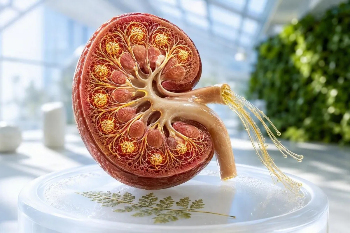
True kidney filtration normally rises by roughly 40% to 50% during pregnancy, so creatinine should fall rather than rise. A reported eGFR is often unreliable in pregnancy; the more useful signals are creatinine trend, blood pressure, and urine protein.


- True GFR usually rises by about 40% to 50% by early-to-mid pregnancy, often reaching roughly 120-150 mL/min/1.73 m² in a person with previously normal kidneys.
- eGFR in pregnancy is not validated by standard CKD-EPI or MDRD equations, so a printed eGFR should not be used alone to stage kidney disease.
- Creatinine during pregnancy is commonly about 0.4-0.7 mg/dL (35-62 µmol/L); a value at or above 0.85 mg/dL (75 µmol/L) merits clinical review.
- Preeclampsia renal criterion includes serum creatinine above 1.1 mg/dL (97 µmol/L) or a doubling from baseline when no other kidney explanation exists.
- Proteinuria of 300 mg or more in 24 hours, or a urine protein-to-creatinine ratio of 0.3 mg/mg or higher, is clinically significant when hypertension is present.
- A small rise matters because pregnancy should lower creatinine; a rise of 0.2 mg/dL (18 µmol/L) from an individual nadir can be more informative than a single lab flag.
- Urgent assessment is needed for high blood pressure, severe headache, visual change, upper abdominal pain, shortness of breath, reduced urine output, or rapidly worsening swelling.
- Postpartum retesting at about 6-12 weeks helps distinguish temporary pregnancy-related change from underlying chronic kidney disease.
What the normal range for GFR looks like in pregnancy
True GFR rises by approximately 40% to 50% in an uncomplicated pregnancy, beginning within weeks of conception and peaking around the second trimester. In practical terms, a measured GFR around 120-150 mL/min/1.73 m² can be physiologically normal in pregnancy, whereas the same value outside pregnancy would be unusually high.
There is no single universally accepted trimester-specific laboratory interval for measured GFR, partly because formal clearance testing is rarely done in healthy pregnancies. The useful clinical expectation is direction: filtration goes up, serum creatinine goes down, and the change is usually established by 12-16 weeks rather than appearing only late in pregnancy. For context on the components of a renal panel, see our blood biomarker guide.
Pregnancy increases plasma volume and renal plasma flow, which makes the glomeruli filter more water and small solutes. This is also why urea nitrogen often falls to about 7-12 mg/dL, and why a result marked low against a non-pregnant reference interval is usually not a kidney failure signal.
As Dr. Thomas Klein, I tend to ask whether the result fits the trajectory before reacting to a number in isolation. Kantesti is an AI blood test analyzer that places creatinine, urea, electrolytes, blood pressure context, and prior results on the same timeline; that matters because a creatinine of 0.75 mg/dL can be reassuring at booking but unexpectedly high after a personal pregnancy nadir of 0.45 mg/dL.
Creatinine during pregnancy: the result clinicians follow most
Serum creatinine during pregnancy is typically lower than in non-pregnant adults, often around 0.4-0.7 mg/dL (35-62 µmol/L). A creatinine of 0.85 mg/dL (75 µmol/L) or higher should prompt a clinician to check the baseline, repeat the test when appropriate, and assess urine and blood pressure.
The best available systematic review found that the upper limit of normal creatinine in healthy pregnancy is roughly 0.86 mg/dL in the first trimester, 0.81 mg/dL in the second, and 0.87 mg/dL in the third, although assay methods and local populations differ. Wiles and colleagues concluded that creatinine above 77 µmol/L, or 0.87 mg/dL, should trigger investigation rather than being dismissed as normal adult variation (Wiles et al., 2019).
One mg/dL of creatinine equals 88.4 µmol/L. That conversion is useful for people comparing US and UK reports: 0.6 mg/dL is about 53 µmol/L, while 1.1 mg/dL is about 97 µmol/L.
I have seen a 0.82 mg/dL result cause needless panic when it was stable from before conception, and I have seen a rise from 0.42 to 0.68 mg/dL expose early dehydration, urinary obstruction, or evolving hypertensive disease. The lab flag is only a starting point; our creatinine guide for women explains why personal baseline often outranks a generic range.
Why eGFR in pregnancy can mislead
Standard eGFR equations are not validated in pregnancy and can underestimate the true rise in filtration. A laboratory eGFR of 90 mL/min/1.73 m² is not automatically abnormal in pregnancy, but it should never override the actual creatinine trend and the clinical picture.
CKD-EPI and MDRD equations were derived largely in non-pregnant populations with stable creatinine production. During pregnancy, expanded plasma volume, altered tubular handling, changing body size, and lower creatinine concentration break several assumptions built into those calculations.
The UK pregnancy-and-renal-disease guideline specifically advises using serum creatinine rather than eGFR to assess renal function in pregnancy (Wiles et al., 2019). This occasionally frustrates patients who want one neat GFR number, but it is more honest medicine than presenting an unvalidated estimate with false precision.
Kantesti AI is an AI lab test interpretation service that identifies when a printed eGFR is likely to be non-actionable in pregnancy and redirects attention to creatinine, urine findings, and trend direction. Our kidney disease stages guide remains useful after pregnancy, when eGFR again becomes central to chronic kidney disease assessment; our AI technology guide describes how contextual rules are applied.
When a measured GFR or clearance test is actually useful
Measured GFR is not a routine prenatal test, but it may be considered when pre-existing kidney disease, transplant care, or major disagreement between creatinine and clinical status changes management. A 24-hour creatinine clearance is easier to obtain than isotope testing, yet it often overestimates filtration and is vulnerable to collection errors.
Creatinine clearance runs higher than true GFR because the kidney secretes some creatinine into the tubule in addition to filtering it. A reported clearance of 160 mL/min may therefore represent pregnancy physiology, incomplete collection mathematics, or both—not a diagnosis by itself.
When I order a timed collection, I ask practical questions that are surprisingly revealing: Was every urine void captured? Did the container remain cool? Was the collection started after emptying the bladder? Missing even one void can distort a 24-hour result enough to change the apparent clinical story.
For many patients, a repeat serum creatinine plus urine protein measurement is more dependable than one poorly collected clearance. Our detailed BUN and creatinine ratio review explains why urea-based ratios add little in pregnancy unless vomiting, dehydration, gastrointestinal loss, or reduced intake is also present.
How urine protein changes the interpretation of kidney function
Protein in the urine is more concerning when it occurs with high blood pressure, rising creatinine, or new symptoms after 20 weeks. Pregnancy can increase protein excretion modestly, but 300 mg or more in 24 hours remains the conventional threshold for clinically significant proteinuria.
A spot urine protein-to-creatinine ratio of 0.3 mg/mg or more is generally equivalent to 300 mg of protein per day and is accepted by ACOG as a proteinuria threshold in suspected preeclampsia. The result should be interpreted with sample concentration, urinary infection symptoms, and visible blood in urine in mind.
Albumin-to-creatinine ratio and protein-to-creatinine ratio answer slightly different questions. ACR is especially useful for diabetic or chronic kidney disease surveillance, while PCR captures non-albumin proteins and is commonly used in hypertensive pregnancy assessment.
Protein can appear transiently after fever, intense exercise, or a urinary tract infection, so repeat testing is often sensible if the person is clinically well and blood pressure is normal. Our protein in urine guide details the patterns that make a prompt urine culture or kidney work-up more likely.
The kidney pattern that raises concern for preeclampsia
Preeclampsia is suspected when new hypertension after 20 weeks occurs with proteinuria or organ involvement, including renal impairment. A serum creatinine above 1.1 mg/dL (97 µmol/L), or double the known baseline, is a severe-feature criterion when another kidney diagnosis does not explain it.
ACOG defines hypertension in pregnancy as blood pressure of at least 140/90 mmHg on two readings at least 4 hours apart; 160/110 mmHg is severe-range and needs urgent assessment. Renal impairment alone does not diagnose preeclampsia, but it changes the urgency when paired with hypertension, thrombocytopenia, liver abnormalities, neurologic symptoms, or pulmonary symptoms (ACOG, 2020).
The combination I worry about most is not a borderline creatinine alone. It is a meaningful creatinine rise plus new hypertension and rising urine protein, because together they suggest reduced renal reserve or glomerular endothelial injury rather than ordinary pregnancy hyperfiltration.
Thomas Klein, MD, has found that patients often focus on ankle swelling, although swelling by itself is common in late pregnancy and is not a diagnostic criterion. Use a reliable cuff and review our pregnancy blood pressure ranges if readings are repeated at home.
Which GFR-related results need same-day follow-up
Contact your maternity unit the same day for a rising creatinine, new proteinuria with blood pressure of 140/90 mmHg or higher, or reduced urine output. Seek emergency care immediately for blood pressure of 160/110 mmHg or higher, severe headache, visual disturbance, chest symptoms, severe upper abdominal pain, confusion, or breathlessness.
A creatinine increase of 0.2 mg/dL (18 µmol/L) from a documented pregnancy nadir is not a formal preeclampsia definition, but in my experience it deserves attention—particularly if the expected fall in creatinine has reversed. The clinician will usually repeat renal chemistry, check blood pressure, review urine, and assess volume status rather than relying on a single draw.
Kantesti AI can organize a historical result sequence, but it cannot assess fetal movement, blood pressure technique, symptoms, or the urgency visible to an obstetric team. Do not wait for a trend graph if you feel acutely unwell or your home blood pressure is severe-range.
A surprisingly common problem is assuming darker urine proves kidney failure. It far more often reflects concentration from vomiting, heat, or limited intake, but dark urine plus low output and persistent vomiting still merits a clinical call. Our checklist of same-day pregnancy lab flags can help you prepare a concise message for triage.
How pregnancy GFR is assessed with pre-existing kidney disease
In chronic kidney disease, the goal is not to reach a generic pregnancy GFR value; it is to establish an early baseline and detect a sustained creatinine rise, increasing proteinuria, or worsening blood pressure. A person with CKD may not show the same 40% to 50% rise in filtration seen in uncomplicated pregnancy.
A creatinine that remains stable at 1.0 mg/dL may be the patient's known baseline, but it is still higher than expected for a typical pregnancy and should be co-managed with obstetrics and nephrology. The risk discussion also depends on blood pressure, diabetes, kidney scarring, transplant status, and the amount of proteinuria before conception.
Pregnancy does not create chronic kidney disease from one low eGFR printout. CKD requires evidence of kidney abnormality for at least 3 months, and routine eGFR reporting cannot establish that diagnosis during pregnancy.
A useful booking record includes pre-pregnancy creatinine if available, current medication list, ACR or PCR, blood pressure, and prior imaging diagnoses. Patients with established CKD may find our chronic kidney disease overview helpful for understanding the terminology used after delivery.
Albumin-to-creatinine ratio: useful, but not interchangeable with PCR
An elevated urine albumin-to-creatinine ratio can identify glomerular stress, but it does not replace the protein-to-creatinine ratio used in many preeclampsia pathways. In non-pregnant kidney care, ACR of 30 mg/g or higher (3 mg/mmol or higher) is abnormal; pregnancy interpretation needs the clinical setting.
NICE guidance commonly uses a PCR threshold of 30 mg/mmol or an ACR threshold of 8 mg/mmol to quantify significant proteinuria in pregnant people with hypertension. Units can be confusing: 30 mg/mmol is not the same unit as 0.3 mg/mg, although both are used in clinical reporting systems.
A first-morning urine can reduce random dilution variation, but it is not mandatory for acute preeclampsia assessment. Heavy vaginal discharge, menstrual contamination, urinary infection, and gross hematuria can all affect dipstick or ratio results.
Do not try to lower a protein ratio by drinking excessive water before a retest; that can make you unwell without resolving the underlying issue. Our ACR preparation guide explains how to collect a useful sample while avoiding preventable laboratory noise.
The best kidney-function tests at the first prenatal visit
A booking renal assessment is most useful when it includes creatinine, electrolytes, urinalysis or urine protein quantification when indicated, and blood pressure. Establishing these values before 12 weeks makes later rises easier to recognize and can uncover previously silent kidney disease.
People with diabetes, chronic hypertension, lupus, a prior kidney injury, recurrent kidney stones, or a previous hypertensive pregnancy benefit most from an early creatinine and urine protein baseline. A normal creatinine at 8 weeks does not remove later preeclampsia risk, but it gives clinicians a much better reference point.
Cystatin C is not currently a routine substitute for creatinine in pregnancy. Levels tend to rise in later pregnancy despite increased measured filtration, likely from placental and inflammatory influences, so standard cystatin-C eGFR equations can also mislead.
Kantesti can retain a dated baseline alongside later results, which is especially helpful when reports come from different laboratories or countries. If you are planning conception, our pre-pregnancy blood test guide outlines the wider checks worth discussing with a clinician.
Diabetes, dehydration, and medicines that can shift kidney results
Vomiting, dehydration, hyperglycemia, urinary obstruction, and some medicines can raise creatinine during pregnancy without proving preeclampsia. The correct response is still timely assessment because dehydration and preeclampsia can coexist, and treatment choices are pregnancy-specific.
Persistent vomiting can concentrate serum creatinine and increase urea relative to creatinine, while high glucose can cause osmotic diuresis and volume depletion. Glucose should not be assumed to explain everything: diabetes also increases the chance of albuminuria and hypertensive complications.
Do not start, stop, or use over-the-counter anti-inflammatory medicines to manage a creatinine result without obstetric advice. NSAID exposure later in pregnancy can affect fetal renal physiology, and herbal mixtures marketed as kidney cleanses have unpredictable ingredients.
For gestational diabetes screening, a 75-g oral glucose tolerance test is commonly performed at 24-28 weeks in higher-risk settings; its result and kidney markers should be read separately, then together when symptoms suggest dehydration. Our pregnancy glucose tolerance guide covers practical preparation.
Why trend matters more than one GFR or creatinine result
A rising creatinine over days or weeks is more clinically meaningful in pregnancy than a single value inside a broad adult reference range. Ideally, compare results from the same laboratory, in the same units, alongside gestational age, blood pressure, and urine protein.
Normal analytical and biological variation can move creatinine by a few hundredths of a mg/dL. A shift from 0.48 to 0.52 mg/dL is often noise; a sequence of 0.46, 0.58, and 0.72 mg/dL over 3 weeks is a pattern worth discussing even if 0.72 remains near a lab cutoff.
Laboratory method changes matter more at low creatinine concentrations. Enzymatic and Jaffé assays can differ, and a sample drawn after substantial intravenous fluid can look temporarily more reassuring than the patient's usual physiology.
Kantesti's trend view can place mixed-unit results into one sequence, but clinical decisions remain with the treating team. For a practical explanation of meaningful versus trivial shifts, read our blood test variation guide.
Using AI interpretation safely for pregnancy kidney results
AI can help identify a creatinine rise, unit mismatch, or an eGFR report that should not be over-read in pregnancy, but it cannot diagnose preeclampsia or replace obstetric assessment. Any concerning kidney pattern needs confirmation through a clinician who can examine you and assess maternal and fetal wellbeing.
Kantesti is an AI biomarker interpretation platform that reads renal results against pregnancy physiology and highlights combinations such as rising creatinine, reported proteinuria, and abnormal potassium. It should be used to improve the questions you ask, not to decide whether a worrying symptom can wait.
Before uploading a report, check the collection date, gestational week, units, whether the result is serum or urine, and whether the laboratory printed an adult eGFR. A single OCR error can turn 0.60 into 0.80 mg/dL, which is why source-image verification is a clinical safety step rather than an administrative detail.
Our medical validation standards describe the clinical oversight applied to interpretation logic. For a patient-facing checklist on checking AI-generated lab explanations, see our lab report accuracy checklist.
A practical postpartum plan after abnormal kidney results
Creatinine and urine protein should be rechecked after delivery when pregnancy kidney results were abnormal, often at 6-12 weeks postpartum. Persistent creatinine elevation, albuminuria, or hypertension after that point raises the possibility of underlying kidney disease and usually warrants primary-care or nephrology follow-up.
A normal postnatal result is reassuring, but a history of preeclampsia remains relevant to long-term cardiovascular and kidney risk. Keep a copy of the highest blood pressure, peak creatinine, urine protein ratio, medication changes, and discharge plan; these details are often missing from later primary-care records.
As of August 11, 2026, the reasonable next step after a concerning renal result is not to chase a target GFR at home. It is to arrange the right follow-up interval, repeat the tests under comparable conditions, and ask whether urine microscopy, renal ultrasound, or nephrology review would change management.
Dr. Thomas Klein recommends bringing a one-page trend summary to the postnatal visit, particularly after hypertension or proteinuria. Kantesti's clinical content is reviewed with input from our Medical Advisory Board, but your obstetric and renal clinicians remain the people who can tailor follow-up to your individual pregnancy.
Frequently Asked Questions
What is a normal GFR in pregnancy?
True GFR normally increases by about 40% to 50% during an uncomplicated pregnancy, often reaching approximately 120-150 mL/min/1.73 m² by the second trimester in someone with previously normal kidney function. There is no single validated trimester-specific GFR reference range used by all laboratories. Standard calculated eGFR values are unreliable in pregnancy, so clinicians usually follow serum creatinine, which commonly falls to about 0.4-0.7 mg/dL (35-62 µmol/L).
Is an eGFR of 90 normal during pregnancy?
An eGFR of 90 mL/min/1.73 m² on a routine laboratory report should not be interpreted as a definitive kidney-function result during pregnancy. CKD-EPI and MDRD eGFR equations are not validated for pregnancy and may fail to reflect the expected rise in true filtration. A creatinine trend, blood pressure, urine protein result, and symptoms carry more clinical weight than an isolated eGFR of 90.
What creatinine level is too high in pregnancy?
A creatinine at or above about 0.85 mg/dL (75 µmol/L) is higher than expected in pregnancy and generally deserves clinical review, particularly if it is increasing. ACOG considers creatinine above 1.1 mg/dL (97 µmol/L) or doubling from baseline a severe renal feature in suspected preeclampsia when another cause is not present. A smaller increase of 0.2 mg/dL (18 µmol/L) can still matter because creatinine should usually decrease during pregnancy.
Can pregnancy make GFR go up?
Yes. Pregnancy normally increases renal plasma flow and true GFR by roughly 40% to 50%, beginning early and often peaking in mid-pregnancy. This physiologic hyperfiltration lowers creatinine and urea compared with non-pregnant values. A high measured GFR without proteinuria, hypertension, or a rising creatinine is usually expected physiology rather than kidney disease.
Does low eGFR in pregnancy mean preeclampsia?
A low printed eGFR does not by itself mean preeclampsia because routine eGFR equations are inaccurate during pregnancy. Preeclampsia is assessed through new hypertension after 20 weeks plus proteinuria or organ involvement, including creatinine above 1.1 mg/dL (97 µmol/L) or a doubling from baseline. New blood pressure of 140/90 mmHg or higher with rising creatinine or urine protein should be reviewed promptly by a maternity clinician.
When should kidney tests be repeated after pregnancy?
Kidney tests are commonly repeated around 6-12 weeks postpartum after elevated creatinine, proteinuria, hypertension, preeclampsia, or known kidney disease. The recheck usually includes creatinine, eGFR once pregnancy has ended, blood pressure, and a urine ACR or PCR when protein was present. Persistent proteinuria, hypertension, or reduced eGFR beyond 3 months requires assessment for chronic kidney disease rather than being attributed to pregnancy alone.
Get AI-Powered Blood Test Analysis Today
Join over 2 million users worldwide who trust Kantesti for instant, accurate lab test analysis. Upload your blood test results and receive comprehensive interpretation of 15,000+ biomarkers in seconds.
📚 Referenced Research Publications
Klein, T., Mitchell, S., & Weber, H. (2026). BUN/Creatinine Ratio Explained: Kidney Function Test Guide. Kantesti AI Medical Research.
Klein, T., Mitchell, S., & Weber, H. (2026). Urobilinogen in Urine Test: Complete Urinalysis Guide 2026. Kantesti AI Medical Research.
📖 External Medical References
⚕️ Medical Disclaimer
This article is for educational purposes only and does not constitute medical advice. Always consult a qualified healthcare provider for diagnosis and treatment decisions.
E-E-A-T Trust Signals
Experience
Physician-led clinical review of lab interpretation workflows.
Expertise
Laboratory medicine focus on how biomarkers behave in clinical context.
Authoritativeness
Written by Dr. Thomas Klein with review by Dr. Sarah Mitchell and Prof. Dr. Hans Weber.
Trustworthiness
Evidence-based interpretation with clear follow-up pathways to reduce alarm.