What Does High MCHC Mean? Causes, Artifacts and Clues

カテゴリー
記事
CBCの読み方 血液学 2026年の更新 患者さん向け

A high MCHC is uncommon and often more useful as a clue than as a diagnosis. The practical question is whether red cells are genuinely dense and spherical, or whether the sample distorted the calculation.

📖 約11分 📅
📝 公開: 🩺 医学的監修: ✅ エビデンスに基づく
⚡ 簡単な概要 v1.0 —
  1. MCHC range is usually 32-36 g/dL (320-360 g/L) in adults, although the laboratory’s own range takes priority.
  2. High MCHC above 36 g/dL is unusual and should prompt a check for spherocytes, hemolysis, burns, or analytical interference.
  3. Spherocytes are small, dense red cells without central pallor and can occur in hereditary spherocytosis or autoimmune hemolytic anemia.
  4. 冷式凝集素 can falsely raise MCHC by clumping red cells, lowering the measured RBC count, and distorting calculated indices.
  5. Lipemia can overestimate hemoglobin on some analyzers, creating an elevated MCHC even when red-cell shape is normal.
  6. Burn injury can produce true high MCHC through red-cell membrane loss, usually alongside anemia and laboratory evidence of hemolysis.
  7. Hemolysis clues include raised reticulocytes, indirect bilirubin and LDH, with low haptoglobin when red cells are breaking down in the body.
  8. 再検査 with a freshly collected, properly handled EDTA sample is often the first sensible step when MCHC is isolated and only mildly high.

High MCHC on a CBC: the direct answer

High MCHC usually means red cells are unusually dense, but it can also mean the analyzer was misled by the sample. In adults, an MCHC above about 36 g/dL deserves context: true elevations most often point toward spherocytes or significant thermal injury, while cold agglutinins, lipemia, and sample hemolysis are frequent laboratory explanations. As of August 23, 2026, I would not diagnose a blood disorder from this index alone.

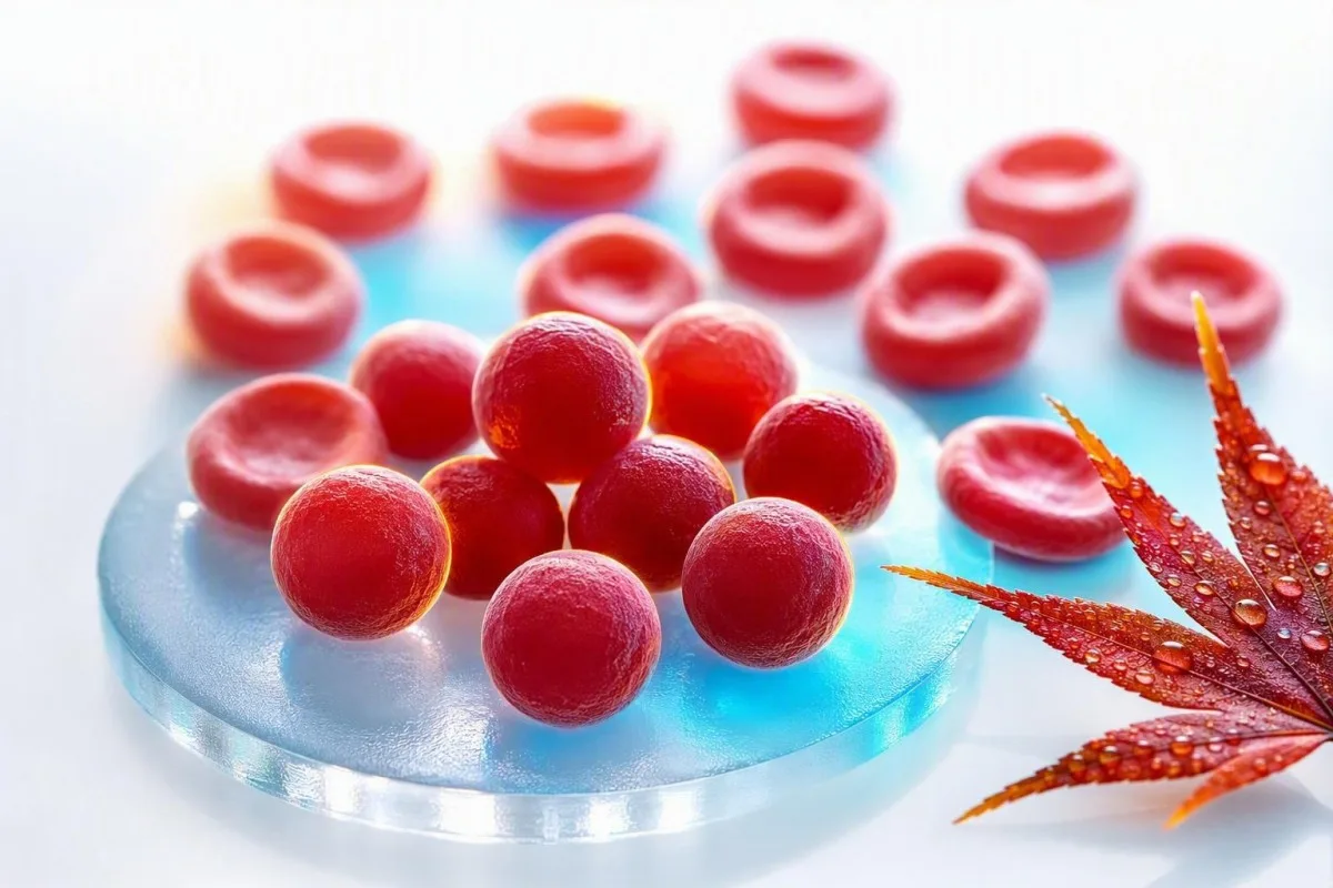
What does high MCHC mean shown by dense spherical red cellular elements in a clinical illustration
図1: Dense spherical cellular elements illustrate the core meaning of a truly raised MCHC.

MCHC is calculated as hemoglobin divided by hematocrit, multiplied by 100, so it is a concentration rather than a direct cell measurement. A hemoglobin of 15.0 g/dL and hematocrit of 40% produces an MCHC of 37.5 g/dL; the number can rise because hemoglobin is over-read, hematocrit is under-read, or both. Our CBC biomarker guide is useful when several indices are flagged at once.

When I review a panel with MCHC 36.5-37.5 g/dL but normal hemoglobin, bilirubin, reticulocytes, and RDW, an artifact is often more likely than disease. An MCHC of 39-42 g/dL is physiologically difficult to sustain in intact circulating red cells, so the laboratory should actively consider interference before anyone panics.

カンテスティは AI血液検査分析装置 that reads MCHC alongside hemoglobin, hematocrit, MCV, RDW, bilirubin, LDH, and prior results rather than treating one red flag as a diagnosis. Dr. Thomas Klein’s clinical rule is simple: verify the arithmetic and the specimen before ordering an expansive work-up; our detailed RDWおよび赤血球指数ガイド explains why this pattern-based approach matters.

からレビュー用にアップロードできます。 32〜36 g/dL Expected hemoglobin concentration within red-cell volume.
軽度に高い 36.1-37.5 g/dL Review the full CBC, sample quality, and prior values.
明らかに高い 37.6-39.0 g/dL Consider spherocytes, hemolysis, cold agglutinins, or lipemia.
著しく高い >39.0 g/dL Analyzer or specimen interference is especially likely; request laboratory review.

MCHC is not MCH or MCV

MCHC describes hemoglobin concentration inside the packed red-cell volume, whereas MCH describes hemoglobin amount per cell and MCV describes cell size. Confusing these three indices is one reason a routine CBC flag can sound more alarming than it is.

What does high MCHC mean compared with cell size and hemoglobin concentration in cellular elements
図2: Cell size, hemoglobin amount, and concentration are separate CBC measurements.

MCH is commonly 27-33 pg per cell, while MCV is commonly 80-100 fL in adults; neither range defines MCHC. Large red cells can carry more hemoglobin and therefore raise MCH, yet their MCHC may remain normal because their volume increased proportionally. See our practical comparison of MCVとMCH if both values are flagged.

A genuine high MCHC often pairs with a normal or low-normal MCV because spherocytes have lost membrane surface area and become rounder, not because they contain limitless hemoglobin. That geometry matters: a sphere holds less surface area for its volume, so it has no pale center on a well-prepared cell sample slide.

The thing is, an elevated MCHC blood test can be mathematically real while biologically misleading. A slightly underestimated hematocrit after red-cell clumping will inflate the ratio even if the patient’s red cells are not dense at all; this is why a full blood count review should precede conclusions.

Spherocytes: the classic true high-MCHC clue

Spherocytes are the most recognizable cause of a true high MCHC because membrane loss makes red cells compact and densely hemoglobinized. They are seen in hereditary spherocytosis and autoimmune hemolytic anemia, but a smear finding must be interpreted with symptoms and hemolysis markers.

What does high MCHC mean with spherical red cellular elements lacking central pallor on a cell slide
図3: Spherical cellular elements can explain a genuine MCHC elevation.

Hereditary spherocytosis often produces MCHC values above 36 g/dL, reticulocytosis, intermittent jaundice, gallstones, splenomegaly, or a family history of anemia. The 2011 British Society for Haematology guideline notes that MCHC supports the diagnosis but is not sufficiently sensitive or specific to confirm it by itself (Bolton-Maggs et al., 2012).

In autoimmune hemolytic anemia, antibodies mark red cells for partial membrane removal in the spleen, leaving spherocytes behind. A direct antiglobulin test, reticulocyte count, bilirubin, LDH, and haptoglobin are more informative together than any isolated index; our guide to low haptoglobin beyond hemolysis covers an easy-to-miss limitation.

One clinical wrinkle: recent transfusion can dilute or mask a patient’s native spherocyte pattern for several weeks. Christensen et al. describe eosin-5-maleimide binding by flow cytometry as a useful test for suspected hereditary spherocytosis, although a hematologist should select and time testing carefully (Christensen et al., 2015).

How to tell if high MCHC comes with hemolysis

High MCHC becomes more clinically meaningful when it accompanies anemia, a raised reticulocyte count, indirect bilirubin elevation, LDH elevation, and low haptoglobin. That cluster indicates increased red-cell turnover, although no single marker is perfect.

What does high MCHC mean alongside bilirubin LDH and reticulocyte laboratory pattern elements
図4: Hemolysis is identified by a pattern of turnover markers, not MCHC alone.

A reticulocyte percentage above 2% can still be inadequate in significant anemia, so clinicians often calculate an absolute reticulocyte count or reticulocyte production index instead. For example, a 4% reticulocyte count with hematocrit 24% may represent only a modest marrow response after correction; our article on high reticulocyte clues walks through the distinction.

Unconjugated bilirubin rises when heme is processed faster than the liver can conjugate it, while LDH rises because red cells contain abundant LDH. Haptoglobin below the laboratory lower limit supports intravascular hemolysis, but it may appear normal during inflammation because haptoglobin is an acute-phase protein. Barcellini and Fattizzo discuss this diagnostic trap in their review of autoimmune hemolytic anemia (Barcellini & Fattizzo, 2015).

Dark urine, rapidly increasing fatigue, breathlessness at rest, chest pain, fainting, or yellow eyes with fever merit same-day clinical assessment. Fragmented cells are a different and more urgent smear clue than spherocytes; our explanation of シストサイトが緊急に見直しを必要とする場合 helps clarify why.

Why burns can cause a genuinely elevated MCHC

Extensive thermal injury can cause true high MCHC because heat damages red-cell membranes, producing membrane loss, spherocyte-like cells, and hemolysis. This is generally a hospital finding after clinically apparent burns, not an explanation for an incidental outpatient CBC result.

What does high MCHC mean in thermal membrane injury shown as dense altered cellular elements
図5: Thermal membrane injury can create dense cells and accelerate red-cell turnover.

Thermal damage may create microspherocytes within hours of major burns, and the MCHC can rise as the membrane-to-volume ratio falls. Hemoglobin can also fall over the following days from fluid shifts, procedures, phlebotomy, and ongoing hemolysis, so a single MCHC cannot quantify injury severity.

I have occasionally seen patients focus on an MCHC of 37.1 g/dL after an emergency admission while overlooking much more actionable abnormalities: falling hemoglobin, rising creatinine, potassium changes, or lactate. In that setting, clinicians manage the person and burn physiology, not the index; our post-hospital test timeline explains why values can move quickly.

Kantesti AIは、 AIバイオマーカー解釈プラットフォーム that can flag a high MCHC alongside dynamic hemoglobin and kidney results, but acute burns require direct hospital assessment and cannot be safely managed from a report interpretation. A result after a severe burn should be compared against the prior 24-72 hours, not against a generic population average.

Cold agglutinins: the high-MCHC artifact doctors look for

Cold agglutinins can produce a spurious high MCHC by causing red cells to clump at room temperature before analysis. The analyzer may count several attached cells as one particle, leading to a falsely low RBC count, falsely high MCV, and an inflated calculated MCHC.

What does high MCHC mean when cold-related cellular clumping affects automated CBC analysis
図6: Cold-related clumping can distort several calculated CBC indices at once.

The classic cold-agglutinin pattern is a low RBC count with disproportionately high MCV, high MCH, and high MCHC, often without a matching clinical story. Warming the EDTA tube to 37°C and rerunning the CBC commonly corrects the values, which is far more informative than repeating a random test weeks later.

Cold agglutinins can occur transiently after some respiratory infections or persist in cold agglutinin disease and certain lymphoproliferative disorders. Symptoms such as color change or discomfort in fingers with cold exposure add clinical weight, but the laboratory pattern itself can be the first clue; a repeat test after hemolysis offers practical redraw questions.

Do not warm yourself aggressively or try to manipulate the result at home. Ask whether the laboratory saw red-cell agglutination, whether the specimen was warmed and rerun, and whether a manual hematocrit was performed; those three answers often settle the issue.

Lipemia and sample hemolysis can distort MCHC

Lipemia and in-vitro sample hemolysis can falsely raise MCHC because many analyzers measure hemoglobin photometrically after red cells are lysed. Turbid lipids or free hemoglobin in the sample can increase optical absorbance and make hemoglobin appear higher than it really is.

What does high MCHC mean when a cloudy laboratory sample changes hemoglobin optical measurement
図7: Sample turbidity can interfere with optical hemoglobin measurement and inflate MCHC.

Lipemia is most often visible after very high triglycerides, usually above about 400-500 mg/dL, though interference depends on the instrument and assay wavelength. A laboratory may use a plasma blank, alternative wavelength, or plasma replacement approach rather than simply accepting the automated hemoglobin result.

In-vitro hemolysis happens after collection or handling, not inside the body, and it can affect more than MCHC. Potassium, LDH, AST, and phosphate may rise in a damaged sample, which is why an isolated high MCHC alongside an unexpected high potassium needs a specimen-quality check; see our guide to hemolysis-related potassium errors.

A visibly pink or red plasma layer is a laboratory clue, not proof of hemolytic anemia in the patient. The practical follow-up is a fresh collection with calm handling and prompt processing, especially if the original report includes a hemolysis index or a comment that results may be affected.

Collection, storage, and tube problems that change CBC indices

Poor sample mixing, underfilling, clotting, and delayed analysis can alter CBC indices, although delayed storage more often lowers rather than raises MCHC by allowing cells to swell. A high MCHC should therefore be read with the laboratory’s specimen comments, not merely the number on the patient portal.

What does high MCHC mean when EDTA sample handling and analyzer preparation require verification
図8: Correct EDTA collection and prompt processing protect the reliability of CBC indices.

CBC samples are generally collected in EDTA tubes, and an underfilled tube can have an excessive anticoagulant-to-sample ratio that changes cellular measurements. Small clots can selectively remove cells from analysis and make the automated count internally inconsistent; our overview of blood tube additives explains why tube type is not a trivial detail.

Storage at room temperature for prolonged periods causes red cells to take up water, raising MCV and tending to decrease MCHC. That direction of change is useful: a very high MCHC after a delayed sample is not automatically explained by age alone, so cold agglutination or optical interference may still be present.

A capillary sample can be appropriate in some settings but is more prone to collection variability than a well-collected venous EDTA specimen. If a high MCHC has no matching symptoms or related abnormalities, I usually prefer one clean venous redraw before treating it as a new chronic diagnosis.

Conditions commonly blamed on MCHC but rarely responsible

Dehydration, iron deficiency, vitamin B12 deficiency, and ordinary stress do not usually cause a truly high MCHC. They can change other CBC values, but they should not be used as convenient explanations for an MCHC of 38 g/dL without confirming the specimen.

What does high MCHC mean compared with common anemia patterns on a clinical cellular illustration
図10: Common nutrient-related anemia patterns usually do not create high MCHC.

Iron deficiency typically lowers MCH and MCHC and often lowers MCV once deficiency is established. Early iron deficiency may have a normal MCV, but it does not explain hyperchromic indices; our explanation of トランスフェリン飽和度の低下 helps separate iron availability from CBC concentration.

Vitamin B12 or folate deficiency more often causes macrocytosis, with MCV above 100 fL, rather than high MCHC. If MCV and MCH are both high but MCHC is normal, that is a different diagnostic direction; see high MCV follow-up clues.

G6PD deficiency causes episodic hemolysis after specific oxidative triggers in susceptible people, but MCHC is not a dependable screening test for it. During an acute episode, the smear and turnover markers can be more revealing, while enzyme testing may need repeating after recovery because young red cells contain more G6PD.

The most useful follow-up tests after elevated MCHC

The best next step for an isolated high MCHC is usually a repeat CBC with laboratory review, followed by a peripheral smear and hemolysis panel only if the elevation persists or other clues are present. Ordering every anemia test immediately can create more noise than answers.

What does high MCHC mean when a repeat CBC smear and hemolysis workup are arranged
図11: Repeat testing and a cell sample slide clarify whether high MCHC is genuine.

A focused first-line review includes repeat CBC, cell sample slide review, reticulocyte count, total and indirect bilirubin, LDH, haptoglobin, and direct antiglobulin testing when autoimmune hemolysis is plausible. The smear can identify spherocytes, agglutination, fragments, polychromasia, or a normal morphology that makes an artifact more likely.

If hereditary spherocytosis remains likely, a hematologist may consider eosin-5-maleimide binding, osmotic fragility testing, or genetic testing. These tests have different false-negative risks after transfusion and during acute hemolysis, which is why the test order should follow a clinical history rather than a checkbox list; our G6PD testing guide illustrates the broader timing problem.

Kantesti AI interprets high MCHC by checking the calculation against related CBC and hemolysis markers, then identifying questions to take to a clinician. It does not replace cell morphology review, particularly when the laboratory has issued an analyzer flag or when anemia is worsening.

When high MCHC needs urgent medical assessment

High MCHC itself is not an emergency threshold, but high MCHC with signs of acute hemolysis, severe anemia, or a serious burn can require same-day care. Symptoms and the rate of change determine urgency far better than whether the flag is marked H.

What does high MCHC mean when urgent anemia warning symptoms require clinical assessment
図12: Urgency depends on symptoms and hemolysis patterns rather than MCHC alone.

Seek urgent assessment for new shortness of breath at rest, chest pain, fainting, confusion, rapidly worsening weakness, tea-colored urine, or yellowing of the eyes with fever. These symptoms can accompany significant anemia or hemolysis, especially when hemoglobin has fallen by more than 2 g/dL from a known baseline.

Contact the clinician who ordered the test within days if MCHC remains above 36 g/dL on a repeat sample, anemia is present, or the report mentions agglutination or spherocytes. In a stable, well person with MCHC 36.2 g/dL and otherwise normal results, a non-urgent redraw is often reasonable; our ビリルビンのパターンガイド can help frame the next discussion.

Do not start iron, folate, steroids, or supplements simply to correct MCHC. None treats the main true causes, and steroids in particular can obscure the assessment of autoimmune hemolysis if begun without clinical supervision.

How Kantesti places MCHC in clinical context

Kantesti reads a high MCHC as a pattern-recognition problem: true dense-cell disorders and sample artifacts produce different combinations of CBC, chemistry, flags, and prior trends. The aim is to make the next clinical question clearer, not to label a rare disease from one calculated index.

What does high MCHC mean interpreted through connected CBC and hemolysis laboratory patterns
図13: Contextual reading separates an isolated index flag from a coherent red-cell pattern.

カンテスティは AI搭載の血液検査解析ツール used by more than 2 million people across 127 countries to organize laboratory results in context. For high MCHC, our AI checks whether hemoglobin and hematocrit mathematically support the result, whether MCV is unexpectedly high, and whether bilirubin, LDH, reticulocytes, or haptoglobin point toward red-cell turnover.

Our platform also looks for internal contradictions: MCHC 40 g/dL with a normal smear comment and no anemia may warrant an artifact prompt, while MCHC 37 g/dL with spherocytes, reticulocytosis, and indirect bilirubin elevation deserves medical follow-up. The technical approach and limits are described in our AIテクノロジーガイド.

I advise patients to save the lab PDF, collection date, fasting status, recent illness, transfusion history, and any laboratory comments. That small record often explains an apparent discrepancy faster than repeating broad testing, especially when results are compared months later.

Questions that make a high-MCHC appointment more productive

Ask whether the MCHC was repeated, whether the lab saw spherocytes or agglutination, and whether anemia or hemolysis markers are present. These questions shift the conversation from “why is one number high?” to a concrete, testable clinical pathway.

What does high MCHC mean during a clinician discussion reviewing CBC trends and sample quality
図14: Focused questions help turn a CBC flag into an efficient clinical plan.

Useful wording includes: “Was the sample hemolyzed or lipemic?”, “Did the analyzer flag red-cell agglutination?”, and “Can we compare this result with my previous MCHC?” A manual smear review is particularly valuable when MCHC is repeatedly above 36 g/dL or anemia is unexplained.

Dr. Thomas Klein recommends taking a list of medicines, recent infections, cold-triggered symptoms, family history of gallstones or anemia, and transfusions within the previous 3 months. That history may point toward hereditary spherocytosis, autoimmune hemolysis, or a transient cold agglutinin far faster than a generic online search.

Kantesti’s medical content is reviewed with physician oversight, but your own clinician can examine you, request a warmed rerun, and speak directly with the laboratory. Readers who want to understand our clinical review framework can meet the 医療諮問委員会を, while persistent or symptomatic abnormalities should always be discussed in person.

Research, uncertainty, and the limits of a single MCHC

MCHC is a useful screening clue, not a standalone diagnostic test for hereditary spherocytosis, autoimmune hemolysis, burns, or cold agglutinin disease. Its value rises sharply when a repeat sample, cell morphology, turnover markers, and clinical history all tell the same story.

No universal MCHC cutoff diagnoses hereditary spherocytosis because analyzer methods, age, reticulocytosis, and coexisting iron deficiency can alter sensitivity. The BSH guideline supports combining family history, smear findings, reticulocytes, bilirubin, and confirmatory testing where needed rather than relying on a single threshold (Bolton-Maggs et al., 2012).

Kantesti supports informed follow-up by identifying result patterns and uncertainty, not by issuing a definitive hematology diagnosis. Our 臨床的妥当性の検証アプローチ explains why unusual values, analyzer flags, and emergency symptoms remain reasons for human laboratory and clinician review.

For related hematology context, see the research publication B陰性血液型、LDH血液検査、網赤血球数ガイド and the broader clinical communication resource 断食後の下痢、便の黒い斑点、消化管ガイド2026. Both are supplementary educational publications rather than substitutes for a peripheral smear or specialist assessment.

よくある質問

What does a high MCHC level mean on a blood test?

A high MCHC level usually means the hemoglobin concentration within red-cell volume is above the laboratory range, commonly above 36 g/dL in adults. True elevations occur with spherocytes from hereditary spherocytosis, autoimmune hemolytic anemia, or major thermal injury. Cold agglutinins, lipemia, and sample hemolysis can also create a false high result. A repeat CBC, cell sample slide review, and hemolysis markers clarify which explanation fits.

Is high MCHC dangerous?

High MCHC is not dangerous by itself; it is a calculated CBC index rather than a disease. An MCHC of 36.2 g/dL with normal hemoglobin and no symptoms is often handled with a routine repeat test, while high MCHC plus falling hemoglobin, jaundice, dark urine, or breathlessness needs prompt assessment. Values above 39 g/dL are especially likely to reflect specimen or analyzer interference. The clinical risk comes from the underlying cause, such as significant hemolysis, not the MCHC number alone.

Can dehydration cause high MCHC?

Dehydration does not usually cause a truly high MCHC because it concentrates plasma volume more than it changes hemoglobin concentration inside individual red cells. Dehydration can raise hemoglobin and hematocrit together, leaving MCHC near its usual 32-36 g/dL range. If MCHC is 37-40 g/dL, clinicians should consider spherocytes, cold agglutination, lipemia, or a measurement issue instead. A repeat well-collected CBC is more useful than trying to correct the value with extra fluids.

Do spherocytes always make MCHC high?

Spherocytes often raise MCHC above 36 g/dL, but they do not always do so and high MCHC does not prove spherocytes are present. Hereditary spherocytosis may be masked by iron deficiency, recent transfusion, or mild disease, while autoimmune hemolysis may show variable findings depending on timing. A cell sample slide reviewed by an experienced laboratory professional is needed to identify spherocytes. Reticulocyte count, bilirubin, LDH, haptoglobin, and direct antiglobulin testing add diagnostic context.

Can cold agglutinins cause high MCHC?

Cold agglutinins can falsely raise MCHC by clumping red cells before the automated CBC is measured. The pattern often includes a low RBC count, high MCV, high MCH, and an MCHC above 36 g/dL that does not fit the patient’s symptoms. Warming the EDTA specimen to 37°C and rerunning it can normalize the counts. Persistent cold agglutination or hemolysis symptoms should be assessed by a clinician or hematologist.

What tests should I ask for after high MCHC?

After a persistent high MCHC, the usual next tests are a repeat CBC, cell sample slide review, reticulocyte count, bilirubin fractions, LDH, haptoglobin, and sometimes a direct antiglobulin test. A clinician may request eosin-5-maleimide binding or other specialized testing if hereditary spherocytosis is suspected. MCHC above 36 g/dL with normal repeat testing and no anemia may need no extensive investigation. The best test sequence depends on symptoms, prior CBC values, family history, and laboratory flags.

今日、AIによる血液検査分析を

いますぐ利用しませんか。即時で正確な検査分析を提供するKantestiを信頼する、世界中の200万人以上のユーザーに参加してください。血液検査結果をアップロードすると、15,000+のバイオマーカーについて数秒で包括的な解釈が得られます。.

📚 Referenced Research Publications

1

Klein, T., Mitchell, S., & Weber, H. (2026). B型(Rh陰性)血液型、LDH血液検査、網赤血球数ガイド.。 Kantesti AI Medical Research.

2

Klein, T., Mitchell, S., & Weber, H. (2026). 断食後の下痢、便の黒い斑点、消化管ガイド2026.。 Kantesti AI Medical Research.

📖 外部の医学的参考文献

3

Bolton-Maggs PHB ほか(2012年)。. 遺伝性球状赤血球症の診断と管理のためのガイドライン—2011年の更新.。 英国血液学雑誌。.

4

Christensen RD et al. (2015). The diagnostic protocol for hereditary spherocytosis—2013 update.。.

5

Barcellini W, Fattizzo B(2015)。. The diagnosis and management of autoimmune hemolytic anemia in adults. Transfusion Medicine and Hemotherapy.

200万以上分析されたテスト
127+
75+言語

⚕️ 医療免責事項

E-E-A-T 信頼性シグナル

経験

医師主導による、検査結果解釈ワークフローの臨床レビュー。.

📋

専門知識

臨床的な文脈においてバイオマーカーがどのように振る舞うかに焦点を当てた検査医学。.

👤

権威

トーマス・クライン博士が執筆し、サラ・ミッチェル博士およびハンス・ヴェーバー教授によるレビュー。.

🛡️

信頼性

アラームを減らすための明確なフォローアップ経路を備えた、エビデンスに基づく解釈。.

🏢 カンテスティ株式会社 イングランドおよびウェールズに登録 · 会社番号. 17090423 ロンドン、イギリス · kantesti.net
blank
Prof. Dr. Thomas Kleinによる

トーマス・クライン博士は、Kantesti AIにおける最高医療責任者(CMO)を務める、ボード認定の臨床血液専門医です。検査医学における15年以上の経験に加え、「血液検査結果」のAI支援による解釈に強い関心を持ち、新しい技術を日常の臨床実践につなげることに取り組んでいます。関心領域には、バイオマーカー解析、臨床意思決定支援の研究、集団特異的な基準範囲の最適化が含まれます。CMOとして、同プラットフォームの内部ベンチマークに対する臨床的インプットを提供し、Kantestiの教育レポートの医療品質に関する臨床的監督を行います。.

コメントを残す

メールアドレスが公開されることはありません。 が付いている欄は必須項目です
