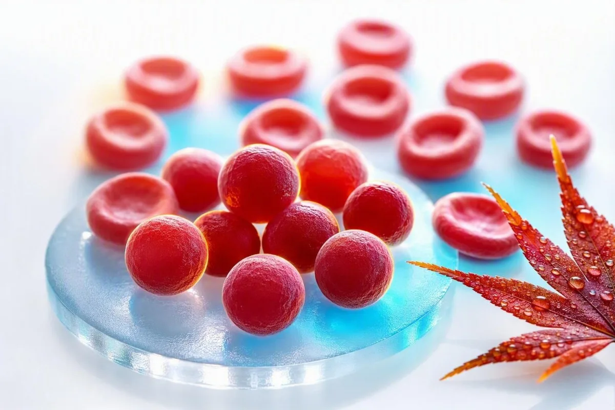
Un MCHC alto es poco común y a menudo es más útil como una pista que como un diagnóstico. La pregunta práctica es si los glóbulos rojos son genuinamente densos y esféricos, o si la muestra distorsionó el cálculo.


- Rango de MCHC suele ser de 32-36 g/dL (320-360 g/L) en adultos, aunque el rango propio del laboratorio tiene prioridad.
- MCHC alto por encima de 36 g/dL es inusual y debe provocar una búsqueda de esferocitos, hemólisis, quemaduras o interferencia analítica.
- Esferocitos son glóbulos rojos pequeños y densos sin palidez central y pueden ocurrir en la esferocitosis hereditaria o la anemia hemolítica autoinmune.
- aglutininas frías pueden elevar falsamente el MCHC al agrupar los glóbulos rojos, disminuir el recuento de RBC medido y distorsionar los índices calculados.
- Lipemia puede sobreestimar la hemoglobina en algunos analizadores, creando un MCHC elevado incluso cuando la forma del glóbulo rojo es normal.
- Quemadura puede producir un MCHC verdaderamente alto debido a la pérdida de membrana de los glóbulos rojos, generalmente junto con anemia y evidencia de laboratorio de hemólisis.
- Pistas de hemólisis incluyen reticulocitos elevados, bilirrubina indirecta y LDH, con haptoglobina baja cuando los glóbulos rojos se descomponen en el cuerpo.
- Repetir la prueba con una muestra de EDTA recién recolectada y manejada adecuadamente es a menudo el primer paso sensato cuando el MCHC está aislado y solo ligeramente alto.
MCHC alto en un CBC: la respuesta directa
Un MCHC alto generalmente significa que los glóbulos rojos son inusualmente densos, pero también puede significar que el analizador fue engañado por la muestra. En adultos, un MCHC superior a aproximadamente 36 g/dL merece contexto: las elevaciones verdaderas apuntan con mayor frecuencia a esferocitos o a una lesión térmica significativa, mientras que los aglutininos fríos, la lipemia y la hemólisis de la muestra son explicaciones frecuentes en laboratorio. A partir del 23 de agosto de 2026, no diagnosticaría un trastorno sanguíneo basándome solo en este índice.
El MCHC se calcula como hemoglobina dividida por hematocrito, multiplicado por 100, por lo que es una concentración en lugar de una medida celular directa. Una hemoglobina de 15.0 g/dL y un hematocrito de 40% producen un MCHC de 37.5 g/dL; el número puede aumentar porque la hemoglobina se lee en exceso, el hematocrito se lee en defecto, o ambos. Nuestra guía de biomarcadores de CBC es útil cuando varios índices se marcan a la vez.
Cuando reviso un panel con MCHC de 36.5-37.5 g/dL pero hemoglobina, bilirrubina, reticulocitos y RDW normales, un artefacto es a menudo más probable que una enfermedad. Un MCHC de 39-42 g/dL es fisiológicamente difícil de mantener en glóbulos rojos intactos en circulación, por lo que el laboratorio debe considerar activamente la interferencia antes de que alguien entre en pánico.
Kantesti es un Analizador de sangre con inteligencia artificial que lee el MCHC junto con la hemoglobina, el hematocrito, el MCV, el RDW, la bilirrubina, la LDH y los resultados previos en lugar de tratar una señal de alerta como un diagnóstico. La regla clínica del Dr. Thomas Klein es simple: verificar la aritmética y la muestra antes de solicitar un estudio exhaustivo; nuestro detallado Guía de RDW y de índices eritrocitarios explica por qué este enfoque basado en patrones es importante.
MCHC no es MCH ni MCV
El MCHC describe la concentración de hemoglobina dentro del volumen de glóbulos rojos empaquetados, mientras que el MCH describe la cantidad de hemoglobina por célula y el MCV describe el tamaño de la célula. Confundir estos tres índices es una razón por la que una bandera de rutina de CBC puede sonar más alarmante de lo que es.
El MCH suele ser de 27-33 pg por célula, mientras que el MCV suele ser de 80-100 fL en adultos; ningún rango define el MCHC. Los glóbulos rojos grandes pueden transportar más hemoglobina y, por lo tanto, aumentar el MCH, pero su MCHC puede permanecer normal porque su volumen aumentó proporcionalmente. Ver nuestra comparación práctica de MCV y MCH si ambos valores están marcados.
Un MCHC alto genuino a menudo se asocia con un MCV normal o bajo-normal porque los esferocitos han perdido área de superficie de membrana y se han vuelto más redondos, no porque contengan hemoglobina ilimitada. Esa geometría importa: una esfera contiene menos área de superficie para su volumen, por lo que no tiene un centro pálido en una muestra celular bien preparada.
El caso es que un análisis de sangre de MCHC elevado puede ser matemáticamente real y biológicamente engañoso. Un hematocrito ligeramente subestimado después de la aglutinación de glóbulos rojos inflará la relación incluso si los glóbulos rojos del paciente no son densos en absoluto; es por eso que una revisión completa del recuento sanguíneo debe preceder a las conclusiones.
Esferocitos: la pista clásica de MCHC alto real
Los esferocitos son la causa más reconocible de un MCHC alto verdadero porque la pérdida de membrana hace que los glóbulos rojos sean compactos y densamente hemoglobínicos. Se observan en la esferocitosis hereditaria y la anemia hemolítica autoinmune, pero un hallazgo de frotis debe interpretarse con los síntomas y los marcadores de hemólisis.
La esferocitosis hereditaria a menudo produce valores de MCHC superiores a 36 g/dL, reticulocitosis, ictericia intermitente, cálculos biliares, esplenomegalia o antecedentes familiares de anemia. La guía de la British Society for Haematology de 2011 señala que el MCHC apoya el diagnóstico pero no es lo suficientemente sensible ni específico para confirmarlo por sí solo (Bolton-Maggs et al., 2012).
En la anemia hemolítica autoinmune, los anticuerpos marcan los glóbulos rojos para la eliminación parcial de la membrana en el bazo, dejando esferocitos. Una prueba de antiglobulina directa, recuento de reticulocitos, bilirrubina, LDH y haptoglobina son más informativos juntos que cualquier índice aislado; nuestra guía sobre haptoglobina baja más allá de la hemólisis cubre una limitación que se pasa por alto fácilmente.
Una complicación clínica: una transfusión reciente puede diluir o enmascarar el patrón de esferocitos nativos de un paciente durante varias semanas. Christensen et al. describen la unión de eosin-5-maleimida mediante citometría de flujo como una prueba útil para la esferocitosis hereditaria sospechada, aunque un hematólogo debe seleccionar y programar las pruebas cuidadosamente (Christensen et al., 2015).
Cómo saber si el MCHC alto viene con hemólisis
Un MCHC alto se vuelve más clínicamente significativo cuando acompaña a anemia, un recuento de reticulocitos elevado, elevación de bilirrubina indirecta, elevación de LDH y haptoglobina baja. Ese grupo indica un aumento de la rotación de glóbulos rojos, aunque ningún marcador único es perfecto.
Un porcentaje de reticulocitos superior al 2% aún puede ser inadecuado en anemia significativa, por lo que los médicos a menudo calculan un recuento absoluto de reticulocitos o un índice de producción de reticulocitos en su lugar. Por ejemplo, un recuento de reticulocitos de 4% con un hematocrito de 24% puede representar solo una respuesta medular modesta después de la corrección; nuestro artículo sobre pistas de reticulocitos altos explica la distinción.
La bilirrubina no conjugada aumenta cuando el hemo se procesa más rápido de lo que el hígado puede conjugarlo, mientras que la LDH aumenta porque los glóbulos rojos contienen abundante LDH. La haptoglobina por debajo del límite inferior del laboratorio apoya la hemólisis intravascular, pero puede parecer normal durante la inflamación porque la haptoglobina es una proteína de fase aguda. Barcellini y Fattizzo analizan esta trampa diagnóstica en su revisión de la anemia hemolítica autoinmune (Barcellini y Fattizzo, 2015).
La orina oscura, la fatiga de rápido aumento, la disnea en reposo, el dolor en el pecho, el desmayo o los ojos amarillos con fiebre merecen una evaluación clínica el mismo día. Las células fragmentadas son una pista de frotis diferente y más urgente que los esferocitos; nuestra explicación de cuándo los esquistocitos necesitan revisión urgente ayuda a aclarar por qué.
Por qué las quemaduras pueden causar un MCHC genuinamente elevado
Las quemaduras térmicas extensas pueden causar MCHC alto verdadero porque el calor daña las membranas de los glóbulos rojos, produciendo pérdida de membrana, células similares a esferocitos y hemólisis. Este es generalmente un hallazgo hospitalario después de quemaduras clínicamente aparentes, no una explicación para un resultado incidental de CBC en pacientes ambulatorios.
El daño térmico puede crear microsferocitos en cuestión de horas después de quemaduras importantes, y el MCHC puede aumentar a medida que la relación membrana/volumen disminuye. La hemoglobina también puede disminuir en los días siguientes debido a cambios de líquidos, procedimientos, flebotomía y hemólisis en curso, por lo que un solo MCHC no puede cuantificar la gravedad de la lesión.
Ocasionalmente he visto a pacientes centrarse en un MCHC de 37.1 g/dL después de una admisión de emergencia, pasando por alto anomalías mucho más accionables: disminución de la hemoglobina, aumento de la creatinina, cambios en el potasio o lactato. En ese entorno, los médicos manejan a la persona y la fisiología de la quemadura, no el índice; nuestro cronograma de pruebas post-hospitalarias explica por qué los valores pueden moverse rápidamente.
Kantesti AI es un plataforma de interpretación de biomarcadores con IA que puede señalar un MCHC alto junto con resultados dinámicos de hemoglobina y riñón, pero las quemaduras agudas requieren evaluación hospitalaria directa y no se pueden manejar de forma segura a partir de la interpretación de un informe. Un resultado después de una quemadura grave debe compararse con las 24-72 horas anteriores, no con un promedio genérico de la población.
Aglutininas frías: el artefacto de MCHC alto que los médicos buscan
Los aglutinantes en frío pueden producir un MCHC alto falso al hacer que los glóbulos rojos se agrupen a temperatura ambiente antes del análisis. El analizador puede contar varias células adheridas como una partícula, lo que lleva a un recuento de RBC falsamente bajo, un MCV falsamente alto y un MCHC calculado inflado.
El patrón clásico de aglutininas frías es un recuento bajo de RBC con un MCV desproporcionadamente alto, MCH alto y MCHC alto, a menudo sin una historia clínica que coincida. Calentar el tubo de EDTA a 37 °C y repetir el CBC comúnmente corrige los valores, lo que es mucho más informativo que repetir una prueba aleatoria semanas después.
Las aglutininas frías pueden ocurrir transitoriamente después de algunas infecciones respiratorias o persistir en la enfermedad de aglutininas frías y ciertos trastornos linfoproliferativos. Síntomas como cambio de color o incomodidad en los dedos con la exposición al frío añaden peso clínico, pero el patrón de laboratorio en sí mismo puede ser la primera pista; una repetición de la prueba después de la hemólisis ofrece preguntas prácticas de repetición de extracción.
No se caliente agresivamente ni intente manipular el resultado en casa. Pregunte si el laboratorio vio aglutinación de glóbulos rojos, si la muestra se calentó y se repitió, y si se realizó un hematocrito manual; esas tres respuestas a menudo resuelven el problema.
La lipemia y la hemólisis de la muestra pueden distorsionar el MCHC
La lipemia y la hemólisis de la muestra in-vitro pueden elevar falsamente el MCHC porque muchos analizadores miden la hemoglobina fotométricamente después de que los glóbulos rojos se lisan. Los lípidos turbios o la hemoglobina libre en la muestra pueden aumentar la absorbancia óptica y hacer que la hemoglobina parezca más alta de lo que realmente es.
La lipemia es más visible después de triglicéridos muy altos, generalmente por encima de unos 400-500 mg/dL, aunque la interferencia depende del instrumento y la longitud de onda del ensayo. Un laboratorio puede utilizar un blanco de plasma, una longitud de onda alternativa o un enfoque de reemplazo de plasma en lugar de simplemente aceptar el resultado automatizado de hemoglobina.
La hemólisis in vitro ocurre después de la recolección o manipulación, no dentro del cuerpo, y puede afectar más que la MCHC. El potasio, la LDH, la AST y el fosfato pueden aumentar en una muestra dañada, por lo que una MCHC alta aislada junto con un potasio alto inesperado requiere una verificación de la calidad de la muestra; consulte nuestra guía de errores de potasio relacionados con la hemólisis.
Una capa de plasma visiblemente rosada o roja es una pista de laboratorio, no una prueba de anemia hemolítica en el paciente. El seguimiento práctico es una nueva recolección con manipulación cuidadosa y procesamiento rápido, especialmente si el informe original incluye un índice de hemólisis o un comentario de que los resultados pueden verse afectados.
Problemas de recolección, almacenamiento y tubos que cambian los índices del CBC
La mala mezcla de la muestra, el llenado insuficiente, la coagulación y el análisis retrasado pueden alterar los índices de CBC, aunque el almacenamiento retrasado con mayor frecuencia disminuye en lugar de aumentar la MCHC al permitir que las células se hinchen. Por lo tanto, una MCHC alta debe leerse con los comentarios de la muestra del laboratorio, no solo con el número en el portal del paciente.
Las muestras de CBC generalmente se recolectan en tubos de EDTA, y un tubo con llenado insuficiente puede tener una proporción excesiva de anticoagulante a muestra que altera las mediciones celulares. Los coágulos pequeños pueden eliminar selectivamente células del análisis y hacer que el recuento automatizado sea internamente inconsistente; nuestra descripción general de aditivos para tubos de sangre explica por qué el tipo de tubo no es un detalle trivial.
El almacenamiento a temperatura ambiente durante períodos prolongados hace que los glóbulos rojos absorban agua, aumentando el MCV y tendiendo a disminuir la MCHC. Esa dirección de cambio es útil: una MCHC muy alta después de una muestra retrasada no se explica automáticamente solo por la edad, por lo que la aglutinación en frío o la interferencia óptica aún pueden estar presentes.
Una muestra capilar puede ser apropiada en algunos entornos, pero es más propensa a la variabilidad en la recolección que una muestra venosa de EDTA bien recolectada. Si una MCHC alta no tiene síntomas coincidentes ni anomalías relacionadas, generalmente prefiero una nueva extracción venosa limpia antes de tratarla como un nuevo diagnóstico crónico.
Use CBC anteriores para decidir si el cambio es real
Un aumento repentino en la MCHC desde una línea base estable de 33-35 g/dL a 38 g/dL es más sugestivo de un problema de la muestra o del analizador que de una esferocitosis hereditaria que evoluciona lentamente. Las tendencias no reemplazan un frotis, pero son una poderosa pista de control de calidad.
La mayoría de las personas tienen un rango personal de MCHC relativamente estrecho con el tiempo, que a menudo varía menos de 1 g/dL cuando las condiciones de prueba son comparables. Una verificación delta de laboratorio compara un resultado nuevo con uno anterior para identificar cambios implausibles antes de que se publiquen los resultados; nuestro artículo sobre comprobaciones de delta muestra cómo funciona esa red de seguridad.
Kantesti es un servicio de interpretación de pruebas de laboratorio de IA que compara los valores actuales y anteriores de CBC, ayudando a distinguir una señal persistente de un valor atípico único. En nuestro flujo de trabajo de revisión, un cambio en la MCHC importa más cuando viaja con la hemoglobina, el MCV, la bilirrubina y los reticulocitos en una dirección biológicamente coherente.
Hay excepciones. La hemólisis autoinmune nueva, una quemadura importante, una reacción transfusional o una aglutinación en frío marcada pueden cambiar genuinamente los resultados rápidamente, por lo que un valor histórico estable es tranquilizador pero nunca es una razón para ignorar ictericia nueva, orina oscura o anemia.
Condiciones comúnmente atribuidas al MCHC pero raramente responsables
La deshidratación, la deficiencia de hierro, la deficiencia de vitamina B12 y el estrés ordinario no suelen causar una MCHC verdaderamente alta. Pueden cambiar otros valores de CBC, pero no deben usarse como explicaciones convenientes para una MCHC de 38 g/dL sin confirmar la muestra.
La deficiencia de hierro típicamente reduce el MCH y el MCHC y a menudo reduce el MCV una vez que la deficiencia se establece. La deficiencia temprana de hierro puede tener un MCV normal, pero no explica los índices hipercromicos; nuestra explicación de saturación de transferrina baja ayuda a separar la disponibilidad de hierro de la concentración del CBC.
La deficiencia de vitamina B12 o folato causa más a menudo macrocitosis, con MCV por encima de 100 fL, en lugar de MCHC alto. Si el MCV y el MCH son ambos altos pero el MCHC es normal, esa es una dirección diagnóstica diferente; ver pistas de seguimiento de MCV alto.
La deficiencia de G6PD causa hemólisis episódica después de desencadenantes oxidativos específicos en personas susceptibles, pero el MCHC no es una prueba de detección confiable para ello. Durante un episodio agudo, el frotis y los marcadores de recambio pueden ser más reveladores, mientras que las pruebas de enzimas pueden necesitar repetirse después de la recuperación porque los glóbulos rojos jóvenes contienen más G6PD.
Las pruebas de seguimiento más útiles después de un MCHC elevado
El mejor siguiente paso para un MCHC alto aislado es generalmente un CBC repetido con revisión de laboratorio, seguido de un frotis periférico y un panel de hemólisis solo si la elevación persiste o hay otras pistas presentes. Solicitar todas las pruebas de anemia de inmediato puede crear más ruido que respuestas.
Una revisión de primera línea enfocada incluye CBC repetido, revisión de muestras de células, recuento de reticulocitos, bilirrubina total e indirecta, LDH, haptoglobina y prueba de antiglobulina directa cuando la hemólisis autoinmune es plausible. El frotis puede identificar esferocitos, aglutinación, fragmentos, policromasia o una morfología normal que hace que un artefacto sea más probable.
Si la esferocitosis hereditaria sigue siendo probable, un hematólogo puede considerar la unión de eosin-5-maleimida, la prueba de fragilidad osmótica o pruebas genéticas. Estas pruebas tienen diferentes riesgos de falsos negativos después de la transfusión y durante la hemólisis aguda, razón por la cual el orden de las pruebas debe seguir una historia clínica en lugar de una lista de verificación; nuestra guía de prueba de G6PD ilustra el problema de tiempo más amplio.
Kantesti AI interpreta MCHC alto verificando el cálculo frente a los marcadores de CBC y hemólisis relacionados, luego identificando preguntas para llevar a un médico. No reemplaza la revisión de la morfología celular, particularmente cuando el laboratorio ha emitido una bandera del analizador o cuando la anemia está empeorando.
Cuándo un MCHC alto necesita una evaluación médica urgente
El MCHC alto en sí mismo no es un umbral de emergencia, pero el MCHC alto con signos de hemólisis aguda, anemia severa o una quemadura grave puede requerir atención el mismo día. Los síntomas y la tasa de cambio determinan la urgencia mucho mejor que si la bandera está marcada como H.
Busque evaluación urgente para dificultad para respirar nueva en reposo, dolor en el pecho, desmayos, confusión, debilidad que empeora rápidamente, orina color té o amarilleo de los ojos con fiebre. Estos síntomas pueden acompañar a anemia o hemólisis significativas, especialmente cuando la hemoglobina ha caído más de 2 g/dL desde una línea de base conocida.
Póngase en contacto con el médico que solicitó la prueba en unos días si el MCHC permanece por encima de 36 g/dL en una muestra repetida, hay anemia presente o el informe menciona aglutinación o esferocitos. En una persona estable y bien con MCHC 36.2 g/dL y resultados por lo demás normales, un nuevo análisis no urgente a menudo es razonable; nuestro guía del patrón de bilirrubina puede ayudar a enmarcar la próxima discusión.
No comience con hierro, folato, esteroides o suplementos simplemente para corregir el MCHC. Ninguno trata las causas verdaderas principales, y los esteroides en particular pueden oscurecer la evaluación de la hemólisis autoinmune si se inician sin supervisión clínica.
Cómo el Kantesti sitúa el MCHC en contexto clínico
Kantesti lee un MCHC alto como un problema de reconocimiento de patrones: los verdaderos trastornos de células densas y los artefactos de muestra producen diferentes combinaciones de CBC, química, banderas y tendencias previas. El objetivo es aclarar la siguiente pregunta clínica, no etiquetar una enfermedad rara a partir de un índice calculado.
Kantesti es un Herramienta de análisis de pruebas de sangre con IA utilizado por más de 2 millones de personas en 127 países para organizar los resultados de laboratorio en contexto. Para MCHC alto, nuestra IA verifica si la hemoglobina y el hematocrito respaldan matemáticamente el resultado, si el MCV es inesperadamente alto y si la bilirrubina, la LDH, los reticulocitos o la haptoglobina apuntan hacia la renovación de los glóbulos rojos.
Nuestra plataforma también busca contradicciones internas: un MCHC de 40 g/dL con un comentario de frotis normal y sin anemia puede justificar una indicación de artefacto, mientras que un MCHC de 37 g/dL con esferocitos, reticulocitosis y elevación de bilirrubina indirecta merece seguimiento médico. El enfoque técnico y los límites se describen en nuestro Guía de tecnología de IA.
Aconsejo a los pacientes que guarden el PDF del laboratorio, la fecha de recolección, el estado de ayuno, la enfermedad reciente, el historial de transfusiones y cualquier comentario de laboratorio. Ese pequeño registro a menudo explica una discrepancia aparente más rápido que repetir pruebas amplias, especialmente cuando los resultados se comparan meses después.
Preguntas que hacen una cita por MCHC alto más productiva
Pregunte si se repitió el MCHC, si el laboratorio observó esferocitos o aglutinación, y si están presentes marcadores de anemia o hemólisis. Estas preguntas cambian la conversación de “¿por qué un número es alto?” a una vía clínica concreta y comprobable.
Las formulaciones útiles incluyen: “¿La muestra estaba hemolizada o lipémica?”, “¿El analizador detectó aglutinación de glóbulos rojos?” y “¿Podemos comparar este resultado con mi MCHC anterior?” La revisión manual del frotis es particularmente valiosa cuando el MCHC está repetidamente por encima de 36 g/dL o la anemia no está explicada.
El Dr. Thomas Klein recomienda llevar una lista de medicamentos, infecciones recientes, síntomas desencadenados por el frío, antecedentes familiares de cálculos biliares o anemia, y transfusiones en los 3 meses anteriores. Ese historial puede apuntar hacia esferocitosis hereditaria, hemólisis autoinmune o un aglutinante en frío transitorio mucho más rápido que una búsqueda genérica en línea.
El contenido médico de Kantesti es revisado con supervisión médica, pero su propio médico puede examinarlo, solicitar una repetición con calentamiento y hablar directamente con el laboratorio. Los lectores que deseen comprender nuestro marco de revisión clínica pueden conocer al consejo médico asesor, mientras que las anomalías persistentes o sintomáticas siempre deben discutirse en persona.
Investigación, incertidumbre y los límites de un solo MCHC
El MCHC es una pista útil para el cribado, no una prueba diagnóstica independiente para la esferocitosis hereditaria, la hemólisis autoinmune, las quemaduras o la enfermedad del aglutinante en frío. Su valor aumenta drásticamente cuando una muestra repetida, la morfología celular, los marcadores de recambio y el historial clínico cuentan la misma historia.
Ningún valor de corte universal de MCHC diagnostica la esferocitosis hereditaria porque los métodos del analizador, la edad, la reticulocitosis y la deficiencia de hierro coexistente pueden alterar la sensibilidad. La guía BSH apoya la combinación de antecedentes familiares, hallazgos de frotis, reticulocitos, bilirrubina y pruebas confirmatorias cuando sea necesario, en lugar de depender de un solo umbral (Bolton-Maggs et al., 2012).
Kantesti apoya el seguimiento informado identificando patrones de resultados e incertidumbre, no emitiendo un diagnóstico hematológico definitivo. Nuestro enfoque de validación clínica explica por qué los valores inusuales, las señales del analizador y los síntomas de emergencia siguen siendo motivos para la revisión por parte del laboratorio y el clínico.
Para obtener contexto hematológico relacionado, consulte la publicación de investigación Tipo de sangre B negativo, guía de prueba de LDH y recuento de reticulocitos y el recurso de comunicación clínica más amplio Diarrea después del ayuno, manchas negras en las heces y guía gastrointestinal 2026. Ambas son publicaciones educativas complementarias y no sustitutos de un frotis periférico o una evaluación especializada.
Preguntas frecuentes
¿Qué significa un nivel alto de MCHC en un análisis de sangre?
Un nivel alto de MCHC generalmente significa que la concentración de hemoglobina dentro del volumen de glóbulos rojos está por encima del rango de laboratorio, comúnmente por encima de 36 g/dL en adultos. Elevaciones verdaderas ocurren con esferocitos por esferocitosis hereditaria, anemia hemolítica autoinmune o quemaduras térmicas importantes. Los anticuerpos fríos, la lipemia y la hemólisis de la muestra también pueden crear un resultado falsamente alto. Una repetición del CBC, la revisión de la muestra celular en portaobjetos y los marcadores de hemólisis aclaran qué explicación se ajusta.
¿Es peligroso un MCHC alto?
Una MCHC alta no es peligrosa por sí sola; es un índice calculado de CBC, no una enfermedad. Una MCHC de 36.2 g/dL con hemoglobina normal y sin síntomas a menudo se maneja con una repetición de prueba de rutina, mientras que una MCHC alta junto con disminución de la hemoglobina, ictericia, orina oscura o dificultad para respirar requiere una evaluación rápida. Los valores superiores a 39 g/dL tienen especial probabilidad de reflejar interferencia de la muestra o del analizador. El riesgo clínico proviene de la causa subyacente, como una hemólisis significativa, no del número de MCHC solo.
¿La deshidratación puede causar un MCHC alto?
La deshidratación no suele causar una MCHC verdaderamente alta porque concentra el volumen plasmático más de lo que cambia la concentración de hemoglobina dentro de los glóbulos rojos individuales. La deshidratación puede elevar la hemoglobina y el hematocrito juntos, dejando la MCHC cerca de su rango habitual de 32-36 g/dL. Si la MCHC es de 37-40 g/dL, los médicos deben considerar esferocitos, aglutinación fría, lipemia o un problema de medición en su lugar. Una repetición de CBC bien recolectada es más útil que intentar corregir el valor con líquidos adicionales.
¿Los esferocitos siempre hacen que la MCHC sea alta?
Los esferocitos a menudo elevan la MCHC por encima de 36 g/dL, pero no siempre lo hacen y una MCHC alta no demuestra la presencia de esferocitos. La esferocitosis hereditaria puede enmascararse por deficiencia de hierro, transfusión reciente o enfermedad leve, mientras que la hemólisis autoinmune puede mostrar hallazgos variables dependiendo del momento. Se necesita una muestra celular en portaobjetos revisada por un profesional de laboratorio con experiencia para identificar esferocitos. El recuento de reticulocitos, la bilirrubina, la LDH, la haptoglobina y la prueba de antiglobulina directa añaden contexto diagnóstico.
¿Pueden los aglutininas frías causar MCHC alto?
Los aglutinantes en frío pueden elevar falsamente el MCHC al agrupar los glóbulos rojos antes de que se mida el CBC automatizado. El patrón a menudo incluye un recuento bajo de RBC, un MCV alto, un MCH alto y un MCHC superior a 36 g/dL que no concuerda con los síntomas del paciente. Calentar la muestra de EDTA a 37 °C y repetirla puede normalizar los recuentos. La aglutinación en frío persistente o los síntomas de hemólisis deben ser evaluados por un médico o hematólogo.
¿Qué pruebas debo solicitar después de un MCHC alto?
Después de una MCHC alta persistente, las siguientes pruebas habituales son un hemograma completo (CBC) repetido, revisión de la lámina de muestra celular, recuento de reticulocitos, fracciones de bilirrubina, LDH, haptoglobina y, a veces, una prueba de antiglobulina directa. Un clínico puede solicitar la tinción de eosinófilo-5-maleimida u otras pruebas especializadas si se sospecha esferocitosis hereditaria. Una MCHC superior a 36 g/dL con pruebas repetidas normales y sin anemia puede no requerir una investigación exhaustiva. La mejor secuencia de pruebas depende de los síntomas, los valores previos del hemograma completo (CBC), los antecedentes familiares y las alertas del laboratorio.
Obtén hoy un análisis de sangre con IA
Únete a más de 2 millones de usuarios en todo el mundo que confían en Kantesti para el análisis instantáneo y preciso de pruebas de laboratorio. Sube tus resultados de análisis de sangre y recibe una interpretación completa de los biomarcadores de 15,000+ en segundos.
📚 Publicaciones de investigación citadas
Klein, T., Mitchell, S., & Weber, H. (2026). Guía de tipo de sangre B negativo, prueba de LDH y recuento de reticulocitos. Investigación médica con IA de Kantesti.
Klein, T., Mitchell, S., & Weber, H. (2026). Diarrea después del ayuno, manchas negras en las heces y guía gastrointestinal 2026. Investigación médica con IA de Kantesti.
📖 Referencias médicas externas
⚕️ Descargo de responsabilidad médica
Este artículo es solo con fines educativos y no constituye asesoramiento médico. Consulta siempre a un profesional sanitario cualificado para decisiones de diagnóstico y tratamiento.
Señales de confianza E-E-A-T
Experiencia
Revisión clínica dirigida por un médico de los flujos de interpretación de análisis.
Pericia
Enfoque en medicina de laboratorio sobre cómo se comportan los biomarcadores en el contexto clínico.
Autoridad
Escrito por el Dr. Thomas Klein, con revisión de la Dra. Sarah Mitchell y el Prof. Dr. Hans Weber.
Integridad
Interpretación basada en la evidencia con vías de seguimiento claras para reducir la alarma.