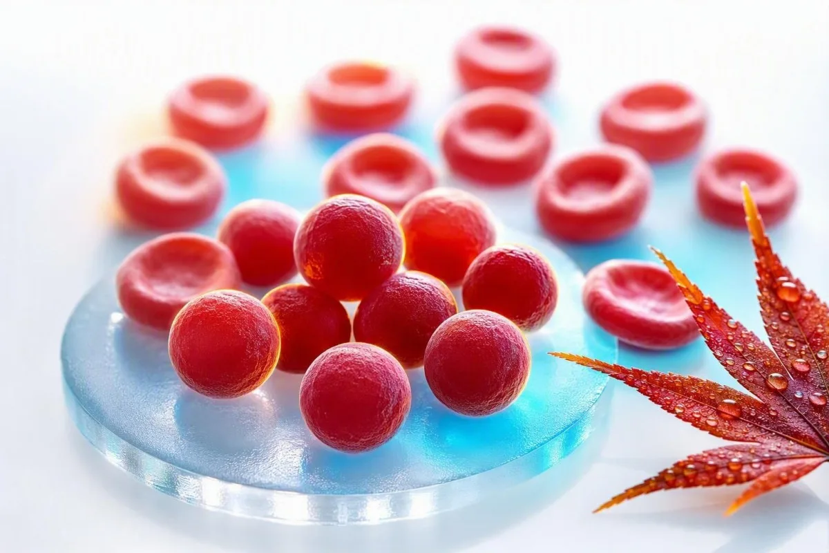
Um MCHC elevado é incomum e muitas vezes mais útil como uma pista do que como um diagnóstico. A questão prática é se as células vermelhas são genuinamente densas e esféricas, ou se a amostra distorceu o cálculo.


- Faixa de MCHC geralmente é de 32-36 g/dL (320-360 g/L) em adultos, embora a faixa do próprio laboratório tenha prioridade.
- MCHC elevado acima de 36 g/dL é incomum e deve levar à verificação de esferócitos, hemólise, queimaduras ou interferência analítica.
- Esferócitos são células vermelhas pequenas e densas sem palidez central e podem ocorrer em esferocitose hereditária ou anemia hemolítica autoimune.
- Aglutininas frias podem elevar falsamente o MCHC por aglutinar as células vermelhas, diminuir a contagem de RBC medida e distorcer os índices calculados.
- Lipemia pode superestimar a hemoglobina em alguns analisadores, criando um MCHC elevado mesmo quando a forma das células vermelhas é normal.
- Queimadura pode produzir MCHC verdadeiramente elevado através de perda da membrana dos glóbulos vermelhos, geralmente acompanhada de anemia e evidências laboratoriais de hemólise.
- Pistas de hemólise incluem reticulócitos elevados, bilirrubina indireta e LDH, com haptoglobina baixa quando os glóbulos vermelhos estão a ser destruídos no corpo.
- Repetir o teste com uma amostra de EDTA recém-recolhida e devidamente manuseada é muitas vezes o primeiro passo sensato quando o MCHC está isolado e apenas ligeiramente elevado.
MCHC elevado em um CBC: a resposta direta
MCHC elevado geralmente significa que os glóbulos vermelhos são anormalmente densos, mas também pode significar que o analisador foi enganado pela amostra. Em adultos, um MCHC acima de cerca de 36 g/dL merece contexto: elevações verdadeiras apontam mais frequentemente para esferócitos ou lesão térmica significativa, enquanto aglutininas frias, lipemia e hemólise da amostra são explicações laboratoriais frequentes. A partir de 23 de agosto de 2026, eu não diagnosticaria uma doença sanguínea apenas com este índice.
O MCHC é calculado como hemoglobina dividida pelo hematócrito, multiplicado por 100, pelo que é uma concentração em vez de uma medição direta da célula. Uma hemoglobina de 15,0 g/dL e um hematócrito de 40% produzem um MCHC de 37,5 g/dL; o número pode aumentar porque a hemoglobina é lida em excesso, o hematócrito é lido em menos, ou ambos. O nosso Guia de biomarcadores de CBC é útil quando vários índices são sinalizados ao mesmo tempo.
Quando reviso um painel com MCHC de 36,5-37,5 g/dL, mas com hemoglobina, bilirrubina, reticulócitos e RDW normais, um artefato é frequentemente mais provável do que doença. Um MCHC de 39-42 g/dL é fisiologicamente difícil de sustentar em glóbulos vermelhos circulantes intactos, pelo que o laboratório deve considerar ativamente a interferência antes que alguém entre em pânico.
Kantesti é um Analisador de teste de sangue de IA que lê o MCHC juntamente com a hemoglobina, o hematócrito, o MCV, o RDW, a bilirrubina, o LDH e os resultados anteriores, em vez de tratar um sinal de alerta como um diagnóstico. A regra clínica do Dr. Thomas Klein é simples: verificar a aritmética e a amostra antes de encomendar uma investigação expansiva; a nossa detalhada Guia de RDW e índices de hemácias explica porque esta abordagem baseada em padrões é importante.
MCHC não é MCH nem MCV
O MCHC descreve a concentração de hemoglobina dentro do volume de glóbulos vermelhos compactados, enquanto o MCH descreve a quantidade de hemoglobina por célula e o MCV descreve o tamanho da célula. Confundir esses três índices é uma razão pela qual um sinal de rotina do CBC pode soar mais alarmante do que realmente é.
O MCH é comumente de 27-33 pg por célula, enquanto o MCV é comumente de 80-100 fL em adultos; nenhum intervalo define o MCHC. Células vermelhas grandes podem carregar mais hemoglobina e, portanto, aumentar o MCH, mas seu MCHC pode permanecer normal porque seu volume aumentou proporcionalmente. Veja nossa comparação prática de MCV e MCH se ambos os valores forem sinalizados.
Um MCHC alto genuíno geralmente se associa a um MCV normal ou baixo-normal porque os esferócitos perderam área de superfície da membrana e se tornaram mais redondos, e não porque contêm hemoglobina ilimitada. Essa geometria importa: uma esfera retém menos área de superfície para seu volume, de modo que não tem um centro pálido em uma lâmina de amostra de células bem preparada.
A questão é que um exame de sangue com MCHC elevado pode ser matematicamente real, mas biologicamente enganoso. Um hematócrito ligeiramente subestimado após agregação de glóbulos vermelhos inflará a proporção, mesmo que os glóbulos vermelhos do paciente não estejam densos; é por isso que uma revisão completa da contagem sanguínea deve preceder as conclusões.
Esferócitos: a pista clássica de MCHC elevado verdadeiro
Esferócitos são a causa mais reconhecível de um MCHC alto verdadeiro porque a perda de membrana torna os glóbulos vermelhos compactos e densamente hemoglobinizados. Eles são vistos na esferocitose hereditária e na anemia hemolítica autoimune, mas um achado no esfregaço deve ser interpretado com sintomas e marcadores de hemólise.
A esferocitose hereditária frequentemente produz valores de MCHC acima de 36 g/dL, reticulocitose, icterícia intermitente, cálculos biliares, esplenomegalia ou histórico familiar de anemia. A diretriz de 2011 da British Society for Haematology observa que o MCHC apoia o diagnóstico, mas não é sensível ou específico o suficiente para confirmá-lo por si só (Bolton-Maggs et al., 2012).
Na anemia hemolítica autoimune, anticorpos marcam os glóbulos vermelhos para remoção parcial da membrana no baço, deixando esferócitos para trás. Um teste antiglobulina direto, contagem de reticulócitos, bilirrubina, LDH e haptoglobina são mais informativos juntos do que qualquer índice isolado; nosso guia para baixa haptoglobina além da hemólise cobre uma limitação de fácil esquecimento.
Uma complicação clínica: transfusão recente pode diluir ou mascarar o padrão de esferócitos nativos do paciente por várias semanas. Christensen et al. descrevem a ligação da eosina-5-maleimida por citometria de fluxo como um teste útil para suspeita de esferocitose hereditária, embora um hematologista deva selecionar e programar os testes cuidadosamente (Christensen et al., 2015).
Como saber se o MCHC elevado vem com hemólise
MCHC alto torna-se clinicamente mais significativo quando acompanha anemia, contagem de reticulócitos elevada, elevação da bilirrubina indireta, elevação de LDH e baixa haptoglobina. Esse aglomerado indica aumento da renovação de glóbulos vermelhos, embora nenhum marcador isolado seja perfeito.
Uma porcentagem de reticulócitos acima de 2% ainda pode ser inadequada em anemia significativa, portanto, os médicos geralmente calculam uma contagem absoluta de reticulócitos ou um índice de produção de reticulócitos. Por exemplo, uma contagem de reticulócitos de 4% com hematócrito de 24% pode representar apenas uma resposta medular modesta após correção; nosso artigo sobre pistas de reticulócitos altos discute a distinção.
A bilirrubina não conjugada aumenta quando o heme é processado mais rapidamente do que o fígado consegue conjugá-lo, enquanto a LDH aumenta porque as células vermelhas contêm LDH em abundância. A haploglobina abaixo do limite inferior do laboratório suporta hemólise intravascular, mas pode parecer normal durante a inflamação porque a haploglobina é uma proteína de fase aguda. Barcellini e Fattizzo discutem essa armadilha diagnóstica em sua revisão de anemia hemolítica autoimune (Barcellini & Fattizzo, 2015).
Urina escura, fadiga em rápida progressão, falta de ar em repouso, dor no peito, desmaio ou olhos amarelados com febre merecem avaliação clínica no mesmo dia. Células fragmentadas são uma pista de esfregaço diferente e mais urgente do que esferócitos; nossa explicação de quando esquistócitos precisam de revisão urgente ajuda a esclarecer por quê.
Por que queimaduras podem causar um MCHC genuinamente elevado
Lesão térmica extensa pode causar MCHC verdadeira e alta porque o calor danifica as membranas das células vermelhas, produzindo perda de membrana, células semelhantes a esferócitos e hemólise. Este é geralmente um achado hospitalar após queimaduras clinicamente aparentes, não uma explicação para um resultado incidental de CBC ambulatorial.
O dano térmico pode criar micrósferos dentro de horas de queimaduras graves, e o MCHC pode aumentar à medida que a relação membrana/volume diminui. A hemoglobina também pode cair nos dias seguintes devido a alterações de fluidos, procedimentos, flebotomia e hemólise contínua, portanto, um único MCHC não pode quantificar a gravidade da lesão.
Ocasionalmente, vi pacientes se concentrarem em um MCHC de 37,1 g/dL após uma admissão de emergência, enquanto negligenciam anormalidades muito mais acionáveis: hemoglobina em queda, creatinina em alta, alterações de potássio ou lactato. Nesse cenário, os clínicos gerenciam a pessoa e a fisiologia da queimadura, não o índice; nosso cronograma de testes pós-hospitalares explica por que os valores podem se mover rapidamente.
Kantesti AI é uma plataforma de interpretação de biomarcadores por IA que pode sinalizar um MCHC alto juntamente com resultados dinâmicos de hemoglobina e rins, mas queimaduras agudas exigem avaliação hospitalar direta e não podem ser geridas com segurança a partir de uma interpretação de relatório. Um resultado após uma queimadura grave deve ser comparado com as 24-72 horas anteriores, não com uma média genérica da população.
Aglutininas frias: o artefato de MCHC elevado que os médicos procuram
Aglutininas frias podem produzir um MCHC falsamente alto fazendo com que as células vermelhas se aglutinem à temperatura ambiente antes da análise. O analisador pode contar várias células ligadas como uma partícula, levando a uma contagem de RBC falsamente baixa, um MCV falsamente alto e um MCHC calculado inflado.
O padrão clássico de aglutinina fria é uma contagem baixa de RBC com MCV desproporcionalmente alto, MCH e MCHC, muitas vezes sem uma história clínica correspondente. Aquecer o tubo de EDTA a 37°C e executar o CBC novamente geralmente corrige os valores, o que é muito mais informativo do que repetir um teste aleatório semanas depois.
Aglutininas frias podem ocorrer transitoriamente após algumas infecções respiratórias ou persistir em doença de aglutininas frias e certos distúrbios linfoproliferativos. Sintomas como mudança de cor ou desconforto nos dedos com exposição ao frio adicionam peso clínico, mas o padrão laboratorial em si pode ser a primeira pista; um teste repetido após hemólise oferece perguntas práticas de nova coleta.
Não se aqueça agressivamente nem tente manipular o resultado em casa. Pergunte se o laboratório viu aglutinação de células vermelhas, se a amostra foi aquecida e repetida, e se um hematócrito manual foi realizado; essas três respostas geralmente resolvem a questão.
Lipemia e hemólise da amostra podem distorcer o MCHC
Lipemia e hemólise in vitro da amostra podem aumentar falsamente o MCHC porque muitos analisadores medem a hemoglobina fotometricamente após as células vermelhas serem lisadas. Lipídios turvos ou hemoglobina livre na amostra podem aumentar a absorbância óptica e fazer com que a hemoglobina pareça maior do que realmente é.
A lipemia é mais frequentemente visível após triglicerídeos muito altos, geralmente acima de cerca de 400-500 mg/dL, embora a interferência dependa do instrumento e do comprimento de onda do ensaio. Um laboratório pode usar um branco de plasma, um comprimento de onda alternativo ou uma abordagem de substituição de plasma em vez de simplesmente aceitar o resultado automatizado da hemoglobina.
A hemólise in vitro ocorre após a coleta ou manuseio, não dentro do corpo, e pode afetar mais do que a MCHC. Potássio, LDH, AST e fosfato podem aumentar em uma amostra danificada, razão pela qual uma MCHC alta isolada ao lado de um potássio alto inesperado requer uma verificação da qualidade da amostra; veja nosso guia de erros de potássio relacionados à hemólise.
Uma camada de plasma visivelmente rosada ou vermelha é uma pista laboratorial, não prova de anemia hemolítica no paciente. O acompanhamento prático é uma nova coleta com manuseio calmo e processamento rápido, especialmente se o laudo original incluir um índice de hemólise ou um comentário de que os resultados podem ser afetados.
Problemas de coleta, armazenamento e tubo que alteram os índices do CBC
Mistura inadequada da amostra, subenchimento, coagulação e análise atrasada podem alterar os índices do CBC, embora o armazenamento atrasado mais frequentemente diminua do que aumente a MCHC ao permitir que as células inchem. Uma MCHC alta deve, portanto, ser lida em conjunto com os comentários da amostra do laboratório, não apenas com o número no portal do paciente.
As amostras de CBC são geralmente coletadas em tubos de EDTA, e um tubo subenchido pode ter uma proporção excessiva de anticoagulante para amostra que altera as medições celulares. Pequenos coágulos podem remover seletivamente células da análise e tornar a contagem automatizada internamente inconsistente; nossa visão geral de aditivos de tubos de sangue explica por que o tipo de tubo não é um detalhe trivial.
O armazenamento em temperatura ambiente por períodos prolongados faz com que as células vermelhas absorvam água, aumentando o MCV e tendendo a diminuir a MCHC. Essa direção de mudança é útil: uma MCHC muito alta após uma amostra atrasada não é automaticamente explicada apenas pela idade, portanto, a aglutinação a frio ou a interferência óptica ainda podem estar presentes.
Uma amostra capilar pode ser apropriada em algumas situações, mas é mais propensa a variabilidade na coleta do que uma amostra venosa de EDTA bem coletada. Se uma MCHC alta não tiver sintomas correspondentes ou anormalidades relacionadas, geralmente prefiro uma nova coleta venosa limpa antes de tratá-la como um novo diagnóstico crônico.
Use CBCs anteriores para decidir se a alteração é real
Um aumento súbito na MCHC de uma linha de base estável de 33-35 g/dL para 38 g/dL é mais sugestivo de um problema na amostra ou no analisador do que esferocitose hereditária em evolução lenta. Tendências não substituem um esfregaço, mas são uma poderosa pista de controle de qualidade.
A maioria das pessoas tem uma faixa pessoal de MCHC relativamente estreita ao longo do tempo, muitas vezes variando em menos de 1 g/dL quando as condições de teste são comparáveis. Um "delta check" laboratorial compara um novo resultado com um anterior para identificar mudanças implausíveis antes que os resultados sejam liberados; nosso artigo sobre verificações de delta mostra como essa rede de segurança funciona.
Kantesti é um serviço de interpretação de testes do laboratório de IA que compara valores atuais e anteriores do CBC, ajudando a distinguir um sinal persistente de um outlier isolado. Em nosso fluxo de trabalho de revisão, uma mudança na MCHC é mais importante quando viaja com hemoglobina, MCV, bilirrubina e reticulócitos em uma direção biologicamente coerente.
Existem exceções. Nova hemólise autoimune, uma queimadura grave, uma reação transfusional ou aglutinação a frio marcada podem genuinamente alterar os resultados rapidamente, portanto, um valor histórico estável é tranquilizador, mas nunca é um motivo para ignorar nova icterícia, urina escura ou anemia.
Condições comumente culpadas pelo MCHC, mas raramente responsáveis
Desidratação, deficiência de ferro, deficiência de vitamina B12 e estresse comum geralmente não causam uma MCHC verdadeiramente alta. Eles podem alterar outros valores do CBC, mas não devem ser usados como explicações convenientes para uma MCHC de 38 g/dL sem confirmar a amostra.
A deficiência de ferro geralmente diminui MCH e MCHC e, frequentemente, diminui MCV assim que a deficiência se estabelece. A deficiência precoce de ferro pode ter um MCV normal, mas não explica índices hipercrômicos; nossa explicação de saturação de transferrina baixa ajuda a separar a disponibilidade de ferro da concentração de CBC.
A deficiência de vitamina B12 ou folato causa mais frequentemente macrocitose, com MCV acima de 100 fL, em vez de MCHC alta. Se MCV e MCH estiverem ambos altos, mas MCHC normal, essa é uma direção diagnóstica diferente; consulte pistas de acompanhamento de MCV alto.
A deficiência de G6PD causa hemólise episódica após gatilhos oxidativos específicos em pessoas suscetíveis, mas MCHC não é um teste de rastreio confiável para isso. Durante um episódio agudo, o esfregaço e os marcadores de renovação podem ser mais reveladores, enquanto os testes enzimáticos podem precisar ser repetidos após a recuperação, pois os glóbulos vermelhos jovens contêm mais G6PD.
Os testes de acompanhamento mais úteis após MCHC elevado
O melhor próximo passo para um MCHC isolado e elevado é geralmente um CBC repetido com revisão laboratorial, seguido por um esfregaço periférico e painel de hemólise apenas se a elevação persistir ou houver outras pistas presentes. Solicitar todos os testes de anemia imediatamente pode criar mais ruído do que respostas.
Uma revisão focada de primeira linha inclui CBC repetido, revisão da lâmina de amostra de células, contagem de reticulócitos, bilirrubina total e indireta, LDH, haptoglobina e teste de antiglobulina direto quando a hemólise autoimune é plausível. O esfregaço pode identificar esferócitos, aglutinação, fragmentos, policromasia ou uma morfologia normal que torna um artefato mais provável.
Se a esferocitose hereditária permanecer provável, um hematologista pode considerar o teste de ligação de eosinofilia-5-maleimida, o teste de fragilidade osmótica ou testes genéticos. Esses testes têm diferentes riscos de falsos negativos após transfusão e durante hemólise aguda, razão pela qual a ordem dos testes deve seguir um histórico clínico em vez de uma lista de verificação; nosso guia de teste G6PD ilustra o problema de temporização mais amplo.
O Kantesti AI interpreta MCHC alto verificando o cálculo em relação a marcadores relacionados de CBC e hemólise, identificando então perguntas para levar a um médico. Ele não substitui a revisão da morfologia celular, particularmente quando o laboratório emitiu um sinalizador do analisador ou quando a anemia está piorando.
Quando MCHC elevado necessita de avaliação médica urgente
MCHC alto em si não é um limiar de emergência, mas MCHC alto com sinais de hemólise aguda, anemia grave ou uma queimadura grave pode exigir atendimento no mesmo dia. Sintomas e a taxa de mudança determinam a urgência muito melhor do que se o sinalizador está marcado como H.
Busque avaliação urgente para nova falta de ar em repouso, dor no peito, desmaio, confusão, fraqueza em rápida piora, urina cor de chá ou icterícia com febre. Esses sintomas podem acompanhar anemia ou hemólise significativas, especialmente quando a hemoglobina caiu mais de 2 g/dL de uma linha de base conhecida.
Entre em contato com o médico que solicitou o exame em poucos dias se o MCHC permanecer acima de 36 g/dL em uma amostra repetida, houver anemia ou o laudo mencionar aglutinação ou esferócitos. Em uma pessoa estável e bem, com MCHC de 36,2 g/dL e resultados normais em outros aspectos, uma nova coleta não urgente é frequentemente razoável; nosso guia do padrão de bilirrubina pode ajudar a orientar a próxima discussão.
Não inicie ferro, folato, esteroides ou suplementos simplesmente para corrigir o MCHC. Nenhum deles trata as principais causas verdadeiras, e os esteroides em particular podem obscurecer a avaliação da hemólise autoimune se iniciados sem supervisão clínica.
Como o Kantesti coloca o MCHC em contexto clínico
O Kantesti lê um MCHC alto como um problema de reconhecimento de padrão: verdadeiros distúrbios de células densas e artefatos de amostra produzem diferentes combinações de CBC, química, sinalizadores e tendências anteriores. O objetivo é tornar a próxima pergunta clínica mais clara, não rotular uma doença rara a partir de um único índice calculado.
Kantesti é um Ferramenta de análise de exames de sangue com IA usado por mais de 2 milhões de pessoas em 127 países para organizar resultados laboratoriais em contexto. Para MCHC elevado, nossa IA verifica se hemoglobina e hematócrito suportam matematicamente o resultado, se o MCV está inesperadamente alto e se bilirrubina, LDH, reticulócitos ou haptoglobina apontam para a renovação de glóbulos vermelhos.
Nossa plataforma também procura contradições internas: MCHC 40 g/dL com comentário de esfregaço normal e sem anemia pode justificar um alerta de artefato, enquanto MCHC 37 g/dL com esferócitos, reticulocitose e elevação da bilirrubina indireta merece acompanhamento médico. A abordagem técnica e os limites são descritos em nosso guia de tecnologia de IA.
Aconselho os pacientes a guardar o PDF do laboratório, data da coleta, status de jejum, doença recente, histórico de transfusão e quaisquer comentários laboratoriais. Esse pequeno registro muitas vezes explica uma discrepância aparente mais rapidamente do que repetir testes amplos, especialmente quando os resultados são comparados meses depois.
Perguntas que tornam uma consulta de MCHC elevado mais produtiva
Pergunte se o MCHC foi repetido, se o laboratório viu esferócitos ou aglutinação e se anemia ou marcadores de hemólise estão presentes. Essas perguntas mudam a conversa de “por que um número está alto?” para um caminho clínico concreto e testável.
Uma redação útil inclui: “A amostra estava hemolisada ou lipêmica?”, “O analisador sinalizou aglutinação de glóbulos vermelhos?” e “Podemos comparar este resultado com meu MCHC anterior?” Uma revisão manual do esfregaço é particularmente valiosa quando o MCHC está repetidamente acima de 36 g/dL ou a anemia é inexplicada.
O Dr. Thomas Klein recomenda levar uma lista de medicamentos, infecções recentes, sintomas desencadeados pelo frio, histórico familiar de cálculos biliares ou anemia e transfusões nos 3 meses anteriores. Esse histórico pode apontar para esferocitose hereditária, hemólise autoimune ou um aglutinínio frio transitório muito mais rápido do que uma busca genérica online.
O conteúdo médico do Kantesti é revisado com supervisão médica, mas seu próprio clínico pode examiná-lo, solicitar uma repetição aquecida e falar diretamente com o laboratório. Leitores que desejam entender nossa estrutura de revisão clínica podem conhecer o conselho consultivo médico, enquanto anormalidades persistentes ou sintomáticas devem sempre ser discutidas pessoalmente.
Pesquisa, incerteza e os limites de um único MCHC
O MCHC é uma pista útil de rastreamento, não um teste diagnóstico isolado para esferocitose hereditária, hemólise autoimune, queimaduras ou doença por aglutinina a frio. Seu valor aumenta acentuadamente quando uma amostra repetida, morfologia celular, marcadores de renovação e histórico clínico contam a mesma história.
Nenhum valor de corte universal de MCHC diagnostica esferocitose hereditária porque métodos de analisadores, idade, reticulocitose e deficiência de ferro coexistente podem alterar a sensibilidade. A diretriz da BSH apoia a combinação de histórico familiar, achados de esfregaço, reticulócitos, bilirrubina e testes confirmatórios onde necessário, em vez de depender de um único limiar (Bolton-Maggs et al., 2012).
O Kantesti apoia o acompanhamento informado identificando padrões de resultados e incertezas, não emitindo um diagnóstico hematológico definitivo. Nosso abordagem de validação clínica explica por que valores incomuns, alertas de analisador e sintomas de emergência permanecem razões para revisão laboratorial e clínica humana.
Para contexto hematológico relacionado, veja a publicação de pesquisa Tipo Sanguíneo B Negativo, Exame de Sangue de LDH e Guia de Contagem de Reticulócitos e o recurso mais amplo de comunicação clínica Diarreia após jejum, pontos pretos nas fezes e guia GI 2026. Ambos são publicações educacionais suplementares, não substitutos para um esfregaço periférico ou avaliação especializada.
Perguntas frequentes
O que significa um nível elevado de MCHC em um exame de sangue?
Um nível elevado de MCHC geralmente significa que a concentração de hemoglobina dentro do volume de glóbulos vermelhos está acima da faixa laboratorial, comumente acima de 36 g/dL em adultos. Elevações verdadeiras ocorrem com esferócitos de esferocitose hereditária, anemia hemolítica autoimune ou lesão térmica grave. Aglutininas frias, lipemia e hemólise da amostra também podem criar um resultado falso alto. Um CBC repetido, revisão da lâmina da amostra celular e marcadores de hemólise esclarecem qual explicação se aplica.
A MCHC elevada é perigosa?
MCHC alto não é perigoso por si só; é um índice calculado do hemograma completo (CBC) e não uma doença. Uma MCHC de 36,2 g/dL com hemoglobina normal e sem sintomas geralmente é tratada com um teste de repetição de rotina, enquanto MCHC alto mais queda de hemoglobina, icterícia, urina escura ou falta de ar requer avaliação imediata. Valores acima de 39 g/dL são especialmente propensos a refletir interferência na amostra ou no analisador. O risco clínico advém da causa subjacente, como hemólise significativa, não apenas do número da MCHC.
A desidratação pode causar um MCHC elevado?
A desidratação geralmente não causa um MCHC verdadeiramente elevado porque concentra o volume plasmático mais do que altera a concentração de hemoglobina dentro de eritrócitos individuais. A desidratação pode elevar a hemoglobina e o hematócrito juntos, deixando o MCHC próximo de sua faixa usual de 32-36 g/dL. Se o MCHC for de 37-40 g/dL, os clínicos devem considerar esferócitos, aglutinação a frio, lipemia ou um problema de medição em vez disso. Uma nova CBC bem coletada é mais útil do que tentar corrigir o valor com fluidos extras.
Esferócitos sempre aumentam o MCHC?
Esferócitos frequentemente elevam a MCHC acima de 36 g/dL, mas nem sempre o fazem e MCHC alta não prova que esferócitos estão presentes. A esferocitose hereditária pode ser mascarada por deficiência de ferro, transfusão recente ou doença leve, enquanto a hemólise autoimune pode mostrar achados variáveis dependendo do momento. Uma lâmina de amostra de células revisada por um profissional de laboratório experiente é necessária para identificar esferócitos. A contagem de reticulócitos, bilirrubina, LDH, haptoglobina e teste de antiglobulina direto adicionam contexto diagnóstico.
Aglutininas frias podem causar MCHC alto?
Aglutininas frias podem elevar falsamente o MCHC aglutinando as hemácias antes que o CBC automatizado seja medido. O padrão frequentemente inclui uma contagem baixa de RBC, MCV alto, MCH alto e um MCHC acima de 36 g/dL que não se encaixa nos sintomas do paciente. Aquecer a amostra de EDTA a 37°C e repetir a análise pode normalizar as contagens. Sintomas persistentes de aglutinação a frio ou hemólise devem ser avaliados por um clínico ou hematologista.
Quais exames devo pedir após MCHC alto?
Após uma MCHC persistentemente alta, os testes subsequentes habituais são uma repetição do hemograma completo (CBC), revisão da lâmina de amostra celular, contagem de reticulócitos, frações de bilirrubina, LDH, haploglobina e, às vezes, um teste de antiglobulina direto. Um clínico pode solicitar a ligação de eosina-5-maleimida ou outros testes especializados se houver suspeita de esferocitose hereditária. MCHC acima de 36 g/dL com repetição normal dos testes e sem anemia pode não necessitar de investigação extensiva. A melhor sequência de testes depende dos sintomas, valores prévios do hemograma completo (CBC), histórico familiar e flags laboratoriais.
Faça hoje a análise de exame de sangue com IA
Junte-se a mais de 2 milhões de usuários no mundo todo que confiam na Kantesti para análise instantânea e precisa de exames laboratoriais. Envie seus resultados de exame de sangue e receba uma interpretação abrangente de biomarcadores 15,000+ em segundos.
📚 Publicações de pesquisa referenciadas
Klein, T., Mitchell, S., & Weber, H. (2026). Guia de tipo sanguíneo B negativo, teste de sangue de LDH e contagem de reticulócitos. Pesquisa Médica por IA da Kantesti.
Klein, T., Mitchell, S., & Weber, H. (2026). Diarreia após jejum, pontos pretos nas fezes e guia GI 2026. Pesquisa Médica por IA da Kantesti.
📖 Referências Médicas Externas
⚕️ Aviso Médico
Este artigo é apenas para fins educacionais e não constitui aconselhamento médico. Consulte sempre um profissional de saúde qualificado para decisões de diagnóstico e tratamento.
Sinais de confiança E-E-A-T
Experiência
Revisão clínica orientada por médicos dos fluxos de interpretação de exames laboratoriais.
Especialização
Foco em medicina laboratorial sobre como os biomarcadores se comportam no contexto clínico.
Autoridade
Escrito pelo Dr. Thomas Klein, com revisão da Dra. Sarah Mitchell e do Prof. Dr. Hans Weber.
Confiabilidade
Interpretação baseada em evidências, com caminhos de acompanhamento claros para reduzir alarmes.