Un MCHC ridicat este neobișnuit și adesea mai util ca un indiciu decât ca un diagnostic. Întrebarea practică este dacă celulele roșii sunt cu adevărat dense și sferice, sau dacă proba a distorsionat calculul.


- Interval MCHC este de obicei 32-36 g/dL (320-360 g/L) la adulți, deși intervalul propriu al laboratorului are prioritate.
- MCHC ridicat peste 36 g/dL este neobișnuit și ar trebui să determine verificarea sferocitelor, hemolizei, arsurilor sau interferențelor analitice.
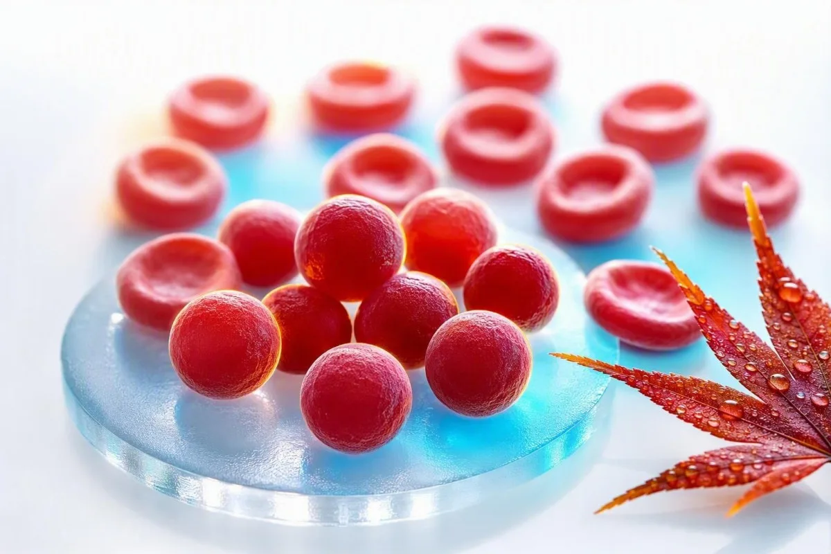
- Sferocite sunt celule roșii mici, dense, fără paloare centrală și pot apărea în sferocitoza ereditară sau anemia hemolitică autoimună.
- hiperglicemie pot crește fals MCHC prin aglutinarea celulelor roșii, scăderea numărului măsurat de RBC și distorsionarea indicilor calculați.
- Lipemie poate supraestima hemoglobina pe anumiți analizori, creând un MCHC crescut chiar și atunci când forma globulelor roșii este normală.
- Arsura termică poate produce o MCHC ridicată reală prin pierderea membranei globulare, de obicei însoțită de anemie și dovezi de laborator de hemoliză.
- Indicii de hemoliză includ reticulocite crescute, bilirubină indirectă și LDH, cu haptoglobină scăzută atunci când globulele roșii se distrug în organism.
- Repetarea testării cu un eșantion EDTA proaspăt recoltat și manipulat corespunzător este adesea primul pas logic atunci când MCHC este izolată și doar ușor crescută.
MCHC ridicat pe un CBC: răspunsul direct
O MCHC ridicată înseamnă de obicei că globulele roșii sunt neobișnuit de dense, dar poate însemna și că analizorul a fost indus în eroare de eșantion. La adulți, o MCHC peste aproximativ 36 g/dL necesită context: creșterile reale indică cel mai frecvent sferocite sau leziuni termice semnificative, în timp ce aglutininele la rece, lipemia și hemoliza eșantionului sunt explicații frecvente de laborator. Începând cu 23 august 2026, nu aș diagnostica o boală a sângelui doar pe baza acestui indice.
MCHC se calculează ca hemoglobină împărțită la hematocrit, înmulțit cu 100, deci este o concentrație mai degrabă decât o măsurătoare directă a celulei. O hemoglobină de 15,0 g/dL și un hematocrit de 40% produc o MCHC de 37,5 g/dL; numărul poate crește deoarece hemoglobina este supraestimată, hematocritul este subestimat sau ambele. Ghidul nostru de biomarkeri CBC este util atunci când mai mulți indici sunt marcați simultan.
Când revizuiesc un panou cu MCHC 36,5-37,5 g/dL, dar hemoglobină, bilirubină, reticulocite și RDW normale, un artefact este adesea mai probabil decât o boală. O MCHC de 39-42 g/dL este fiziologic dificil de susținut în globulele roșii intacte în circulație, așa că laboratorul ar trebui să ia în considerare activ interferența înainte ca cineva să intre în panică.
Kantesti este o Analizor de teste de sânge AI care citește MCHC alături de hemoglobină, hematocrit, MCV, RDW, bilirubină, LDH și rezultate anterioare, mai degrabă decât să trateze o singură alertă ca diagnostic. Regula clinică a Dr. Thomas Klein este simplă: verificați aritmetica și specimenul înainte de a comanda o investigație extinsă; ghidul nostru detaliat RDW și ghidul indicilor eritrocitari explică de ce această abordare bazată pe modele este importantă.
MCHC nu este MCH sau MCV
MCHC descrie concentrația de hemoglobină în volumul de globule roșii compactate, în timp ce MCH descrie cantitatea de hemoglobină per celulă și MCV descrie dimensiunea celulei. Confuzionarea acestor trei indici este un motiv pentru care un flag de rutină la CBC poate suna mai alarmant decât este.
MCH este în mod obișnuit 27-33 pg per celulă, în timp ce MCV este în mod obișnuit 80-100 fL la adulți; niciun interval nu definește MCHC. Celulele roșii mari pot transporta mai multă hemoglobină și, prin urmare, pot crește MCH, totuși MCHC-ul lor poate rămâne normal, deoarece volumul lor a crescut proporțional. Vedeți comparația noastră practică MCV și MCH dacă ambele valori sunt marcate.
Un MCHC ridicat autentic se asociază adesea cu un MCV normal sau la limita inferioară a normalului, deoarece sferocitele și-au pierdut suprafața membranei și au devenit mai rotunde, nu pentru că ar conține hemoglobină nelimitată. Această geometrie contează: o sferă reține mai puțină suprafață pentru volumul său, deci nu are centru palid pe o lamă de frotiu celular bine pregătită.
Ideea este că un test sanguin MCHC crescut poate fi matematic real, dar biologic înșelător. Un hematocrit ușor subestimat după aglomerarea globulelor roșii va umfla raportul, chiar dacă globulele roșii ale pacientului nu sunt deloc dense; acesta este motivul pentru care o revizuire completă a numărătorii de sânge ar trebui să preceadă concluziile.
Sferocite: indiciul clasic pentru MCHC ridicat real
Sferocitele sunt cauza cea mai recunoscută a unui MCHC ridicat real, deoarece pierderea membranei face ca globulele roșii să fie compacte și dens hemoglobinizate. Ele sunt observate în sferocitoza ereditară și anemia hemolitică autoimună, dar o constatare la frotiu trebuie interpretată împreună cu simptomele și markerii de hemoliză.
Sferocitoza ereditară produce adesea valori MCHC peste 36 g/dL, reticulocitoză, icter intermitent, calculi biliari, splenomegalie sau un istoric familial de anemie. Ghidul din 2011 al Societății Britanice de Hematologie notează că MCHC susține diagnosticul, dar nu este suficient de sensibil sau specific pentru a-l confirma de la sine (Bolton-Maggs et al., 2012).
În anemia hemolitică autoimună, anticorpii marchează globulele roșii pentru îndepărtarea parțială a membranei în splină, lăsând în urmă sferocite. Un test antiglobulinic direct, numărul de reticulocite, bilirubina, LDH și haptoglobina sunt mai informative împreună decât orice indice izolat; ghidul nostru despre haptoglobină scăzută dincolo de hemoliză acoperă o limitare ușor de ratat.
O particularitate clinică: transfuzia recentă poate dilua sau masca modelul nativ de sferocite al pacientului timp de câteva săptămâni. Christensen et al. descriu legarea de eozină-5-maleimidă prin citometrie în flux ca un test util pentru sferocitoza ereditară suspectată, deși un hematolog ar trebui să selecteze și să programeze testarea cu atenție (Christensen et al., 2015).
Cum să afli dacă MCHC ridicat este însoțit de hemoliză
MCHC ridicat devine mai relevant clinic atunci când însoțește anemia, un număr crescut de reticulocite, o creștere a bilirubinei indirecte, o creștere a LDH și o haptoglobină scăzută. Acest grup indică o creștere a turnover-ului globulelor roșii, deși niciun marker individual nu este perfect.
Un procent de reticulocite peste 2% poate fi totuși inadecvat în anemia semnificativă, așa că medicii calculează adesea un număr absolut de reticulocite sau un indice de producție a reticulocitelor în schimb. De exemplu, un număr de reticulocite de 4% cu un hematocrit de 24% poate reprezenta doar un răspuns modest al măduvei osoase după corecție; articolul nostru despre indicii de reticulocite ridicate explică distincția.
Bilirubina neconjugată crește atunci când hemul este procesat mai rapid decât îl poate conjuga ficatul, în timp ce LDH crește deoarece celulele roșii din sânge conțin LDH abundent. Haptoglobina sub limita inferioară a laboratorului susține hemoliza intravasculară, dar poate apărea normală în timpul inflamației, deoarece haptoglobina este o proteină de fază acută. Barcellini și Fattizzo discută această capcană diagnostică în recenzia lor despre anemia hemolitică autoimună (Barcellini & Fattizzo, 2015).
Urina închisă la culoare, oboseala cu creștere rapidă, respirația dificilă în repaus, durerea în piept, leșinul sau ochii galbeni cu febră necesită evaluare clinică în aceeași zi. Celulele fragmentate sunt un indiciu la frotiu diferit și mai urgent decât sfero citocele; explicația noastră despre când schistocitele necesită evaluare urgentă ajută la clarificarea motivului.
De ce arsurile pot cauza un MCHC ridicat real
Leziunea termică extinsă poate cauza MCHC ridicat real, deoarece căldura deteriorează membranele celulelor roșii din sânge, producând pierdere de membrană, celule asemănătoare sferocitelor și hemoliză. Aceasta este, în general, o constatare spitalicească după arsuri clinic aparente, nu o explicație pentru un rezultat incidental al CBC la ambulatoriu.
Leziunea termică poate crea microsferocite în decurs de câteva ore de la arsuri majore, iar MCHC poate crește pe măsură ce raportul membrană/volum scade. Hemoglobina poate, de asemenea, să scadă în zilele următoare din cauza deplasărilor de lichide, procedurilor, flebotomiei și hemolizei continue, deci un singur MCHC nu poate cuantifica severitatea leziunii.
Am văzut ocazional pacienți care se concentrează pe un MCHC de 37,1 g/dL după o internare de urgență, în timp ce ignoră anomalii mult mai acționabile: scăderea hemoglobinei, creșterea creatininei, modificări ale potasiului sau lactat. În acest context, medicii gestionează persoana și fiziologia arsurilor, nu indicatorul; al nostru calendar de teste post-spitalizare explică de ce valorile se pot mișca rapid.
Kantesti AI este un platformă de interpretare a biomarkerilor de către AI care poate semnala un MCHC ridicat, alături de rezultate dinamice ale hemoglobinei și rinichilor, dar arsurile acute necesită evaluare directă în spital și nu pot fi gestionate în siguranță doar pe baza interpretării unui raport. Un rezultat după o arsură severă trebuie comparat cu cele 24-72 de ore anterioare, nu cu o medie generică a populației.
Aglutinine la rece: artefactul MCHC ridicat pe care medicii îl caută
Aglutininele la rece pot produce un MCHC ridicat fals, determinând aglutinarea celulelor roșii din sânge la temperatura camerei înainte de analiză. Analizorul poate număra mai multe celule atașate ca o singură particulă, ducând la un număr RBC fals scăzut, un MCV fals ridicat și un MCHC calculat umflat.
Modelul clasic al aglutininelor la rece este un număr RBC scăzut cu un MCV disproporționat de ridicat, MCH ridicat și MCHC ridicat, adesea fără o poveste clinică potrivită. Încălzirea eprubetei cu EDTA la 37°C și reanalizarea CBC corectează de obicei valorile, ceea ce este mult mai informativ decât repetarea unui test aleatoriu săptămâni mai târziu.
Aglutininele la rece pot apărea tranzitoriu după unele infecții respiratorii sau pot persista în boala aglutininelor la rece și anumite tulburări limfoproliferative. Simptomele precum schimbarea culorii sau disconfortul în degete la expunerea la frig adaugă greutate clinică, dar modelul de laborator în sine poate fi primul indiciu; un test repetat după hemoliză oferă întrebări practice de retragere a probei.
Nu vă încălziți agresiv și nu încercați să manipulați rezultatul acasă. Întrebați dacă laboratorul a observat aglutinare a celulelor roșii, dacă proba a fost încălzită și reanalizată și dacă a fost efectuat un hematocrit manual; aceste trei răspunsuri rezolvă adesea problema.
Lipemia și hemoliza probei pot distorsiona MCHC
Lipemia și hemoliza probei in-vitro pot crește fals MCHC, deoarece mulți analizori măsoară hemoglobina fotometric după ce celulele roșii din sânge sunt lizate. Lipidele tulburi sau hemoglobina liberă din probă pot crește absorbția optică și pot face ca hemoglobina să pară mai mare decât este de fapt.
Lipemia este cel mai adesea vizibilă după trigliceride foarte ridicate, de obicei peste aproximativ 400-500 mg/dL, deși interferența depinde de instrument și de lungimea de undă a testului. Un laborator poate utiliza un blanc de plasmă, o lungime de undă alternativă sau o abordare de înlocuire a plasmei, în loc să accepte pur și simplu rezultatul automat al hemoglobinei.
Hemoliza in vitro are loc după recoltare sau manipulare, nu în interiorul corpului, și poate afecta mai mult decât MCHC. Potasiul, LDH, AST și fosfatul pot crește într-o probă deteriorată, motiv pentru care un MCHC ridicat izolat, alături de un potasiu ridicat neașteptat, necesită o verificare a calității specimenului; consultați ghidul nostru despre erori legate de potasiu cauzate de hemoliză.
Un strat de plasmă vizibil roz sau roșu este un indiciu de laborator, nu o dovadă a anemiei hemolitice la pacient. Urmărirea practică este o nouă recoltare cu o manipulare calmă și procesare promptă, mai ales dacă raportul inițial include un indice de hemoliză sau un comentariu conform căruia rezultatele pot fi afectate.
Probleme legate de recoltare, depozitare și tuburi care modifică indicii CBC
Amestecarea deficitară a probei, umplerea insuficientă, coagularea și analiza întârziată pot modifica indicii CBC, deși depozitarea întârziată duce mai des la scăderea decât la creșterea MCHC, permițând celulelor să se umfle. Prin urmare, un MCHC ridicat trebuie citit împreună cu comentariile laboratorului referitoare la specimene, nu doar cu numărul de pe portalul pacientului.
Probele CBC sunt, în general, colectate în tuburi cu EDTA, iar un tub sub-umplut poate avea un raport anticoagulant-probă excesiv care modifică măsurătorile celulare. Cheagurile mici pot elimina selectiv celulele din analiză și pot face ca numărătoarea automată să fie intern inconsistentă; prezentarea noastră generală a aditivilor din tuburile de sânge explică de ce tipul tubului nu este un detaliu trivial.
Depozitarea la temperatura camerei pentru perioade prelungite face ca globulele roșii să absoarbă apă, crescând MCV și având tendința de a scădea MCHC. Această direcție de modificare este utilă: un MCHC foarte ridicat după o probă întârziată nu este explicat automat doar de vârstă, deci aglutinarea la rece sau interferența optică pot fi încă prezente.
O probă capilară poate fi adecvată în anumite situații, dar este mai predispusă la variabilitate la recoltare decât un specimen venos EDTA recoltat corect. Dacă un MCHC ridicat nu are simptome corespondente sau anomalii conexe, prefer de obicei o nouă recoltare venoasă curată înainte de a o trata ca pe un nou diagnostic cronic.
Utilizați CBC anterioare pentru a decide dacă modificarea este reală
O creștere bruscă a MCHC de la o linie de bază stabilă de 33-35 g/dL la 38 g/dL sugerează mai degrabă o problemă a specimenului sau a analizorului decât o sferocitoză ereditară care evoluează lent. Tendințele nu înlocuiesc un frotiu, dar sunt un indiciu puternic de control al calității.
Majoritatea oamenilor au un interval personal de MCHC relativ restrâns în timp, adesea variind cu mai puțin de 1 g/dL atunci când condițiile de testare sunt comparabile. O verificare delta de laborator compară un rezultat nou cu unul anterior pentru a identifica schimbări implauzibile înainte ca rezultatele să fie eliberate; articolul nostru despre verificări delta arată cum funcționează acea plasă de siguranță.
Kantesti este o serviciul de interpretare a testelor de laborator AI care compară valorile CBC actuale și anterioare, ajutând la deosebirea unui semnal persistent de un outlier unic. În fluxul nostru de lucru de revizuire, o schimbare a MCHC contează mai mult atunci când se deplasează împreună cu hemoglobina, MCV, bilirubina și reticulocitele într-o direcție biologic coerentă.
Există excepții. Hemoliza autoimună nouă, o arsură majoră, o reacție transfuzională sau o aglutinare la rece marcată pot schimba cu adevărat rezultatele rapid, deci o valoare istorică stabilă este liniștitoare, dar niciodată un motiv pentru a ignora icterul nou, urina închisă la culoare sau anemia.
Afecțiuni frecvent învinovățite pe MCHC, dar rareori responsabile
Deshidratarea, deficitul de fier, deficitul de vitamina B12 și stresul obișnuit nu cauzează de obicei un MCHC cu adevărat ridicat. Acestea pot schimba alte valori CBC, dar nu ar trebui utilizate ca explicații convenabile pentru un MCHC de 38 g/dL fără a confirma specimenul.
Deficiența de fier scade, de obicei, MCH și MCHC și adesea scade MCV odată ce deficiența este stabilită. Deficiența timpurie de fier poate avea un MCV normal, dar nu explică indicii hipercromice; explicația noastră saturație scăzută a transferinei ajută la separarea disponibilității fierului de concentrația CBC.
Deficiența de vitamina B12 sau de folat determină mai frecvent macrocitoză, cu un MCV peste 100 fL, mai degrabă decât MCHC ridicat. Dacă MCV și MCH sunt ambele ridicate, dar MCHC este normal, aceasta este o direcție diagnostică diferită; consultați indicii de urmărire pentru MCV ridicat.
Deficiența de G6PD cauzează hemoliză episodică după declanșatori oxidativi specifici la persoanele susceptibile, dar MCHC nu este un test de screening fiabil pentru aceasta. În timpul unei crize acute, frotiul și markerii de turnover pot fi mai relevanți, în timp ce testarea enzimatică poate necesita repetare după recuperare, deoarece celulele roșii tinere conțin mai mult G6PD.
Cele mai utile teste de urmărire după MCHC crescut
Cel mai bun pas următor pentru un MCHC ridicat izolat este, de obicei, repetarea CBC cu revizuirea de laborator, urmată de un frotiu periferic și un panou de hemoliză doar dacă nivelul crescut persistă sau sunt prezente alte indicii. Solicitarea imediată a tuturor testelor pentru anemie poate crea mai mult zgomot decât răspunsuri.
O revizuire inițială focalizată include repetarea CBC, revizuirea lamei de probă celulară, numărarea reticulocitelor, bilirubina totală și indirectă, LDH, haptoglobina și testarea directă cu antiglobulină atunci când hemoliza autoimună este plauzibilă. Frotiul poate identifica sfero cite, aglutinare, fragmente, policromazie sau o morfologie normală care face mai probabil un artefact.
Dacă sferocitoza ereditară rămâne probabilă, un hematolog poate lua în considerare legarea de eozină-5-maleimidă, testarea fragilității osmotice sau testarea genetică. Aceste teste au riscuri diferite de rezultate fals negative după transfuzie și în timpul hemolizei acute, motiv pentru care ordinea testelor ar trebui să urmeze un istoric clinic mai degrabă decât o listă de bifat; ghidul nostru pentru testarea G6PD ilustrează problema mai largă a momentului.
Kantesti AI interpretează MCHC ridicat prin verificarea calculului în raport cu markeri CBC și de hemoliză relevanți, apoi identifică întrebări pentru un medic. Acesta nu înlocuiește revizuirea morfologiei celulare, în special atunci când laboratorul a emis un semnal de alarmă al analizorului sau când anemia se agravează.
Când MCHC ridicat necesită evaluare medicală urgentă
MCHC ridicat în sine nu este un prag de urgență, dar MCHC ridicat cu semne de hemoliză acută, anemie severă sau arsuri grave poate necesita îngrijire în aceeași zi. Simptomele și rata de schimbare determină urgența mult mai bine decât dacă indicatorul este marcat H.
Solicitați evaluare urgentă pentru dificultăți noi de respirație în repaus, dureri toracice, leșin, confuzie, slăbiciune care se agravează rapid, urină de culoare ceai sau îngălbenirea ochilor cu febră. Aceste simptome pot însoți anemia sau hemoliza semnificativă, în special atunci când hemoglobina a scăzut cu mai mult de 2 g/dL față de un nivel de bază cunoscut.
Contactați medicul care a comandat testul în câteva zile dacă MCHC rămâne peste 36 g/dL pe o probă repetată, este prezentă anemia sau raportul menționează aglutinare sau sfero cite. La o persoană stabilă, sănătoasă, cu MCHC 36,2 g/dL și rezultate altfel normale, o re-recoltare neurgentă este adesea rezonabilă; pentru tiparul bilirubinei ne poate ajuta să conturăm următoarea discuție.
Nu începeți fier, folat, steroizi sau suplimente doar pentru a corecta MCHC. Niciunul nu tratează cauzele principale reale, iar steroizii în special pot estompa evaluarea hemolizei autoimune dacă sunt administrați fără supraveghere clinică.
Cum plasează CE MCHC în context clinic
Kantesti citește MCHC ridicat ca o problemă de recunoaștere a tiparelor: tulburările reale ale celulelor dense și artefactele de probă produc combinații diferite de CBC, chimie, semnale de alarmă și tendințe anterioare. Scopul este de a face următoarea întrebare clinică mai clară, nu de a eticheta o boală rară dintr-un singur indice calculat.
Kantesti este o Instrument de analiză a analizelor de sânge bazat pe AI folosit de peste 2 milioane de oameni din 127 de țări pentru a organiza rezultatele de laborator în context. Pentru MCHC ridicat, AI-ul nostru verifică dacă hemoglobina și hematocritul susțin matematic rezultatul, dacă MCV este neașteptat de ridicat și dacă bilirubina, LDH, reticulocitele sau haptoglobina indică turnover-ul globulelor roșii.
Platforma noastră caută, de asemenea, contradicții interne: MCHC 40 g/dL cu o mențiune normală a frotiului și fără anemie poate justifica o alertă de artefact, în timp ce MCHC 37 g/dL cu sferocite, reticulocitoză și creșterea bilirubinei indirecte necesită urmărire medicală. Abordarea tehnică și limitele sunt descrise în Ghid pentru tehnologia AI.
Îi sfătuiesc pe pacienți să salveze PDF-ul de laborator, data colectării, starea de post, boala recentă, istoricul transfuziilor și orice comentarii de laborator. Această mică înregistrare explică adesea o discrepanță aparentă mai rapid decât repetarea unor teste extinse, mai ales când rezultatele sunt comparate după luni de zile.
Întrebări care fac o programare pentru MCHC ridicat mai productivă
Întrebați dacă MCHC a fost repetat, dacă laboratorul a observat sferocite sau aglutinare și dacă sunt prezente markeri de anemie sau hemoliză. Aceste întrebări mută conversația de la “de ce este un număr mare?” la o cale clinică concretă, testabilă.
Formulări utile includ: “A fost proba hemolizată sau lipemică?”, “A semnalat analizorul aglutinarea globulelor roșii?” și “Putem compara acest rezultat cu MCHC-ul meu anterior?” O revizuire manuală a frotiului este deosebit de valoroasă atunci când MCHC este în mod repetat peste 36 g/dL sau când anemia este inexplicabilă.
Dr. Thomas Klein recomandă să luați o listă de medicamente, infecții recente, simptome declanșate de frig, istoric familial de calculi biliari sau anemie și transfuzii în ultimele 3 luni. Acest istoric poate indica spre sferocitoză ereditară, hemoliză autoimună sau un aglutinin rece tranzitoriu mult mai rapid decât o căutare generică online.
Conținutul medical al Kantesti este revizuit sub supraveghere medicală, dar propriul dumneavoastră medic vă poate examina, solicita o reanalizare la cald și poate discuta direct cu laboratorul. Cititorii care doresc să înțeleagă cadrul nostru de revizuire clinică pot întâlni consiliul medical consultativ, în timp ce anomaliile persistente sau simptomatice trebuie întotdeauna discutate în persoană.
Cercetare, incertitudine și limitele unui singur MCHC
MCHC este un indiciu util de screening, nu un test diagnostic independent pentru sferocitoză ereditară, hemoliză autoimună, arsuri sau boală cauzată de aglutinine la rece. Valoarea sa crește brusc atunci când o probă repetată, morfologia celulară, markerii de turnover și istoricul clinic spun aceeași poveste.
Niciun prag MCHC universal nu diagnostichează sferocitoza ereditară, deoarece metodele analizoarelor, vârsta, reticulocitoza și deficiența de fier coexistente pot altera sensibilitatea. Ghidul BSH susține combinarea istoricului familial, a constatărilor frotiului, a reticulocitelor, a bilirubinei și a testării confirmative acolo unde este necesar, mai degrabă decât bazarea pe un singur prag (Bolton-Maggs et al., 2012).
Kantesti sprijină urmărirea informată prin identificarea modelelor de rezultate și a incertitudinii, nu prin emiterea unui diagnostic hematologic definitiv. Nostrul abordare de validare clinică explică de ce valorile neobișnuite, semnalele analizorului și simptomele de urgență rămân motive pentru revizuirea umană de către laborator și clinician.
Pentru context hematologic conex, vezi publicația de cercetare Tip de sânge B negativ, ghid pentru analiza LDH și numărul de reticulocite și resursa mai largă de comunicare clinică Diaree după post, pete negre în scaun și ghid gastrointestinal 2026. Ambele sunt publicații educaționale suplimentare, mai degrabă decât substitute pentru un frotiu periferic sau o evaluare de specialist.
Întrebări frecvente
Ce înseamnă un nivel MCHC crescut la un test de sânge?
Un nivel ridicat de MCHC înseamnă, de obicei, că concentrația de hemoglobină în volumul globulelor roșii depășește intervalul de laborator, frecvent peste 36 g/dL la adulți. Creșteri reale apar în cazul sferocitelor din sferocitoza ereditară, anemie hemolitică autoimună sau leziuni termice majore. Aglutininele la rece, lipemia și hemoliza probei pot, de asemenea, crea un rezultat fals ridicat. Un nou CBC, examinarea frotiului sanguin și markeri de hemoliză clarifică ce explicație este cea corectă.
Este MCHC ridicat periculos?
MCHC ridicat nu este periculos în sine; este un indice CBC calculat, mai degrabă decât o boală. Un MCHC de 36,2 g/dL cu hemoglobină normală și fără simptome este adesea gestionat cu un test de repetiție de rutină, în timp ce MCHC ridicat plus scăderea hemoglobinei, icter, urină închisă la culoare sau dificultăți de respirație necesită o evaluare promptă. Valorile peste 39 g/dL sunt în special susceptibile de a reflecta interferențe ale specimenului sau ale analizorului. Riscul clinic provine de la cauza subiacentă, cum ar fi hemoliza semnificativă, nu de la numărul MCHC singur.
Deshidratarea poate cauza un MCHC ridicat?
Deshidratarea nu provoacă de obicei o valoare MCHC cu adevărat ridicată, deoarece concentrează volumul plasmatic mai mult decât modifică concentrația hemoglobinei în interiorul globulelor roșii individuale. Deshidratarea poate crește hemoglobina și hematocritul împreună, lăsând MCHC în intervalul său obișnuit de 32-36 g/dL. Dacă MCHC este de 37-40 g/dL, medicii ar trebui să ia în considerare sferocitele, aglutinarea la rece, lipemia sau o problemă de măsurare în schimb. Un CBC repetat, colectat corect, este mai util decât încercarea de a corecta valoarea cu fluide suplimentare.
Sferocitele cresc întotdeauna MCHC?
Sferocitele cresc adesea MCHC peste 36 g/dL, dar nu o fac întotdeauna, iar MCHC ridicat nu dovedește prezența sferocitelor. Sferocitoza ereditară poate fi mascată de deficitul de fier, transfuzie recentă sau boală ușoară, în timp ce hemoliza autoimună poate prezenta rezultate variabile în funcție de moment. Pentru identificarea sferocitelor este necesară o probă de celule examinată de un profesionist de laborator experimentat. Numărul de reticulocite, bilirubina, LDH, haptoglobina și testul direct de antiglobulină adaugă context diagnostic.
Pot aglutininele reci să cauzeze MCHC ridicat?
Aglutininele la rece pot crește fals MCHC prin aglutinarea globulelor roșii înainte ca CBC automatizat să fie măsurat. Modelul include adesea un număr scăzut de RBC, MCV crescut, MCH crescut și un MCHC peste 36 g/dL care nu se potrivește cu simptomele pacientului. Încălzirea probei de EDTA la 37°C și rularea din nou o pot normaliza. Aglutinarea la rece persistentă sau simptomele de hemoliză trebuie evaluate de un clinician sau hematolog.
Ce teste ar trebui să cer după un MCHC crescut?
După o valoare persistent ridicată a MCHC, următoarele investigații uzuale includ repetarea CBC, examinarea frotiului de sânge periferic, numărarea reticulocitelor, fracțiuni de bilirubină, LDH, haptoglobină și, uneori, un test Coombs direct. Un clinician poate solicita testul de legare a eozinei-5-maleimidă sau alte analize specializate dacă se suspectează sferocitoza ereditară. O valoare MCHC peste 36 g/dL, cu rezultate normale la repetarea analizelor și fără anemie, poate să nu necesite investigații extensive. Cea mai bună secvență de investigații depinde de simptome, valorile anterioare ale CBC, istoricul familial și anomaliile detectate de laborator.
Obține astăzi analiză de sânge cu AI
Alătură-te a peste 2 milioane de utilizatori din întreaga lume care au încredere în Kantesti pentru analiza instantanee și precisă a analizelor de laborator. Încarcă rezultatele analizelor tale de sânge și primește o interpretare completă a biomarkerilor 15,000+ în câteva secunde.
📚 Publicații de cercetare citate
Klein, T., Mitchell, S., & Weber, H. (2026). Ghid pentru grupa sanguină B negativ, testul de sânge LDH și numărul de reticulocite. Kantesti AI Medical Research.
Klein, T., Mitchell, S., & Weber, H. (2026). Diaree după post, pete negre în scaun și ghid gastrointestinal 2026. Kantesti AI Medical Research.
📖 Referințe medicale externe
⚕️ Declarație medicală
Acest articol are doar scop educațional și nu constituie sfat medical. Consultă întotdeauna un furnizor calificat de servicii medicale pentru decizii privind diagnosticarea și tratamentul.
Semnale de încredere E-E-A-T
Experienţă
Revizuire clinică condusă de medici a fluxurilor de lucru pentru interpretarea analizelor.
Expertiză
Focalizare pe medicina de laborator asupra modului în care biomarkerii se comportă în context clinic.
Autoritate
Scris de dr. Thomas Klein, cu revizuire de dr. Sarah Mitchell și prof. dr. Hans Weber.
Încredere
Interpretare bazată pe dovezi, cu căi clare de urmărire pentru a reduce alarmele.