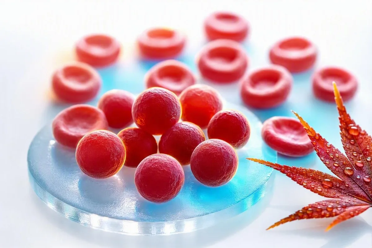
Un MCHC elevato è raro e spesso più utile come indizio che come diagnosi. La domanda pratica è se i globuli rossi siano veramente densi e sferici, o se il campione abbia distorto il calcolo.


- Intervallo MCHC è solitamente 32-36 g/dL (320-360 g/L) negli adulti, sebbene l'intervallo del laboratorio abbia la precedenza.
- MCHC elevato sopra 36 g/dL è insolito e dovrebbe indurre un controllo per sferociti, emolisi, ustioni o interferenze analitiche.
- Sferociti sono globuli rossi piccoli e densi senza pallore centrale e possono verificarsi in caso di sferocitosi ereditaria o anemia emolitica autoimmune.
- iperglicemia possono aumentare falsamente l'MCHC per aggregazione dei globuli rossi, abbassando il numero di RBC misurato e distorcendo gli indici calcolati.
- Lipemia può sovrastimare l'emoglobina su alcuni analizzatori, creando un MCHC elevato anche quando la forma dei globuli rossi è normale.
- Ustione può produrre un MCHC elevato vero attraverso la perdita della membrana dei globuli rossi, di solito associata ad anemia ed evidenze di laboratorio di emolisi.
- Indizi di emolisi includono un aumento dei reticolociti, della bilirubina indiretta e della LDH, con una bassa aptoglobina quando i globuli rossi si distruggono all'interno del corpo.
- Ripetere il test con un campione EDTA appena raccolto e correttamente manipolato è spesso il primo passo sensato quando l'MCHC è isolato e solo lievemente elevato.
MCHC elevato su un CBC: la risposta diretta
Un MCHC elevato di solito significa che i globuli rossi sono insolitamente densi, ma può anche significare che l'analizzatore è stato ingannato dal campione. Negli adulti, un MCHC superiore a circa 36 g/dL merita contesto: le vere elevazioni puntano più spesso a sferociti o a una significativa lesione termica, mentre agglutinine fredde, lipemia ed emolisi del campione sono frequenti spiegazioni di laboratorio. A partire dal 23 agosto 2026, non diagnosticherei un disturbo del sangue basandomi solo su questo indice.
L'MCHC è calcolato come emoglobina divisa per ematocrito, moltiplicato per 100, quindi è una concentrazione piuttosto che una misurazione diretta della cellula. Un'emoglobina di 15,0 g/dL ed ematocrito di 40% producono un MCHC di 37,5 g/dL; il numero può aumentare perché l'emoglobina è sovra-letta, l'ematocrito è sotto-letto, o entrambi. La nostra guida ai biomarcatori CBC è utile quando più indici sono segnalati contemporaneamente.
Quando revisiono un pannello con MCHC 36,5-37,5 g/dL ma emoglobina, bilirubina, reticolociti e RDW normali, un artefatto è spesso più probabile della malattia. Un MCHC di 39-42 g/dL è fisiologicamente difficile da mantenere in globuli rossi circolanti intatti, quindi il laboratorio dovrebbe considerare attivamente l'interferenza prima che qualcuno vada nel panico.
Kantesti è un Analizzatore di analisi del sangue AI che legge l'MCHC insieme all'emoglobina, all'ematocrito, all'MCV, all'RDW, alla bilirubina, alla LDH e ai risultati precedenti piuttosto che trattare un singolo segnale d'allarme come una diagnosi. La regola clinica del Dr. Thomas Klein è semplice: verificare l'aritmetica e il campione prima di ordinare un approfondimento esteso; il nostro dettagliato guida su RDW e indici dei globuli rossi spiega perché questo approccio basato sui pattern è importante.
MCHC non è MCH o MCV
L'MCHC descrive la concentrazione di emoglobina all'interno del volume dei globuli rossi compattati, mentre l'MCH descrive la quantità di emoglobina per cellula e l'MCV descrive la dimensione della cellula. Confondere questi tre indici è uno dei motivi per cui un normale esame CBC (emocromo completo) può sembrare più allarmante di quanto non sia.
L'MCH è comunemente 27-33 pg per cellula, mentre l'MCV è comunemente 80-100 fL negli adulti; nessun intervallo definisce l'MCHC. I globuli rossi grandi possono contenere più emoglobina e quindi aumentare l'MCH, tuttavia il loro MCHC può rimanere normale perché il loro volume è aumentato proporzionalmente. Vedere il nostro confronto pratico di MCV e MCH se entrambi i valori sono segnalati.
Un MCHC elevato genuino si associa spesso a un MCV normale o ai limiti inferiori della norma perché le sferociti hanno perso area superficiale di membrana e sono diventati più rotondi, non perché contengano emoglobina illimitata. Quella geometria è importante: una sfera trattiene meno superficie per il suo volume, quindi non ha un centro pallido su un vetrino di campione cellulare ben preparato.
Il punto è che un esame del sangue MCHC elevato può essere matematicamente reale pur essendo biologicamente fuorviante. Un'ematocrito leggermente sottostimato dopo aggregazione dei globuli rossi gonfierà il rapporto anche se i globuli rossi del paziente non sono affatto densi; ecco perché una revisione completa dell'emocromo dovrebbe precedere le conclusioni.
Sferociti: il classico vero indizio di MCHC elevato
Le sferociti sono la causa più riconoscibile di un vero MCHC elevato perché la perdita di membrana rende i globuli rossi compatti e densamente emoglobinizzati. Si osservano nella sferocitosi ereditaria e nell'anemia emolitica autoimmune, ma un reperto su striscio deve essere interpretato con i sintomi e i marcatori di emolisi.
La sferocitosi ereditaria produce spesso valori di MCHC superiori a 36 g/dL, reticolocitosi, ittero intermittente, calcoli biliari, splenomegalia o una storia familiare di anemia. La linea guida del 2011 della British Society for Haematology rileva che l'MCHC supporta la diagnosi ma non è sufficientemente sensibile o specifico per confermarla da solo (Bolton-Maggs et al., 2012).
Nell'anemia emolitica autoimmune, gli anticorpi contrassegnano i globuli rossi per la rimozione parziale della membrana nella milza, lasciando dietro le sferociti. Un test antiglobulinico diretto, un conteggio dei reticolociti, la bilirubina, l'LDH e l'aptoglobina sono più informativi insieme di qualsiasi singolo indice; la nostra guida per l'aptoglobina bassa oltre l'emolisi copre una limitazione facilmente trascurabile.
Una complicazione clinica: una trasfusione recente può diluire o mascherare il pattern di sferociti nativi del paziente per diverse settimane. Christensen et al. descrivono il legame dell'eosina-5-maleimide mediante citometria a flusso come un test utile per la sospetta sferocitosi ereditaria, sebbene un ematologo debba selezionare e programmare attentamente i test (Christensen et al., 2015).
Come capire se l'MCHC elevato è associato a emolisi
Un MCHC elevato diventa più clinicamente significativo quando accompagna anemia, un aumento del conteggio dei reticolociti, un aumento della bilirubina indiretta, un aumento dell'LDH e un'aptoglobina bassa. Quel raggruppamento indica un aumento del ricambio dei globuli rossi, sebbene nessun singolo marcatore sia perfetto.
Una percentuale di reticolociti superiore a 2% può essere ancora inadeguata in caso di anemia significativa, quindi i medici calcolano spesso invece un conteggio assoluto dei reticolociti o un indice di produzione dei reticolociti. Ad esempio, un conteggio di reticolociti 4% con ematocrito 24% può rappresentare solo una modesta risposta del midollo dopo correzione; il nostro articolo sui indizi di reticolociti elevati illustra la distinzione.
La bilirubina non coniugata aumenta quando l'eme viene processato più velocemente di quanto il fegato possa coniugarlo, mentre la LDH aumenta perché i globuli rossi contengono abbondante LDH. L'aptoglobina al di sotto del limite inferiore di laboratorio supporta l'emolisi intravascolare, ma può apparire normale durante l'infiammazione poiché l'aptoglobina è una proteina della fase acuta. Barcellini e Fattizzo discutono di questa trappola diagnostica nella loro revisione sull'anemia emolitica autoimmune (Barcellini & Fattizzo, 2015).
Urina scura, affaticamento in rapido aumento, affanno a riposo, dolore toracico, svenimento o occhi gialli con febbre meritano una valutazione clinica in giornata. Le cellule frammentate sono un indizio sullo striscio diverso e più urgente delle sferociti; la nostra spiegazione di quando gli schistociti richiedono una revisione urgente aiuta a chiarire perché.
Perché le ustioni possono causare un MCHC genuinamente elevato
Estese lesioni termiche possono causare un MCHC elevato reale perché il calore danneggia le membrane dei globuli rossi, producendo perdita di membrana, cellule simili a sferociti ed emolisi. Questo è generalmente un reperto ospedaliero dopo ustioni clinicamente evidenti, non una spiegazione per un risultato incidentale di CBC ambulatoriale.
Il danno termico può creare microsferociti entro ore da gravi ustioni, e l'MCHC può aumentare man mano che il rapporto membrana/volume diminuisce. L'emoglobina può anche diminuire nei giorni successivi a causa di spostamenti di liquidi, procedure, salasso ed emolisi in corso, quindi un singolo MCHC non può quantificare la gravità della lesione.
Ho occasionalmente visto pazienti concentrarsi su un MCHC di 37,1 g/dL dopo un ricovero d'urgenza trascurando anomalie molto più gestibili: emoglobina in calo, creatinina in aumento, alterazioni del potassio o lattato. In tale contesto, i medici gestiscono la persona e la fisiologia dell'ustione, non l'indice; il nostro cronologia dei test post-ospedalieri spiega perché i valori possono cambiare rapidamente.
Kantesti AI è un piattaforma di interpretazione dei biomarcatori AI che può segnalare un MCHC elevato insieme a emoglobina e risultati renali dinamici, ma le ustioni acute richiedono una valutazione ospedaliera diretta e non possono essere gestite in sicurezza dall'interpretazione di un referto. Un risultato dopo una grave ustione dovrebbe essere confrontato con le 24-72 ore precedenti, non con una media della popolazione generica.
Agglutinine a freddo: l'artefatto di MCHC elevato che i medici cercano
Le agglutinine fredde possono produrre un MCHC falsamente elevato facendo aggregare i globuli rossi a temperatura ambiente prima dell'analisi. L'analizzatore può contare diverse cellule attaccate come una singola particella, portando a un conteggio RBC falsamente basso, un MCV falsamente alto e un MCHC calcolato gonfiato.
Il classico schema delle agglutinine fredde è un basso conteggio RBC con un MCV sproporzionatamente alto, MCH alto e MCHC alto, spesso senza una storia clinica corrispondente. Il riscaldamento della provetta EDTA a 37°C e la ripetizione del CBC correggono comunemente i valori, il che è molto più informativo che ripetere un test casuale settimane dopo.
Le agglutinine fredde possono verificarsi transitoriamente dopo alcune infezioni respiratorie o persistere nella malattia da agglutinine fredde e in alcuni disturbi linfoproliferativi. Sintomi come cambiamento di colore o disagio alle dita con l'esposizione al freddo aggiungono peso clinico, ma il pattern di laboratorio stesso può essere il primo indizio; un test ripetuto dopo emolisi offre domande pratiche per il prelievo.
Non scaldarsi aggressivamente o cercare di manipolare il risultato a casa. Chiedere se il laboratorio ha riscontrato agglutinazione dei globuli rossi, se il campione è stato riscaldato e rianalizzato, e se è stato eseguito un ematocrito manuale; queste tre risposte spesso risolvono la questione.
La lipemia e l'emolisi del campione possono distorcere l'MCHC
La lipemia e l'emolisi in vitro del campione possono aumentare falsamente l'MCHC perché molti analizzatori misurano l'emoglobina fotometricamente dopo la lisi dei globuli rossi. I lipidi torbidi o l'emoglobina libera nel campione possono aumentare l'assorbanza ottica e far apparire l'emoglobina più alta di quanto non sia in realtà.
La lipemia è più spesso visibile dopo livelli molto elevati di trigliceridi, solitamente superiori a circa 400-500 mg/dL, sebbene l'interferenza dipenda dallo strumento e dalla lunghezza d'onda del saggio. Un laboratorio può utilizzare un bianco al plasma, una lunghezza d'onda alternativa o un approccio di sostituzione del plasma anziché accettare semplicemente il risultato automatizzato dell'emoglobina.
L'emolisi in vitro avviene dopo la raccolta o la manipolazione, non all'interno del corpo, e può interessare più della MCHC. Potassio, LDH, AST e fosfato possono aumentare in un campione danneggiato, motivo per cui un'elevata MCHC isolata insieme a un inaspettato aumento del potassio richiede un controllo della qualità del campione; vedere la nostra guida a errori correlati all'emolisi del potassio.
Uno strato di plasma visibilmente rosa o rosso è un indizio di laboratorio, non una prova di anemia emolitica nel paziente. Il follow-up pratico è una nuova raccolta con manipolazione calma e processamento tempestivo, specialmente se il referto originale include un indice di emolisi o un commento che i risultati potrebbero essere influenzati.
Problemi di raccolta, conservazione e provetta che modificano gli indici del CBC
Una miscelazione inadeguata del campione, un riempimento insufficiente, la coagulazione e l'analisi ritardata possono alterare gli indici della CBC, sebbene lo stoccaggio ritardato abbassi più spesso che aumenti la MCHC consentendo alle cellule di gonfiarsi. Pertanto, un'elevata MCHC dovrebbe essere letta con i commenti del laboratorio relativi al campione, non semplicemente il numero sul portale del paziente.
I campioni CBC sono generalmente raccolti in provette di EDTA e una provetta sotto-riempita può avere un rapporto anticoagulante/campione eccessivo che altera le misurazioni cellulari. Piccoli coaguli possono rimuovere selettivamente le cellule dall'analisi e rendere il conteggio automatizzato internamente incoerente; la nostra panoramica di additivi per provette ematiche spiega perché il tipo di provetta non è un dettaglio trascurabile.
La conservazione a temperatura ambiente per periodi prolungati fa sì che i globuli rossi assorbano acqua, aumentando l'MCV e tendendo a diminuire la MCHC. Questa direzione del cambiamento è utile: un'MCHC molto alta dopo un campione ritardato non è automaticamente spiegata solo dall'età, quindi la crioagglutinazione o l'interferenza ottica potrebbero essere ancora presenti.
Un campione capillare può essere appropriato in alcune situazioni ma è più incline alla variabilità di raccolta rispetto a un campione venoso EDTA ben raccolto. Se un'elevata MCHC non presenta sintomi corrispondenti o anomalie correlate, preferisco solitamente un prelievo venoso pulito prima di trattarlo come una nuova diagnosi cronica.
Utilizzare i CBC precedenti per decidere se la variazione è reale
Un improvviso aumento della MCHC da una linea di base stabile di 33-35 g/dL a 38 g/dL è più suggestivo di un problema del campione o dell'analizzatore rispetto alla sferocitosi ereditaria a lenta evoluzione. Le tendenze non sostituiscono uno striscio, ma sono un potente indizio di controllo qualità.
La maggior parte delle persone ha un intervallo MCHC personale relativamente ristretto nel tempo, che spesso varia meno di 1 g/dL quando le condizioni di test sono comparabili. Un controllo delta di laboratorio confronta un nuovo risultato con uno precedente per identificare cambiamenti implausibili prima che i risultati vengano rilasciati; il nostro articolo su controlli delta mostra come funziona quella rete di sicurezza.
Kantesti è un servizio di interpretazione dei test del laboratorio di IA che confronta i valori CBC attuali e precedenti, aiutando a distinguere un segnale persistente da un outlier singolo. Nel nostro flusso di lavoro di revisione, un cambiamento di MCHC è più importante quando viaggia insieme a emoglobina, MCV, bilirubina e reticolociti in una direzione biologicamente coerente.
Ci sono eccezioni. Una nuova emolisi autoimmune, una grave ustione, una reazione trasfusionale o una marcata crioagglutinazione possono cambiare genuinamente i risultati rapidamente, quindi un valore storico stabile è rassicurante ma mai un motivo per ignorare un nuovo ittero, urine scure o anemia.
Condizioni comunemente attribuite all'MCHC ma raramente responsabili
La disidratazione, la carenza di ferro, la carenza di vitamina B12 e lo stress ordinario di solito non causano un'MCHC veramente elevata. Possono cambiare altri valori della CBC, ma non dovrebbero essere utilizzati come spiegazioni convenienti per un'MCHC di 38 g/dL senza confermare il campione.
La carenza di ferro tipicamente abbassa MCH e MCHC e spesso abbassa MCV una volta che la carenza è stabilita. La carenza di ferro precoce può avere un MCV normale, ma non spiega indici ipercromici; la nostra spiegazione di saturazione della transferrina bassa aiuta a separare la disponibilità di ferro dalla concentrazione di CBC.
La carenza di vitamina B12 o folati causa più spesso macrocitosi, con MCV superiore a 100 fL, piuttosto che un'elevata MCHC. Se MCV e MCH sono entrambi alti ma MCHC è normale, quella è una diversa direzione diagnostica; vedere indizi di follow-up per MCV elevato.
La carenza di G6PD causa emolisi episodica dopo specifici trigger ossidativi in persone suscettibili, ma la MCHC non è un test di screening affidabile per essa. Durante un episodio acuto, lo striscio e i marcatori di turnover possono essere più rivelatori, mentre i test enzimatici potrebbero dover essere ripetuti dopo il recupero perché i globuli rossi giovani contengono più G6PD.
I test di follow-up più utili dopo un MCHC elevato
Il miglior passo successivo per un'elevata MCHC isolata è di solito un CBC ripetuto con revisione di laboratorio, seguito da uno striscio periferico e un pannello di emolisi solo se l'elevazione persiste o sono presenti altri indizi. Ordinare subito ogni test per l'anemia può creare più rumore che risposte.
Una revisione mirata di prima linea include CBC ripetuto, revisione del vetrino del campione cellulare, conta dei reticolociti, bilirubina totale e indiretta, LDH, aptoglobina e test diretto all'antiglobulina quando l'emolisi autoimmune è plausibile. Lo striscio può identificare sferociti, agglutinazione, frammenti, policromasia o una morfologia normale che rende più probabile un artefatto.
Se la sferocitosi ereditaria rimane probabile, un ematologo può considerare il legame con l'eosin-5-maleimide, il test di fragilità osmotica o il test genetico. Questi test hanno diversi rischi di falsi negativi dopo la trasfusione e durante l'emolisi acuta, motivo per cui l'ordine dei test dovrebbe seguire una storia clinica piuttosto che un elenco di caselle da selezionare; la nostra guida ai test G6PD illustra il problema più ampio della tempistica.
Kantesti AI interpreta un'elevata MCHC controllando il calcolo rispetto ai marcatori CBC ed emolitici correlati, quindi identificando domande da porre a un clinico. Non sostituisce la revisione della morfologia cellulare, in particolare quando il laboratorio ha emesso un flag dell'analizzatore o quando l'anemia sta peggiorando.
Quando un MCHC elevato richiede una valutazione medica urgente
Un'elevata MCHC di per sé non è una soglia di emergenza, ma un'elevata MCHC con segni di emolisi acuta, anemia grave o ustione grave può richiedere cure nello stesso giorno. I sintomi e la velocità di cambiamento determinano l'urgenza molto meglio che se il flag è contrassegnato come H.
Richiedere una valutazione urgente per nuova dispnea a riposo, dolore toracico, svenimento, confusione, debolezza in rapido peggioramento, urine color tè o ingiallimento degli occhi con febbre. Questi sintomi possono accompagnare un'anemia o un'emolisi significativa, specialmente quando l'emoglobina è scesa di oltre 2 g/dL rispetto a un basale noto.
Contattare il clinico che ha ordinato il test entro pochi giorni se la MCHC rimane superiore a 36 g/dL su un campione ripetuto, è presente anemia o il referto menziona agglutinazione o sferociti. In una persona stabile e in buone condizioni con MCHC di 36,2 g/dL e risultati altrimenti normali, un nuovo prelievo non urgente è spesso ragionevole; il nostro guida al pattern della bilirubina può aiutare a inquadrare la prossima discussione.
Non iniziare ferro, folati, steroidi o integratori semplicemente per correggere la MCHC. Nessuno di questi tratta le vere cause principali, e gli steroidi in particolare possono oscurare la valutazione dell'emolisi autoimmune se iniziati senza supervisione clinica.
Come [Kantesti] inserisce l'MCHC nel contesto clinico
Kantesti legge un'elevata MCHC come un problema di riconoscimento di pattern: veri disturbi delle cellule dense e artefatti del campione producono diverse combinazioni di CBC, chimica, flag e trend precedenti. L'obiettivo è rendere la prossima domanda clinica più chiara, non etichettare una malattia rara da un singolo indice calcolato.
Kantesti è un Strumento di analisi degli esami del sangue basato su AI utilizzato da oltre 2 milioni di persone in 127 paesi per organizzare i risultati di laboratorio nel contesto. Per MCHC elevato, la nostra IA verifica se emoglobina ed ematocrito supportano matematicamente il risultato, se MCV è inaspettatamente alto e se bilirubina, LDH, reticolociti o aptoglobina indicano un ricambio dei globuli rossi.
La nostra piattaforma cerca anche contraddizioni interne: MCHC 40 g/dL con commento normale sullo striscio e nessuna anemia può giustificare un avviso di artefatto, mentre MCHC 37 g/dL con sferociti, reticolocitosi e aumento della bilirubina indiretta merita un follow-up medico. L'approccio tecnico e i limiti sono descritti nel nostro Guida alla tecnologia AI.
Consiglio ai pazienti di conservare il PDF del laboratorio, la data del prelievo, lo stato di digiuno, la malattia recente, la storia trasfusionale e qualsiasi commento di laboratorio. Quel piccolo registro spiega spesso una discrepanza apparente più rapidamente che ripetere test ampi, specialmente quando i risultati vengono confrontati mesi dopo.
Domande che rendono un appuntamento per MCHC elevato più produttivo
Chiedere se l'MCHC è stato ripetuto, se il laboratorio ha visto sferociti o agglutinazione e se sono presenti marcatori di anemia o emolisi. Queste domande spostano la conversazione da “perché un numero è alto?” a un percorso clinico concreto e verificabile.
Una formulazione utile include: “Il campione era emolitico o lipemico?”, “Lo strumento ha segnalato agglutinazione dei globuli rossi?” e “Possiamo confrontare questo risultato con il mio MCHC precedente?” Una revisione manuale dello striscio è particolarmente preziosa quando l'MCHC è ripetutamente superiore a 36 g/dL o l'anemia è inspiegabile.
Il Dr. Thomas Klein raccomanda di portare un elenco di farmaci, infezioni recenti, sintomi scatenati dal freddo, storia familiare di calcoli biliari o anemia e trasfusioni negli ultimi 3 mesi. Tale storia può indicare sferocitosi ereditaria, emolisi autoimmune o un agglutinina fredda transitoria molto più velocemente di una generica ricerca online.
Il contenuto medico di Kantesti viene revisionato con supervisione medica, ma il tuo clinico può visitarti, richiedere una ripetizione a caldo e parlare direttamente con il laboratorio. I lettori che desiderano comprendere il nostro quadro di revisione clinica possono incontrare il comitato consultivo medico, mentre le anomalie persistenti o sintomatiche devono sempre essere discusse di persona.
Ricerca, incertezza e limiti di un singolo MCHC
L'MCHC è un utile indizio di screening, non un test diagnostico autonomo per sferocitosi ereditaria, emolisi autoimmune, ustioni o malattia da agglutinine fredde. Il suo valore aumenta notevolmente quando un campione ripetuto, la morfologia cellulare, i marcatori di turnover e la storia clinica raccontano tutti la stessa storia.
Nessun cut-off universale dell'MCHC diagnostica la sferocitosi ereditaria perché i metodi analitici, l'età, la reticolocitosi e la carenza di ferro concomitante possono alterare la sensibilità. La linea guida BSH supporta la combinazione di storia familiare, reperti dello striscio, reticolociti, bilirubina e test di conferma dove necessario piuttosto che affidarsi a una singola soglia (Bolton-Maggs et al., 2012).
Kantesti supporta un follow-up informato identificando schemi di risultati e incertezze, non emettendo una diagnosi ematologica definitiva. Il nostro approccio di validazione clinica spiega perché valori insoliti, flag analitici e sintomi di emergenza rimangono motivi per la revisione da parte del laboratorio e del clinico.
Per un contesto ematologico correlato, vedere la pubblicazione di ricerca Tipo sanguigno B negativo, guida per esame del sangue LDH e conta dei reticolociti e la risorsa più ampia sulla comunicazione clinica Diarrea dopo il digiuno, macchie nere nelle feci e guida gastrointestinale 2026. Entrambi sono pubblicazioni educative supplementari piuttosto che sostituti di uno striscio periferico o di una valutazione specialistica.
Domande frequenti
Cosa significa un livello elevato di MCHC in un esame del sangue?
Un livello elevato di MCHC significa solitamente che la concentrazione di emoglobina all'interno del volume dei globuli rossi è superiore all'intervallo di laboratorio, comunemente superiore a 36 g/dL negli adulti. Vere elevazioni si verificano con sferociti da sferocitosi ereditaria, anemia emolitica autoimmune o grave ustione termica. Agglutinine fredde, lipemia e emolisi del campione possono anche creare un falso risultato elevato. Un CBC ripetuto, la revisione del vetrino del campione cellulare e i marcatori di emolisi chiariscono quale spiegazione si adatta.
L'MCHC alto è pericoloso?
Un valore elevato di MCHC non è di per sé pericoloso; è un indice calcolato del CBC piuttosto che una malattia. Un MCHC di 36,2 g/dL con emoglobina normale e assenza di sintomi viene spesso gestito con un test di controllo di routine, mentre un MCHC elevato associato a calo dell'emoglobina, ittero, urine scure o affanno richiede una valutazione tempestiva. Valori superiori a 39 g/dL riflettono più probabilmente interferenze da parte del campione o dello strumento. Il rischio clinico deriva dalla causa sottostante, come un'emolisi significativa, non dal solo valore di MCHC.
La disidratazione può causare un MCHC elevato?
La disidratazione di solito non causa un MCHC veramente alto perché concentra il volume plasmatico più di quanto modifichi la concentrazione di emoglobina all'interno dei singoli globuli rossi. La disidratazione può aumentare l'emoglobina e l'ematocrito insieme, lasciando l'MCHC nell'intervallo abituale di 32-36 g/dL. Se l'MCHC è 37-40 g/dL, i medici dovrebbero considerare invece sferociti, agglutinazione fredda, lipemia o un problema di misurazione. Una ripetizione di un CBC ben eseguito è più utile che cercare di correggere il valore con liquidi aggiuntivi.
Gli sferociti rendono sempre alto il MCHC?
Gli sferociti spesso aumentano l'MCHC sopra 36 g/dL, ma non sempre lo fanno e un MCHC elevato non dimostra la presenza di sferociti. La sferocitosi ereditaria può essere mascherata da carenza di ferro, trasfusione recente o malattia lieve, mentre l'emolisi autoimmune può mostrare reperti variabili a seconda del momento. È necessario un campione di cellule strisciato rivisto da un professionista esperto di laboratorio per identificare gli sferociti. La conta dei reticolociti, la bilirubina, la LDH, l'aptoglobina e il test diretto dell'antiglobulina aggiungono contesto diagnostico.
Gli agglutinini freddi possono causare un MCHC elevato?
Gli agglutinanti freddi possono falsare verso l'alto l'MCHC raggruppando i globuli rossi prima che venga misurato il CBC automatizzato. Il pattern spesso include un basso numero di RBC, un MCV elevato, un MCH elevato e un MCHC superiore a 36 g/dL che non corrisponde ai sintomi del paziente. Il riscaldamento del campione di EDTA a 37°C e la sua rianalisi possono normalizzare i conteggi. La persistente agglutinazione fredda o i sintomi di emolisi devono essere valutati da un medico o da un ematologo.
Quali test dovrei richiedere dopo un MCHC elevato?
Dopo un MCHC persistentemente elevato, i test successivi usuali sono un CBC ripetuto, revisione dello striscio del campione cellulare, conta dei reticolociti, frazioni della bilirubina, LDH, aptoglobina e talvolta un test diretto dell'antiglobulina. Un clinico può richiedere il legame eosina-5-maleimide o altri test specializzati se si sospetta una sferocitosi ereditaria. Un MCHC superiore a 36 g/dL con test ripetuti normali e assenza di anemia potrebbe non richiedere un'indagine approfondita. La migliore sequenza di test dipende dai sintomi, dai valori precedenti del CBC, dall'anamnesi familiare e dai flag di laboratorio.
Ottieni oggi l’analisi degli esami del sangue con IA (AI-Powered Blood Test Analysis)
Unisciti a oltre 2 milioni di utenti in tutto il mondo che si fidano di Kantesti per un’analisi istantanea e accurata degli esami di laboratorio. Carica i tuoi risultati analisi del sangue e ricevi un’interpretazione completa dei biomarcatori di 15,000+ in pochi secondi.
📚 Pubblicazioni di ricerca di riferimento
Klein, T., Mitchell, S., & Weber, H. (2026). Guida al gruppo sanguigno B negativo, al test del LDH e al conteggio dei reticolociti. Kantesti AI Medical Research.
Klein, T., Mitchell, S., & Weber, H. (2026). Diarrea dopo il digiuno, macchie nere nelle feci e guida gastrointestinale 2026. Kantesti AI Medical Research.
📖 Riferimenti medici esterni
⚕️ Esclusione di responsabilità medica
Questo articolo ha solo scopo educativo e non costituisce consulenza medica. Consulta sempre un operatore sanitario qualificato per decisioni su diagnosi e trattamento.
Segnali di fiducia E-E-A-T
Esperienza
Revisione clinica guidata da un medico dei flussi di lavoro di interpretazione degli esami.
Competenza
Focus sulla medicina di laboratorio su come i biomarcatori si comportano nel contesto clinico.
autorevolezza
Scritto dal dott. Thomas Klein con revisione della dott.ssa Sarah Mitchell e del Prof. Dr. Hans Weber.
Affidabilità
Interpretazione basata su evidenze, con percorsi di follow-up chiari per ridurre l’allarme.