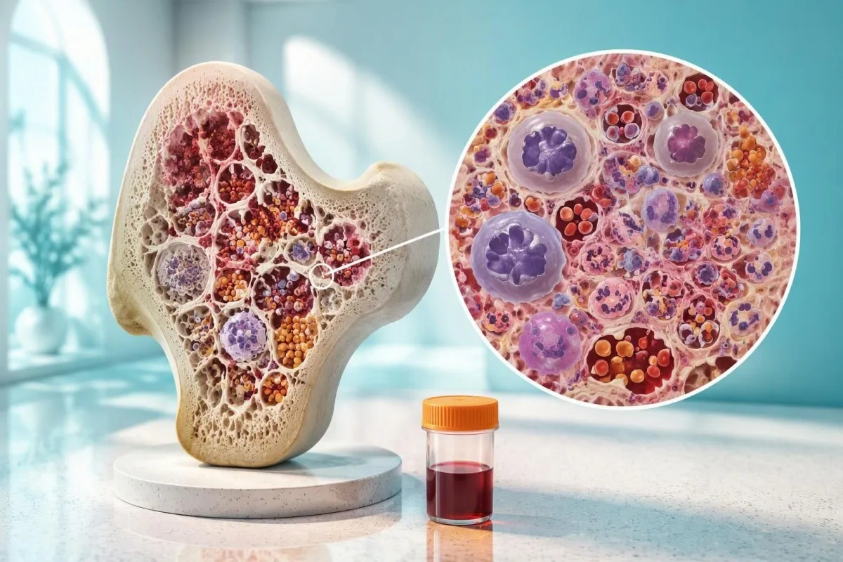
Blood tests can raise suspicion for MDS, but they cannot confirm it. Persistent low counts need an evaluation that connects bone marrow findings with chromosome and mutation results.


- Diagnostic limit: No single myelodysplastic syndrome blood test confirms MDS; diagnosis generally requires bone marrow assessment and exclusion of other causes.
- Persistent cytopenia: Low hemoglobin, neutrophils, or platelets lasting around 4 months can support evaluation, but worsening counts require earlier assessment.
- Macrocytosis: An MCV above 100 fL can accompany MDS, vitamin B12 deficiency, alcohol effects, medications, or thyroid disease.
- Marrow morphology: Dysplasia affecting at least 10% of an assessed marrow cell lineage is a conventional diagnostic threshold, not a finding a routine CBC can measure.
- Clonal findings: A mutation with a variant allele fraction of at least 2% can meet a conventional clonal hematopoiesis threshold; it does not automatically establish MDS.
- Reversible mimics: Vitamin B12, folate, and copper deficiencies can cause cytopenias and marrow changes resembling MDS.
- Urgent assessment: Fever of 38°C or higher with severe neutropenia, particularly an ANC below 0.5 × 10^9/L, needs immediate medical evaluation.
- Classification matters: WHO and ICC terminology differs for some cases with 10–19% blasts; the report should identify which classification was used.
Can a blood test diagnose myelodysplastic syndrome?
A myelodysplastic syndrome blood test cannot confirm MDS by itself. A complete blood count can identify low counts in 1 or more cell lines, but diagnosis generally requires bone marrow examination, exclusion of alternative causes, and interpretation of chromosome or mutation findings.
MDS is a clonal disorder of marrow blood-forming cells, now called myelodysplastic neoplasms in the WHO classification. The marrow may contain plenty of developing cells while producing too few effective circulating cells; that mismatch helps explain why 2 patients with similar hemoglobin can have very different underlying conditions.
A normal-looking white cell count does not cancel an abnormal hemoglobin or platelet trend. Conversely, a single low result does not establish cancer: an isolated platelet count of 125 × 10^9/L can reflect sample clumping, medication effects, liver disease, or several other explanations.
Kantesti is an AI blood test analyzer that helps explain CBC abnormalities and compare results across 2 or more reports, without diagnosing MDS. Our organización e misión clínica distinguish interpretation support from the hematologist's responsibility to establish a marrow diagnosis.
I am Dr. Thomas Klein, Chief Medical Officer at Kantesti, and my approach to a panel showing 2 low cell lines is to ask what connects them—not to label the patient from a red flag. The 2022 WHO framework likewise integrates clinical, morphologic, and genetic evidence rather than treating a CBC pattern as diagnostic proof (Khoury et al., 2022).
Which CBC results can suggest MDS?
Anemia, neutropenia, and thrombocytopenia can suggest MDS when they are unexplained and persistent, particularly when more than 1 cell line is affected. Macrocytosis adds context, but neither a high MCV nor a low count identifies the cause on its own.
The 2022 ICC uses hemoglobin below 13 g/dL in men or 12 g/dL in women, ANC below 1.8 × 10^9/L, and platelets below 150 × 10^9/L as cytopenia thresholds in this diagnostic context. These are assessment thresholds, not boundaries that make MDS likely or unlikely by themselves (Arber et al., 2022).
Older MDS scoring systems often use more pronounced abnormalities, including hemoglobin below 10 g/dL or platelets below 100 × 10^9/L. Those prognostic cutoffs should not become a gatekeeper for referral: a patient can have MDS with milder abnormalities, while another patient with much lower counts has a reversible condition.
An MCV above 100 fL indicates macrocytosis in most adult laboratories, although local ranges differ. Reviewing patróns de MCV límite helps separate a steadily rising value from a small reference-range crossing, especially after treatment, transfusion, or a change in medication.
RDW describes variation in red cell size rather than proving marrow dysplasia. An MCV of 104 fL with elevated RDW could reflect MDS, combined iron and B12 deficiency, or recovery after treatment; our guía de interpretación do RDW explains why averages can conceal mixed cell populations.
How long must low blood counts persist before evaluation?
Unexplained cytopenias persisting for approximately 4 months can support assessment for a clonal marrow disorder, but this is not a mandatory waiting period. Rapid deterioration, circulating blasts, severe cytopenias, or concerning symptoms justify earlier hematology evaluation.
Persistence means a repeated pattern, not merely 2 abnormal tests collected during the same illness. A CBC obtained during a viral episode and another 3 days later may describe one temporary event; an abnormal result months before that illness changes the reasoning substantially.
The direction of travel matters as much as the absolute value. A platelet decline from 190 to 140 to 95 × 10^9/L deserves a different conversation from a stable count around 140 for several years, although neither trajectory alone establishes MDS.
Kantesti can help organize serial CBC reports, but a trend is only useful when the dates and circumstances are correct. Our explanation of cambios entre análises de sangue covers biological variation, measurement variation, and why a small difference between 2 visits may not represent disease progression.
Repeat timing should match severity and suspected cause, not a universal calendar. A clinician may repeat a mild, stable abnormality after recovery over several weeks, whereas a falling ANC near 0.5 × 10^9/L or newly reported blasts calls for prompt action rather than another routine appointment.
What are the early signs of myelodysplastic syndrome?
Early signs of myelodysplastic syndrome are usually consequences of low counts: fatigue or breathlessness from anemia, recurrent infections from neutropenia, and bruising or bleeding from thrombocytopenia. Some patients have no symptoms and first learn about an abnormality through 1 routine CBC.
Fatigue does not distinguish MDS from iron deficiency, kidney disease, or many nonhematologic conditions. Breathlessness with hemoglobin of 9 g/dL might be largely anemia-related, but the same symptom with hemoglobin of 12 g/dL should not automatically be attributed to a mildly abnormal CBC.
Repeated infections are more informative when paired with an absolute neutrophil count, medication history, and infection type. Our guide to probas para infeccións frecuentes explains why 3 mild respiratory illnesses are not equivalent to repeated unusual or severe infections.
Fever of 38°C or higher in someone with known severe neutropenia requires immediate medical assessment, especially when ANC is below 0.5 × 10^9/L. Do not wait for an AI explanation or the next planned repeat test; a low white cell response can make serious infection look deceptively quiet.
Active bleeding, black stools, new neurologic symptoms, chest pain, or breathlessness at rest also warrant urgent care. A platelet count below 10 × 10^9/L is a particularly high-risk result, while decisions around hemoglobin near 7–8 g/dL depend on symptoms, cardiovascular disease, and the clinical setting.
What can a peripheral blood smear show in suspected MDS?
A peripheral smear can reveal abnormal cell shapes or maturation patterns that strengthen suspicion for MDS, including hypogranular neutrophils and unusually segmented nuclei. However, abnormalities in 1 smear are not equivalent to diagnostic marrow dysplasia.
Pseudo-Pelger–Huët neutrophils have reduced nuclear segmentation, sometimes with only 2 prominent lobes. They can occur in MDS, but medications and other illnesses can produce similar appearances; the laboratory comment needs to be connected to the patient's exposures and the proportion of affected cells.
Large or oddly granulated platelets can accompany abnormal marrow production, but platelet size is not a dependable stand-alone discriminator. A platelet count of 80 × 10^9/L with large platelets might reflect increased peripheral turnover, a marrow disorder, or an inherited platelet condition.
Teardrop cells are not specific for MDS, and their context matters more than finding 1 unusual cell. Numerous teardrops accompanied by nucleated red cells and immature myeloid cells can suggest marrow fibrosis or infiltration; our teardrop cell interpretation explains this broader differential.
A report mentioning blasts should be reviewed promptly rather than converted into a diagnosis from the percentage alone. Even 1 newly identified circulating blast may justify clarification of the manual differential and hematology review; an automated analyzer's blast flag can also require confirmation by an experienced laboratory professional.
Which reversible conditions can mimic MDS?
Vitamin B12, folate, and copper deficiencies can mimic MDS by causing cytopenias and abnormal marrow maturation. Alcohol exposure, medications, thyroid disease, and some infections provide additional explanations, so checking reversible causes is part of diagnosis—not an optional step after 1 abnormal CBC.
B12 deficiency can produce macrocytosis, low counts in multiple cell lines, and striking marrow abnormalities. A result around 200–300 pg/mL may need further assessment depending on the assay and symptoms; our guía de seguimento de B12 límite explains methylmalonic acid testing and the confounding effect of reduced kidney function.
Copper deficiency deserves particular attention after bariatric surgery, malabsorption, or prolonged excessive zinc exposure. For example, a person taking 50 mg of elemental zinc daily for months may develop impaired copper absorption; our low copper causes guide explains why anemia plus neutropenia is a useful clue.
An unexpectedly low platelet count can be an artifact rather than a true cytopenia. When platelets read 65 × 10^9/L but the smear shows clumps, the laboratory may request a different collection tube or another counting method; a agregación de plaquetas con EDTA should be excluded before building a marrow-disease narrative.
Improvement after treating a deficiency supports its contribution, but partial recovery does not exclude a second condition. If hemoglobin rises from 9 to 11 g/dL while neutropenia persists, the clinician should reassess the remaining abnormality rather than assuming every count had the same cause.
Which additional blood tests help explain cytopenias?
Additional blood tests look for alternative causes and clarify whether cells are underproduced or being destroyed. A focused evaluation commonly includes a reticulocyte count, B12 and folate, iron studies, kidney and liver tests, and thyroid testing; copper and other investigations depend on the findings in 1 individual patient.
An inadequate reticulocyte response during anemia suggests insufficient effective red cell production, but does not identify MDS specifically. A reticulocyte percentage of 2% can be misleading when hemoglobin is substantially reduced; an absolute count or corrected production estimate better reflects whether the marrow response is appropriate.
Hemolysis assessment may include LDH, bilirubin, haptoglobin, and a direct antiglobulin test when clinically indicated. An LDH result of 500 IU/L is not a marrow-cancer marker: its interpretation depends on the laboratory range, other tissues, sample integrity, and evidence described in our direct Coombs interpretation guide.
Duffy-null–associated neutrophil counts can explain a stable lower ANC without the infection risk expected from marrow failure. An isolated ANC of 1.2 × 10^9/L in an otherwise well person with a longstanding baseline differs from new multilineage cytopenia; see our Duffy-null neutrophil count guide rather than making assumptions from ancestry alone.
Kantesti is an AI blood test interpretation platform that places CBC findings alongside available nutrient, kidney, and liver results, including results from 2 separate visits. Missing tests remain missing information—not normal findings—and no software can rule out MDS because a limited uploaded panel contains no obvious reversible explanation.
Why is an MDS bone marrow biopsy usually necessary?
An MDS bone marrow biopsy, usually accompanied by an aspirate, examines the tissue where blood cells develop. The 2 specimens provide complementary information: the aspirate assesses individual cells and blast proportions, while the core assesses cellularity, architecture, fibrosis, and possible infiltration.
Aspirate and core findings cannot be reconstructed from a routine CBC. A patient with 3 low circulating cell lines may have a hypercellular marrow with ineffective production, a hypocellular marrow, or an infiltrated marrow; those alternatives can lead to very different diagnoses and management.
The usual sampling site is the posterior pelvic bone, not the spinal cord. Local anesthetic is standard, and sedation options vary; patients should ask about the expected 2 components of sampling, pain control, anticoagulants, and whether someone needs to accompany them home.
Aplastic anemia and hypocellular MDS can overlap clinically, making the marrow architecture and supporting studies especially valuable. Our guía de CBC para anemia aplásica explains why low reticulocytes and 2 or 3 cytopenias identify a production problem without naming its cause.
A poor-quality or diluted aspirate can underestimate abnormal cells, while an inadequate core can limit assessment of patchy changes. When the report describes a dry tap or limited material, ask whether the sample answered all diagnostic questions; occasionally a second examination or expert pathology review is more useful than ordering another broad blood panel.
How do marrow dysplasia and blast counts establish MDS?
Marrow dysplasia and blast percentages help establish and classify MDS after competing explanations are addressed. A conventional morphologic threshold is dysplasia in at least 10% of an assessed marrow lineage, although certain genetically defined categories have additional or different requirements.
The 10% dysplasia threshold refers to marrow morphology, not 10% abnormal values on a CBC. Even above that threshold, severe nutritional deficiency or drug effects can complicate interpretation; the pathologist needs clinical information, not just a specimen and a request to exclude MDS.
In relevant low-blast categories, marrow blasts are below 5% and peripheral blood blasts are below 2%, with category-specific exclusions. Those numbers describe classification once the broader evidence is assessed; a patient with marrow blasts of 3% does not have MDS solely because a percentage appears on the report.
WHO and ICC differ in their handling of some cases with 10–19% blasts. The ICC includes an MDS/AML category for appropriate cases, whereas WHO retains MDS with increased blasts-2; AML-defining genetic abnormalities can change the classification, so a universal 20% rule is misleading (Khoury et al., 2022; Arber et al., 2022).
Borderline morphology may justify an expert hematopathology review before a lifelong diagnostic label is applied. Our enfoque de validación clínica explains why interpretation standards and uncertainty reporting matter; an automated explanation of 1 pathology report cannot substitute for direct review of the slides.
What do cytogenetic tests add to an MDS evaluation?
Cytogenetic tests identify acquired chromosome abnormalities that can support MDS classification and prognosis. Conventional karyotyping examines dividing cells, usually from marrow, while FISH looks for selected abnormalities; a normal result from either of these 2 approaches does not exclude MDS.
Deletion of chromosome 5q is associated with a specific MDS category when the complete criteria are met. The chromosome finding can also influence treatment discussions, but del(5q) is not interpreted in isolation from blast count, additional abnormalities, and the rest of the diagnostic work-up.
Conventional chromosome analysis often aims to assess at least 20 metaphases when sufficient dividing cells are available. A report based on fewer evaluable cells may have reduced sensitivity for a small clone; that limitation should appear in the laboratory interpretation rather than being hidden behind the word normal.
FISH is targeted, not a complete chromosome survey. A negative panel examining 4 or 5 selected regions cannot exclude abnormalities elsewhere, and some clonal chromosome findings are less specific for MDS than others; the question is which abnormality was found and whether it satisfies the chosen classification.
Not every hematologic genetic test is an MDS test. For example, BCR-ABL1 result interpretation concerns a different diagnostic pathway; that test may be appropriate for selected competing diagnoses, but a negative BCR-ABL1 result supplies no stand-alone reassurance about MDS.
Can molecular blood testing distinguish MDS from a harmless clone?
Molecular testing can detect clonal hematopoiesis, but a mutation alone does not distinguish MDS from CHIP or CCUS. A conventional variant allele fraction threshold of 2% is used for CHIP and CCUS definitions, while the mutation pattern, clone size, cytopenias, and marrow findings determine its meaning.
Variant allele fraction is the proportion of sequencing reads containing a variant, not a direct percentage of marrow replaced by cancer. A VAF of 10% can arise from approximately 20% heterozygous cells under simplified assumptions, but copy-number changes, sample composition, and other biological factors can alter that relationship.
Common age-associated mutations include DNMT3A, TET2, and ASXL1, while spliceosome mutations and combinations of abnormalities may carry different implications. In a 2017 study of unexplained cytopenias, Malcovati and colleagues found that mutation patterns and clone size improved diagnostic prediction; their results do not justify treating every positive panel as MDS (Malcovati et al., 2017).
Some genetic findings help define MDS categories, including qualifying SF3B1 or TP53 abnormalities, but each category has further requirements. With TP53, 1 reported mutation is not automatically equivalent to biallelic inactivation; the laboratory may need copy-number or loss-of-heterozygosity information to establish the relevant genetic state.
Kantesti can explain terminology in an uploaded laboratory report, but interpreting a negative sequencing panel requires knowing which genes and variant types were tested. Our blood test technology guide describes interpretation support; no AI explanation can infer an unmeasured chromosome change from 1 negative molecular result.
What is the difference between CHIP, CCUS, ICUS, and MDS?
CHIP is clonal hematopoiesis without unexplained cytopenia; CCUS combines a clone with unexplained cytopenia but lacks criteria for a myeloid neoplasm. ICUS describes unexplained cytopenia without established clonality or diagnostic MDS findings, while MDS meets integrated neoplasm criteria; 1 mutation cannot settle these distinctions.
A CHIP mutation may be incidental to an unrelated anemia. Consider an illustrative 72-year-old with a small DNMT3A clone and clearly documented iron deficiency: the clinician must first determine whether iron deficiency explains the cytopenia rather than automatically relabeling the combination as CCUS.
CCUS is not simply early MDS with a different name. Some patients remain stable, while others progress; 2 or more mutations, larger clones, selected higher-risk genes, and worsening counts can influence surveillance, but a single average progression percentage is not a reliable forecast for an individual patient.
ICUS does not mean that symptoms are imaginary or that follow-up is unnecessary. It means the current investigation has not established the cause; repeat assessment may be appropriate if a hemoglobin of 11 g/dL falls further, another cell line becomes low, or new smear abnormalities appear.
Other marrow-failure-associated clones need different tests and interpretation. citometría de fluxo para HPN can identify a distinct type of clone in selected patients; a small PNH clone does not independently diagnose MDS, just as a myeloid sequencing result cannot replace the specialized PNH assay.
What should you do after an abnormal CBC or mutation result?
After an abnormal CBC, confirm the result, review previous counts, assess reversible causes, and agree on referral or monitoring with a clinician. After a positive mutation panel, ask whether the finding explains the cytopenia and whether marrow assessment is needed; do not make treatment decisions from 1 result.
Bring the complete reports rather than screenshots of selected red flags. A useful packet includes at least 2 dated CBCs when available, the differential and smear comments, nutritional results, current medications and supplements, and any history of chemotherapy, radiation, transfusion, or major gastrointestinal surgery.
Three practical questions often clarify the next appointment: which cell lines are affected, what alternatives have been excluded, and what specific finding would trigger marrow examination? A patient with stable mild anemia may reasonably have a different timetable from someone whose platelets have fallen below 50 × 10^9/L.
Kantesti is an AI-powered blood test analysis tool that can organize serial CBC information for a clinician discussion, rather than confirm MDS in approximately 60 seconds. Before sharing 2 or more reports, review our report upload privacy steps and remove identifiers that are unnecessary for interpretation.
Monitoring for CCUS is individualized; clinicians may choose CBC review every 3–6 months for some stable patients, with shorter intervals for concerning changes. Our información sobre privacidade e datos explains platform handling, but neither a follow-up schedule nor a digital alert should delay urgent assessment of fever, bleeding, or worsening breathlessness.
Research publications and the practical limits of MDS testing
The diagnostic evidence for MDS comes from hematology classification standards and integrated clinical studies, not from general biomarker guides. As of October 22, 2026, the 2022 WHO and ICC publications remain the classification sources cited here; the additional 2 Zenodo guides below provide background reading, not diagnostic validation for MDS.
Serum protein results may help investigate selected alternative explanations for anemia, but an albumin/globulin ratio of 1.0 cannot confirm or exclude MDS. Background citation: guía de proteínas séricas: proba de sangue de globulinas, albúmina e relación A/G. (s. d.). Zenodo. https://doi.org/10.5281/zenodo.18316300; related interpretación proteína sérica explains the clinical context.
That serum-protein guide is also discoverable through Busca de publicacións en ResearchGate e Busca de publicacións en Academia; these are search links, not verified copies of the publication. A repository DOI identifies an archived item, but 1 DOI does not establish peer review, guideline endorsement, or clinical diagnostic accuracy.
Complement and ANA testing belongs to a targeted autoimmune differential, not a routine MDS confirmation panel. Background citation: Guía de proba de sangue C3 C4 do complemento e título ANA. (s. d.). Zenodo. https://doi.org/10.5281/zenodo.18353989; o noso guía de complemento e ANA discusses why an ANA titer of 1:80 requires clinical context.
The complement guide has ResearchGate search access e Academia search access, again without implying verified hosted copies. My practical advice as Dr. Thomas Klein, Kantesti's Chief Medical Officer, is to connect 3 elements—persistent counts, marrow evidence, and genetic context—while following our estándares de contido médico; this article is educational and cannot determine an individual's diagnosis.
Preguntas frecuentes
Pode unha análise de sangue CBC diagnosticar a síndrome mielodisplásica?
Un hemograma non pode diagnosticar por si só a síndrome mielodisplásica. Pode revelar anemia, neutropenia ou trombocitopenia que afecten 1 ou máis liñas celulares, o que pode xustificar unha avaliación adicional cando son persistentes e non teñen unha causa explicada. Un MCV superior a 100 fL pode aumentar a sospeita, pero tamén se observa en casos de deficiencia de B12, exposición ao alcohol, uso de medicamentos e outras afeccións. O diagnóstico require, en xeral, unha avaliación da medula ósea e unha interpretación integrada dos achados clínicos, cromosómicos e moleculares.
How is MDS diagnosed?
MDS is diagnosed by integrating persistent unexplained cytopenias, bone marrow findings, and relevant genetic results after alternative causes have been evaluated. A conventional morphologic threshold is dysplasia affecting at least 10% of an assessed marrow lineage, although genetically defined categories have additional requirements. Marrow blast percentages and chromosome or mutation findings help classify the condition. No single CBC value, mutation, or smear comment independently answers every diagnostic question.
Does a high MCV mean I have MDS?
A high MCV does not mean that you have MDS. An MCV above 100 fL indicates macrocytosis in most adult laboratories and can result from B12 or folate deficiency, alcohol exposure, liver disease, thyroid disease, medications, or marrow disorders. Persistent macrocytosis accompanied by unexplained anemia or another low cell line deserves evaluation. The clinician should interpret the trend and reversible causes rather than diagnose MDS from 1 number.
Can a genetic blood test confirm MDS without a bone marrow biopsy?
A positive genetic blood test generally cannot confirm MDS without the broader diagnostic assessment, usually including marrow examination. A mutation with a variant allele fraction of at least 2% may meet a conventional threshold for clonal hematopoiesis, but the finding can represent CHIP or CCUS rather than MDS. Certain genetic abnormalities help define MDS categories only when their additional criteria are satisfied. A negative panel also cannot exclude MDS because test coverage and sensitivity vary.
How long should low blood counts persist before an MDS work-up?
Unexplained cytopenias lasting approximately 4 months can support evaluation for a clonal marrow disorder, but clinicians do not need to wait that long when findings are concerning. Rapidly declining counts, circulating blasts, severe cytopenias, or significant symptoms warrant earlier assessment. Two tests performed during the same short illness may not establish a persistent baseline problem. Repeat timing should be individualized to severity and the suspected cause.
What can look like MDS but be reversible?
B12, folate, and copper deficiencies can cause cytopenias and marrow maturation changes resembling MDS. Copper deficiency is particularly relevant after malabsorption, bariatric surgery, or prolonged excessive zinc use, such as 50 mg of elemental zinc daily for months. Alcohol, medications, thyroid disease, and selected infections can also contribute. Correcting a reversible cause may improve counts, but persistent abnormalities after treatment still require reassessment.
When do low blood counts need emergency care?
Fever of 38°C or higher with known severe neutropenia requires immediate medical assessment, particularly when ANC is below 0.5 × 10^9/L. Active bleeding, chest pain, breathlessness at rest, fainting, or new neurologic symptoms also warrant urgent evaluation. A platelet count below 10 × 10^9/L is a particularly high-risk finding. These situations should not wait for a repeat routine blood test or an online interpretation.
Obtén hoxe unha análise de sangue con IA
Únete a máis de 2 millóns de usuarios en todo o mundo que confían en Kantesti para obter unha análise instantánea e precisa das análises de laboratorio. Carga os teus resultados de análise de sangue e recibe unha interpretación completa de biomarcadores de 15,000+ en segundos.
📚 Publicacións de investigación citadas
Klein, T., Mitchell, S., & Weber, H. (2026). Guía de proteínas séricas: análise de sangue de globulinas, albumina e proporción A/G. Kantesti Investigación médica con IA.
Klein, T., Mitchell, S., & Weber, H. (2026). Guía de proba de sangue de complemento C3 C4 e título de ANA. Kantesti Investigación médica con IA.
📖 Referencias médicas externas
Khoury JD et al. (2022). A 5ª edición da Clasificación de Tumores Hematolinfoides da Organización Mundial da Saúde: Neoplasias Mieloides e Histiocíticas/ Dendríticas. Leukemia.
Arber DA et al. (2022). Clasificación de Consenso Internacional de Neoplasias Mieloides e Leucemias Agudas: integrando datos morfolóxicos, clínicos e xenómicos. Blood.
Malcovati L et al. (2017). Clinical significance of somatic mutation in unexplained blood cytopenia. Blood.
⚕️ Aviso médico
Este artigo é só para fins educativos e non constitúe asesoramento médico. Consulta sempre un/ha profesional sanitario/a cualificado/a para decisións de diagnóstico e tratamento.
Sinais de confianza E-E-A-T
Experiencia
Revisión clínica dirixida por un médico dos fluxos de interpretación de análises.
Experiencia
Foco en medicina de laboratorio sobre como se comportan os biomarcadores no contexto clínico.
Autoridade
Escrito polo Dr. Thomas Klein, con revisión da Dra. Sarah Mitchell e do Prof. Dr. Hans Weber.
Fiabilidade
Interpretación baseada en evidencias con vías de seguimento claras para reducir a alarma.