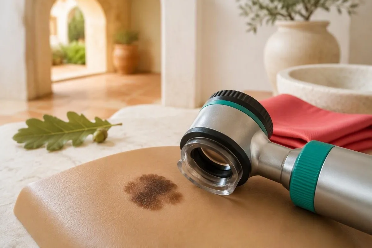
Melanoma symptoms can include a changing mole, an unusual new growth, or a persistent mark on a nail, palm or sole. Look for asymmetry, irregular borders, uneven color and evolution—but do not wait for every warning sign or a diameter above 6 mm.


- ABCDE melanoma rule means asymmetry, border irregularity, color variation, diameter and evolution; one concerning feature can justify assessment.
- Diameter above 6 mm is a warning clue, not a diagnostic cutoff; smaller melanomas occur.
- New growths matter: a 2017 meta-analysis estimated that 70.9% of melanomas arose without an associated pre-existing mole.
- Ugly-duckling sign means a spot looks different from your own usual mole pattern, even when it appears symmetrical.
- Nodular melanoma may be elevated, firm and growing over weeks; pink or skin-colored growths can also be melanoma.
- Nail changes such as a widening dark band, pigment on adjacent skin or unexplained nail distortion need clinical assessment.
- Palms and soles deserve inspection in every skin tone; persistent patches or growths can resemble bruises, warts or pressure damage.
- Bloodwork cannot reliably detect or exclude early melanoma; diagnosis requires clinical examination and usually a skin biopsy.
- Paso seguinte is to contact a clinician promptly for an evolving or unusual spot rather than waiting for a routine blood test.
Which melanoma symptoms should prompt a skin check?
Melanoma symptoms most often involve a visible change: an evolving mole, a new unusual growth, or a persistent abnormal patch. Early melanoma may cause no pain or itching, and a suspicious spot can measure less than 6 mm; clinical assessment, often followed by biopsy, is the route to diagnosis.
A spot does not need all 5 ABCDE features to deserve attention. A small, evenly colored growth that is clearly enlarging can matter more than a large mole that has remained unchanged for years; symptoms should be judged against the person's previous appearance, not a single photograph.
Melanoma can appear on skin that previously looked entirely ordinary. Ask 2 separate questions during a skin check: has an existing spot changed, and has something new appeared that does not fit? Looking only at familiar moles leaves an avoidable blind spot.
My advice as Thomas Klein, MD, Chief Medical Officer at Kantesti's organization, is to make the next step concrete: arrange a skin assessment when a spot is evolving. A reassuring laboratory panel answers a different question and should not postpone examination of that growth.
Kantesti é un Analizador de análises de sangue con IA, not a melanoma screening test; even a report with 0 abnormal flags cannot exclude skin cancer. Our role is to explain laboratory findings within their limits, while a suspicious skin change needs a clinician who can examine it directly.
How does the ABCDE melanoma rule work?
O/A ABCDE melanoma rule describes asymmetry, border irregularity, color variation, diameter and evolution. Diameter greater than 6 mm is a useful warning clue, but evolution—change in size, shape, color or behavior—can justify assessment at any diameter.
A and B concern shape rather than size: 2 halves may look different, or the edge may be scalloped, notched or poorly defined. Neither feature proves melanoma, because benign atypical moles can share them; the clinician also considers history, location and the person's background mole pattern.
C includes uneven shades of brown or black, but also areas of pink, white, red or blue. A spot containing 2 colors is not automatically cancerous, and a uniformly pink growth is not automatically safe; the distribution and evolution of color are more informative than simply counting shades.
D is often explained using a pencil eraser, roughly 6 mm across. That memory aid must not become a permission slip to ignore smaller growths: diameter describes surface width, whereas the pathological measurement that helps stage melanoma is its depth in millimeters.
E includes a new raised area, recurrent crusting or a change noticed across 2 dated photographs. Our estándares de contido médico distinguish warning signs from diagnostic criteria; ABCDE helps people recognize a reason for assessment, not calculate whether a mole is definitely benign.
What does the ugly-duckling sign reveal?
O/A ugly-duckling sign identifies a spot that looks different from the other moles on the same person. It can flag 1 unusually dark, pale, large, small or raised spot even when that growth does not satisfy the usual ABCDE checklist.
Personal comparison matters because normal mole patterns differ substantially. Someone with 20 similar light-brown moles may have a conspicuous new black spot; another person with many dark moles may instead develop a pink growth that stands out. The outlier does not have to be the darkest mark.
Compare nearby spots as well as the overall pattern. A person may have 2 recurring mole patterns—for example, flatter moles on the limbs and raised moles on the trunk—so comparison works best when a clinician considers body site rather than assuming every mole should look identical.
Consider an illustrative scenario: a 42-year-old notices a symmetrical, 4 mm spot unlike every other mole on the shoulder. Its symmetry and small size do not cancel the unusual appearance; a clinician would ask when it appeared, whether it is changing and what dermoscopy shows.
An ugly duckling is a reason to investigate, not a diagnosis or an indication to remove every outlier. If melanoma runs in the family, discuss the actual relatives and diagnoses rather than requesting a generic cancer panel; our guide to family cancer testing limits explains why targeted assessment differs from broad laboratory screening.
Can melanoma start as a new growth?
Melanoma can arise as a new growth without a changing pre-existing mole. Pampena et al. reported that 70.9% of melanomas in their 2017 meta-analysis were de novo, while 29.1% were associated with a pre-existing nevus; these pooled estimates are not an individual's cancer probability.
The meta-analysis combined 38 studies and 20,126 melanomas, giving the new-growth message a stronger basis than an anecdote. Classification depended on the studies and tissue findings, however, so the percentages should not be treated as an exact split for every population or melanoma subtype.
A new spot is not automatically melanoma. Adults commonly develop benign seborrheic keratoses, angiomas and other growths; the useful distinction is whether a new mark persists, evolves or appears unlike the person's usual skin features, especially when it changes over several weeks.
Age changes the context, not the diagnosis. A new mole-like spot in a 65-year-old generally deserves more scrutiny than an otherwise typical new mole in a teenager, but there is no age at which new melanoma becomes impossible or every new growth must be malignant.
Checking only existing moles misses the central lesson of Pampena et al. (2017): surveillance should include previously unremarkable skin. Likewise, routine annual bloodwork is not a substitute for assessing a new growth, even when the last panel was obtained just 1 month ago.
Which melanomas may not look like dark moles?
Nodular melanoma can appear as an elevated, firm growth that enlarges over weeks, while amelanotic melanoma may look pink, red or skin-colored. The 3-part EFG reminder—elevated, firm, growing— complements ABCDE, but neither checklist can reliably exclude melanoma.
A nodule can be symmetrical and have a smooth edge. That is why a growth enlarging over 4–6 weeks should not be dismissed simply because its photograph lacks an irregular outline; vertical growth may produce a different surface appearance from a spreading pigmented patch.
Pink growths have many benign explanations, including irritated follicles and vascular growths. The concerning combination is persistence plus enlargement, spontaneous bleeding or repeated crusting at the same site—not pink color alone. A clinician may need tissue examination because appearance overlaps among several skin conditions.
EFG does not mean every raised mole is dangerous. A soft raised mole unchanged for 10 years is a different clinical problem from a newly firm bump that grows noticeably between photographs; recent behavior supplies the missing context that a static checklist cannot capture.
The distinction between depth and diameter is especially useful here: 1 small surface measurement cannot tell you how deeply a growth extends. Our guide to Comprobacións de fontes de informes de IA explains why a confident-looking summary must never replace the actual examination or pathology report.
Do itching, bleeding or crusting mean melanoma?
Itching, bleeding and crusting can occur with melanoma, but none is specific to it. Persistent symptoms in 1 changing spot, particularly spontaneous or recurrent bleeding, deserve assessment; generalized itching or a clearly traumatized stable mole requires a different clinical interpretation.
Bleeding after catching a mole with a razor has an obvious possible explanation. Bleeding repeatedly without that trigger, or returning after the surface appears healed, is more concerning; record the circumstances of each episode rather than describing every event simply as 'it bled once.'
An itchy mole can reflect friction, eczema or local irritation. Itch affecting 1 evolving growth is more relevant to melanoma assessment than itch affecting both legs with dry skin; our explanation of itching and laboratory limits separates skin examination from tests used for selected systemic causes.
Many itchy bumps appearing over 1–2 weeks suggest a different pattern from a solitary changing growth. Conditions such as scabies and nighttime itching can involve multiple sites and household contacts, but a widespread rash does not automatically explain away an unrelated unusual mole.
Repeatedly scratching or treating a spot makes its original appearance harder to interpret. Take 1 clear photograph before applying a non-prescribed treatment, and avoid home acids, freezing kits or cosmetic removal of a suspicious growth; destroying its surface can delay diagnosis and compromise useful tissue assessment.
What nail changes can signal melanoma?
Nail melanoma may cause a new or widening brown-black longitudinal band, irregular pigmentation, nail distortion or pigment extending onto nearby skin. A changing band affecting 1 nail deserves assessment, although benign pigmentation, trauma and other nail disorders are much more common.
A band's width is not a stand-alone diagnostic test. Width above approximately 3 mm is sometimes used as a warning clue, but a narrower evolving band can still need evaluation; irregular color, widening toward the nail base and change over time add more context than width alone.
Pigment extending onto the cuticle or surrounding nail-fold skin is called the Hutchinson sign. It increases concern but is not exclusive to melanoma; clinicians also distinguish genuine skin pigmentation from pigment visible through a translucent nail fold, sometimes called a pseudo-Hutchinson sign.
A subungual bruise often moves toward the fingertip as the nail grows. Fingernails grow roughly 3 mm per month and toenails more slowly, so dated photographs can help document movement; however, a trauma history or some outward movement cannot independently rule out a persistent underlying growth.
A new unexplained band on 1 nail in an adult should not be managed with repeated antifungal courses without reassessment. Keep previous photographs and reports together using consent-based clinical record sharing, but send nail photographs through the examining clinician's approved system rather than assuming a laboratory app can diagnose them.
How can melanoma appear on palms and soles?
Acral melanoma can develop on palms, soles and within the nail unit, sometimes resembling a bruise, wart or persistent pressure-related patch. A mark that enlarges, changes color or fails to resolve over several weeks needs assessment, even when it is smaller than 6 mm.
Pressure and friction make the sole a particularly confusing site. A person may explain a changing patch as damage from a shoe for 2 months, yet persistence despite reduced friction weakens that explanation; the clinician needs both the mechanical history and an examination of the actual mark.
Dermoscopy can reveal whether pigment follows skin ridges or furrows on palms and soles. A parallel-ridge pattern can raise suspicion, but these specialized patterns are not reliable do-it-yourself rules; small, early or unpigmented acral melanomas may not display a classic pattern.
A persistent foot growth should not automatically be treated as a wart. If 1 area keeps enlarging, changes after treatment or repeatedly breaks down, ask for reassessment before another destructive procedure; photographs taken before treatment can preserve information that the altered surface no longer shows.
Not every slow-healing foot area is melanoma. Diabetes, pressure injury and impaired circulation are alternative explanations, and foot circulation warning signs require their own assessment; more than 1 condition can coexist, so an abnormal vascular test does not explain every pigmented growth.
Who needs to watch for melanoma symptoms?
Melanoma can occur in every skin tone, although overall incidence and the distribution of melanoma subtypes vary. Personal melanoma history, a close family history, numerous atypical moles and ultraviolet exposure increase concern; no skin color makes a changing 1-nail band or sole growth automatically safe.
Darker skin reduces some ultraviolet-related melanoma risk but does not eliminate melanoma. Acral melanoma represents a larger proportion of melanomas diagnosed in some darker-skinned populations, which is not the same as saying its absolute incidence is necessarily higher; percentages need the correct denominator.
A first-degree relative means a parent, sibling or child. If 2 or more close relatives have melanoma, or melanoma occurs alongside certain other cancers in a family, ask whether specialist risk assessment is appropriate; genetic testing is selective, not a routine response to having many moles.
Immunosuppression and a previous melanoma can change the surveillance plan. The relevant details include the medication, duration, previous pathology and number of affected relatives; a generic family-risk score should not determine whether a newly evolving spot receives examination.
At Kantesti, our approach to controis de saúde familiar baseados no risco separates useful laboratory monitoring from examinations that bloodwork cannot replace. Whether the family has 1 affected relative or several, a current suspicious growth needs its own clinical assessment rather than waiting for a risk calculation.
How should you check and photograph your skin?
A useful skin self-check compares your current skin with its previous appearance and includes nails, palms, soles and hard-to-see areas. A monthly check is a practical routine for many people, but clinicians may recommend a different schedule according to individual risk and surveillance needs.
Use good light, a full-length mirror and a hand mirror, with help for the scalp or back if available. Inspect between fingers and toes and remember the soles; checking only sun-exposed skin leaves at least 2 commonly overlooked areas—the nail unit and the underside of the foot.
Take 2 photographs of a concerning spot: an overview showing its location and a close image showing detail. Keep lighting, distance and camera angle as consistent as possible, and place a ruler beside rather than across the spot; image-processing filters can distort the color you are trying to document.
Do not postpone an appointment to collect a longer photo series. A new firmly growing bump or widening nail band warrants assessment now; comparison across 4 weeks may be helpful when requested by a clinician, but it is not a mandatory waiting period.
Photographs and laboratory PDFs are different kinds of medical records. Our medical upload privacy checklist offers transferable privacy principles, but skin images should go only to a service designed and authorized to assess them; 1 attractive interface is not evidence of diagnostic suitability.
How quickly should a suspicious mole be assessed?
Contact a clinician promptly for a clearly evolving mole, a growing firm nodule, unexplained recurrent bleeding or a suspicious nail change. NICE's weighted 7-point checklist recommends a suspected-cancer pathway referral for a suspicious pigmented lesion scoring 3 or more, and also addresses suspected nodular melanoma.
The NICE checklist assigns 2 points each for change in size, irregular shape and irregular color. Diameter of 7 mm or more, inflammation, oozing and altered sensation each score 1 point; this is a referral aid for clinicians, not a home test that guarantees safety below a score of 3.
The 7 mm NICE checklist feature and the 6 mm ABCDE reminder serve different purposes. Neither says that a 5 mm growth is safe, and NICE recommends referral when dermoscopy suggests melanoma or the appearance suggests nodular melanoma regardless of a reassuring self-calculated score (NICE, NG12).
A suspicious mole usually requires urgent outpatient assessment rather than an emergency department visit. If bleeding does not stop after 10–15 minutes of firm direct pressure, or there are separate signs of acute illness, seek urgent medical help for that immediate problem while still arranging assessment of the growth.
When booking, say what changed and over what period: 'This bump has grown over 3 weeks' is more useful than 'I have a mole.' Thomas Klein, MD, recommends clear symptom descriptions rather than a self-diagnosis; our Consello Asesor Médico page identifies the medical governance behind our laboratory education, not a substitute urgent referral service.
Why does melanoma diagnosis usually require a biopsy?
Melanoma diagnosis requires clinical assessment and usually a biopsy because tissue examination determines whether abnormal melanocytes are malignant and how deeply they extend. Dermoscopy improves assessment, but 1 photograph, symptom checklist or blood result cannot supply the microscopic information needed for diagnosis and staging.
The appointment usually combines the growth's history, direct examination and dermoscopy, with a broader skin check when appropriate. Bring 2 useful pieces of information: when the spot first appeared and what has changed; previous photographs may reveal an evolution that is not obvious on the examination day.
When feasible, a suspicious growth is generally removed in its entirety with a narrow diagnostic margin so the pathologist can assess its architecture and depth. Larger growths and difficult sites, including nails and soles, may require a different sampling approach; the clinician chooses a method intended to obtain adequate diagnostic tissue.
Breslow thickness measures melanoma depth in millimeters, not its surface diameter. A report describing 0.8 mm thickness therefore answers a different question from a photograph showing a 6 mm-wide spot; ulceration and other pathological findings can also influence subsequent management.
NICE NG14 recommends trained dermoscopic assessment and pathology-led management rather than relying on routine laboratory screening. Our clinical methodology and validation information concerns laboratory interpretation; even a technically accurate explanation cannot turn an unbiopsied skin growth into a confirmed diagnosis.
Can routine bloodwork detect or rule out melanoma?
Routine bloodwork cannot reliably detect or exclude early melanoma. A complete blood count, liver panel or LDH result may remain normal while melanoma is present; an LDH value such as 180 U/L, if within the reporting laboratory's range, does not clear a suspicious mole.
LDH is a nonspecific marker of cellular injury and has prognostic relevance in some patients with advanced melanoma. Its reference interval varies by laboratory, and an elevated result has many possible explanations; our guide to LDH meaning and limitations explains why 1 abnormal enzyme result does not identify a skin cancer.
A normal white-cell count or CRP does not assess the microscopic structure of a mole. The broader principle appears in our discussion of discordant WBC and CRP: different tests answer different questions, so 2 reassuring laboratory values cannot cancel a concerning examination finding.
Kantesti é un plataforma de interpretación de análise de sangue de IA that can help explain laboratory reports, but it cannot assign a melanoma probability from a routine panel. If a clinician orders kidney or liver tests during treatment planning, those values address organ function or treatment safety—not whether a 4 mm spot is benign.
Selected blood-based technologies, including circulating tumor DNA, are used or studied in particular oncology settings; they are not interchangeable with initial assessment of an undiagnosed mole. Our guide to tumor-marker testing limits illustrates the same boundary: even 1 apparently reassuring marker does not replace the indicated diagnostic procedure.
What happens next, and which sources support this advice?
After assessment, the next step may be reassurance with a specific review plan, clinician-directed photographic monitoring or biopsy followed by pathology-based treatment. As of 14 de outubro de 2026, this article's advice draws on NICE NG12, NICE NG14 and Pampena et al. (2017), rather than a claimed new blood test for melanoma.
If melanoma is confirmed, treatment depends on its stage and pathological features rather than how frightening the photograph looks. A sentinel lymph-node biopsy is a staging procedure, not the initial skin biopsy; NICE NG14 considers it for melanomas thicker than 1.0 mm and selected 0.8–1.0 mm melanomas with additional risk features.
Ask for 3 practical details before leaving an appointment: what the working diagnosis is, when the pathology or review result should arrive, and whom to contact if the growth changes meanwhile. Sun protection reduces ultraviolet exposure but does not treat an existing suspicious growth, and nutrition supplements should not delay its assessment.
Kantesti é un Ferramenta de análise de probas de sangue impulsada por IA for explaining laboratory findings, not examining skin tissue or staging melanoma. Our responsible AI principles support keeping those boundaries explicit: 1 normal report is never a reason to disregard an evolving mole or a clinician's biopsy recommendation.
The 2 Zenodo resources listed below are supplementary laboratory education, not melanoma studies or evidence that bloodwork detects skin cancer. The publicación de interpretación de RDW explains red-cell measurements; the kidney-function ratio publication explains renal laboratory context. ResearchGate and Academia.edu links are title searches, not claims of verified copies or peer review.
Preguntas frecuentes
Cales son os primeiros signos do melanoma?
0 Os primeiros signos de melanoma inclúen un lunar que evoluciona, un novo crecemento inusual ou unha mancha que ten un aspecto diferente ao dos outros lunares da persoa. ABCDE describe a asimetría, a irregularidade dos bordos, a variación da cor, o diámetro e a evolución, pero o melanoma pode medir menos de 6 mm. O melanoma incipiente pode non picar, doer nin sangrar. Un cambio sospeitoso require unha avaliación clínica e, normalmente, unha biopsia se se sospeita de melanoma.
Como se pode distinguir un melanoma dun lunar normal?
0 Un nevo normal adoita permanecer estable e axustarse ao patrón habitual de nevos da persoa, mentres que o melanoma pode evolucionar ou aparecer como un crecemento novo e inusual. Ningunha característica por si soa permite distinguir de maneira fiable un melanoma dun nevo benigno, e unha mancha simétrica de 4 mm pode requirir avaliación se está cambiando. O signo do parrulo feo compara unha mancha cos outros nevos da persoa, e non cunha fotografía de internet. A dermatoscopia e, cando está indicado, a biopsia permiten establecer o diagnóstico de forma máis fiable que a comparación feita na casa.
Pode aparecer un melanoma sen que haxa un lunar previo?
Melanoma can develop on skin without an associated pre-existing mole. A 2017 meta-analysis by Pampena et al. estimated that 70.9% of included melanomas arose de novo and 29.1% were associated with a nevus. Those pooled percentages do not describe the probability that any individual new spot is cancerous. Skin checks should look for new unusual growths as well as changes in familiar moles.
What does melanoma under a nail look like?
Nail melanoma may appear as a new or widening brown-black band, irregular pigment, nail distortion or pigment extending onto adjacent skin. A changing band on 1 nail deserves assessment, although benign bands and bruises are more common. A band wider than about 3 mm can raise concern, but narrower bands are not automatically safe. Diagnosis requires examination and may require a specialist nail-unit biopsy.
Does melanoma show up in routine blood tests?
Routine blood tests do not reliably detect or rule out early melanoma. A normal complete blood count, liver panel or LDH value—even an LDH of 180 U/L within that laboratory's range—cannot establish that a mole is benign. LDH has selected prognostic uses in advanced melanoma but is not a screening test for a suspicious skin growth. Clinical assessment and usually biopsy provide the diagnostic information.
How quickly should I get a changing mole checked?
Contact a clinician promptly when a mole changes, a firm new bump grows over weeks, or a spot repeatedly bleeds without a clear injury. Do not wait for its diameter to exceed 6 mm or for every ABCDE feature to appear. NICE recommends suspected-cancer pathway referral for suspicious pigmented lesions with a weighted 7-point checklist score of 3 or more, while suspected nodular melanoma also warrants referral. The appropriate appointment timing depends on the clinical assessment and local pathway.
Obtén hoxe unha análise de sangue con IA
Únete a máis de 2 millóns de usuarios en todo o mundo que confían en Kantesti para obter unha análise instantánea e precisa das análises de laboratorio. Carga os teus resultados de análise de sangue e recibe unha interpretación completa de biomarcadores de 15,000+ en segundos.
📚 Publicacións de investigación citadas
Klein, T., Mitchell, S., & Weber, H. (2026). Análise de sangue RDW: guía completa de RDW-CV, MCV e MCHC. Kantesti Investigación médica con IA.
Klein, T., Mitchell, S., & Weber, H. (2026). Explicación da proporción BUN/Creatinina: Guía de probas de función renal. Kantesti Investigación médica con IA.
📖 Referencias médicas externas
National Institute for Health and Care Excellence (2015). Cancro sospeitoso: recoñecemento e derivación (NG12). Guía NICE.
National Institute for Health and Care Excellence (2015). Melanoma: assessment and management (NG14). NICE guideline; updated 2022.
Pampena R et al. (2017). A meta-analysis of nevus-associated melanoma: Prevalence and practical implications. Revista da Academia Americana de Dermatoloxía.
⚕️ Aviso médico
Este artigo é só para fins educativos e non constitúe asesoramento médico. Consulta sempre un/ha profesional sanitario/a cualificado/a para decisións de diagnóstico e tratamento.
Sinais de confianza E-E-A-T
Experiencia
Revisión clínica dirixida por un médico dos fluxos de interpretación de análises.
Experiencia
Foco en medicina de laboratorio sobre como se comportan os biomarcadores no contexto clínico.
Autoridade
Escrito polo Dr. Thomas Klein, con revisión da Dra. Sarah Mitchell e do Prof. Dr. Hans Weber.
Fiabilidade
Interpretación baseada en evidencias con vías de seguimento claras para reducir a alarma.