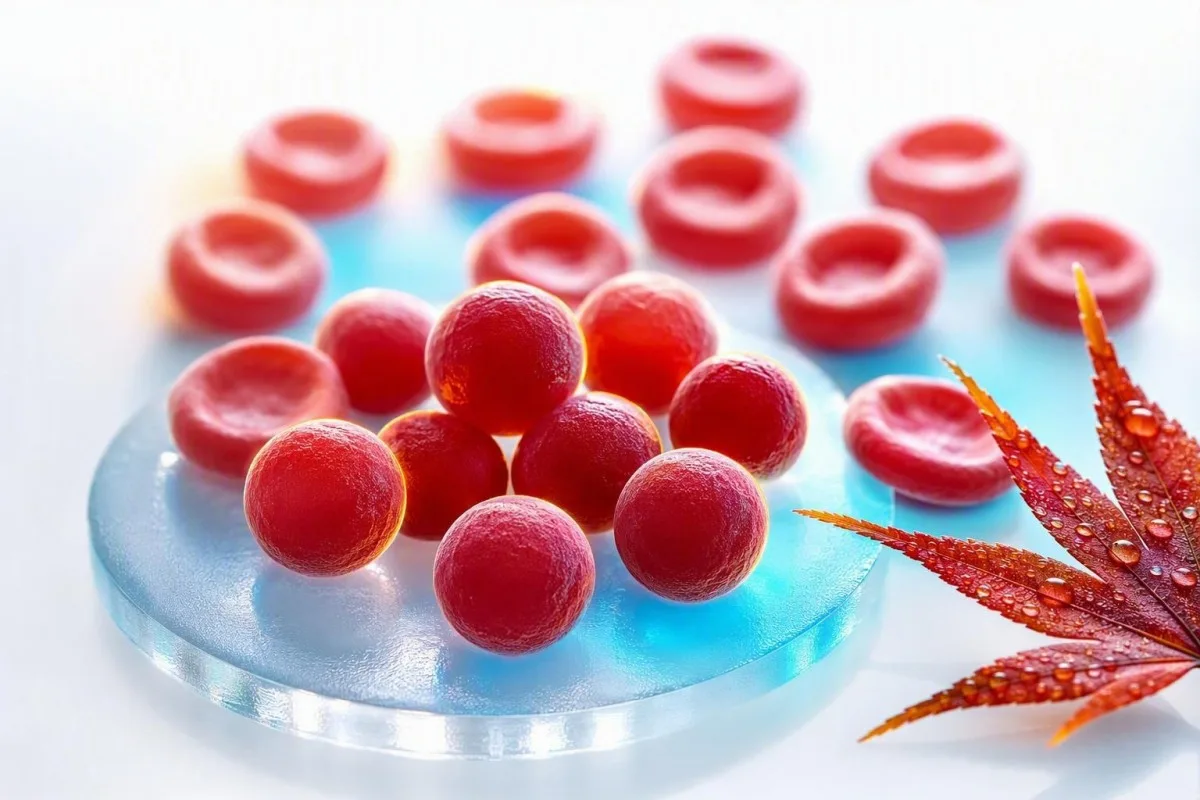
Ein hoher MCHC ist ungewöhnlich und oft eher ein Hinweis als eine Diagnose. Die praktische Frage ist, ob die roten Blutkörperchen tatsächlich dicht und kugelförmig sind oder ob die Probe die Berechnung verzerrt hat.


- MCHC-Bereich liegt normalerweise bei 32-36 g/dL (320-360 g/L) bei Erwachsenen, obwohl der eigene Bereich des Labors Vorrang hat.
- Hoher MCHC über 36 g/dL ist ungewöhnlich und sollte Anlass zur Suche nach Sphärozyten, Hämolyse, Verbrennungen oder analytischen Störungen geben.
- Sphärozyten sind kleine, dichte rote Blutkörperchen ohne zentrale Blässe und können bei hereditärer Sphärozytose oder autoimmuner hämolytischer Anämie auftreten.
- Kälteagglutinine können den MCHC falsch erhöhen, indem sie rote Blutkörperchen verklumpen, die gemessene RBC-Zahl senken und berechnete Indizes verzerren.
- Lipämie kann bei einigen Analysegeräten das Hämoglobin überschätzen und so einen erhöhten MCHC erzeugen, auch wenn die Form der roten Blutkörperchen normal ist.
- Brandverletzung kann einen echten hohen MCHC durch Verlust der roten Blutkörperchen-Membran verursachen, normalerweise zusammen mit Anämie und Laborbefunden einer Hämolyse.
- Hinweise auf Hämolyse umfassen erhöhte Retikulozyten, indirektes Bilirubin und LDH sowie eine niedrige Haptoglobin-Konzentration, wenn rote Blutkörperchen im Körper zerfallen.
- ist oft der richtige nächste Schritt, wenn der TSH-Wert nur leicht abweichend ist und das klinische Bild unklar bleibt. mit einer frisch entnommenen, ordnungsgemäß gehandhabten EDTA-Probe ist oft der erste sinnvolle Schritt, wenn der MCHC isoliert und nur leicht erhöht ist.
Hoher MCHC im CBC: die direkte Antwort
Ein hoher MCHC bedeutet normalerweise, dass die roten Blutkörperchen ungewöhnlich dicht sind, kann aber auch bedeuten, dass das Analysegerät durch die Probe fehlgeleitet wurde. Bei Erwachsenen verdient ein MCHC über etwa 36 g/dL Kontext: Echte Erhöhungen deuten am häufigsten auf Sphärozyten oder signifikante thermische Verletzungen hin, während Kälteagglutinine, Lipämie und Probenhämolyse häufige Laborerklärungen sind. Ab dem 23. August 2026 würde ich allein aufgrund dieses Indexes keine Bluterkrankung diagnostizieren.
MCHC wird berechnet als Hämoglobin geteilt durch Hämatokrit, multipliziert mit 100, daher ist es eine Konzentration und keine direkte Zellmessung. Ein Hämoglobin von 15,0 g/dL und ein Hämatokrit von 40% ergeben einen MCHC von 37,5 g/dL; die Zahl kann steigen, weil das Hämoglobin zu hoch abgelesen wird, der Hämatokrit zu niedrig abgelesen wird oder beides. Unser Leitfaden für CBC-Biomarker ist nützlich, wenn mehrere Indizes gleichzeitig auffällig sind.
Wenn ich ein Panel mit einem MCHC von 36,5–37,5 g/dL, aber normalem Hämoglobin, Bilirubin, Retikulozyten und RDW überprüfe, ist ein Artefakt oft wahrscheinlicher als eine Krankheit. Ein MCHC von 39–42 g/dL ist physiologisch schwer in intakten zirkulierenden roten Blutkörperchen aufrechtzuerhalten, daher sollte das Labor vor Panikstimmung aktiv Interferenzen berücksichtigen.
Kantesti ist ein KI-Bluttestanalysator der MCHC zusammen mit Hämoglobin, Hämatokrit, MCV, RDW, Bilirubin, LDH und früheren Ergebnissen liest, anstatt ein einzelnes Warnsignal als Diagnose zu behandeln. Die klinische Regel von Dr. Thomas Klein ist einfach: Überprüfen Sie die Arithmetik und die Probe, bevor Sie eine umfangreiche Untersuchung anordnen; unser detaillierter RDW- und Erythrozytenindex-Leitfaden erklärt, warum dieser musterbasierte Ansatz wichtig ist.
MCHC ist nicht MCH oder MCV
MCHC beschreibt die Hämoglobinkonzentration im konzentrierten Erythrozytenvolumen, während MCH die Hämoglobinmenge pro Zelle beschreibt und MCV die Zellgröße beschreibt. Das Verwechseln dieser drei Indizes ist ein Grund, warum ein routinemäßiges CBC-Flag alarmierender klingen kann, als es ist.
MCH liegt üblicherweise bei 27-33 pg pro Zelle, während MCV bei Erwachsenen üblicherweise bei 80-100 fL liegt; keiner der Werte definiert MCHC. Große rote Blutkörperchen können mehr Hämoglobin enthalten und dadurch MCH erhöhen, doch ihr MCHC kann normal bleiben, da ihr Volumen proportional zugenommen hat. Sehen Sie unseren praktischen Vergleich von MCV und MCH wenn beide Werte markiert sind.
Ein echtes hohes MCHC geht oft mit einem normalen oder niedrig-normalen MCV einher, da Sphärozyten Oberflächenfläche der Membran verloren haben und runder geworden sind, nicht weil sie grenzenloses Hämoglobin enthalten. Diese Geometrie ist wichtig: eine Kugel hat für ihr Volumen weniger Oberfläche, sodass sie auf einem gut präparierten Zellprobenabstrich kein blasses Zentrum aufweist.
Die Sache ist, dass ein erhöhter MCHC-Bluttest mathematisch real und biologisch irreführend sein kann. Ein leicht unterschätzter Hämatokrit nach der Aggregation von roten Blutkörperchen bläht das Verhältnis auf, selbst wenn die roten Blutkörperchen des Patienten überhaupt nicht dicht sind; deshalb ist eine Überprüfung des vollständigen Blutbildes vor Schlussfolgerungen getroffen werden sollte.
Sphärozyten: der klassische echte Hinweis auf einen hohen MCHC
Sphärozyten sind die erkennbarste Ursache eines echten hohen MCHC, da der Membranverlust rote Blutkörperchen kompakt und dicht mit Hämoglobin macht. Sie treten bei hereditärer Sphärozytose und autoimmuner hämolytischer Anämie auf, aber ein Befund im Ausstrich muss im Zusammenhang mit Symptomen und Hämolyse-Markern interpretiert werden.
Die hereditäre Sphärozytose führt oft zu MCHC-Werten über 36 g/dL, Retikulozytose, intermittierender Gelbsucht, Gallensteinen, Splenomegalie oder einer familiären Anämieanamnese. Die Leitlinie der British Society for Haematology von 2011 stellt fest, dass MCHC die Diagnose unterstützt, aber nicht empfindlich oder spezifisch genug ist, um sie allein zu bestätigen (Bolton-Maggs et al., 2012).
Bei autoimmuner hämolytischer Anämie markieren Antikörper rote Blutkörperchen für eine teilweise Membranentfernung in der Milz, wodurch Sphärozyten zurückbleiben. Ein direkter Antiglobulin-Test, die Retikulozytenzahl, Bilirubin, LDH und Haptoglobin sind zusammen informativer als jeder isolierte Index; unser Leitfaden zu niedrigem Haptoglobin jenseits der Hämolyse behandelt eine leicht zu übersehende Einschränkung.
Eine klinische Komplikation: eine kürzlich erfolgte Transfusion kann das native Sphärozytenmuster eines Patienten über mehrere Wochen verdünnen oder maskieren. Christensen et al. beschreiben die Bindung von Eosin-5-Maleimid mittels Durchflusszytometrie als nützlichen Test für Verdacht auf hereditäre Sphärozytose, obwohl ein Hämatologe die Testung sorgfältig auswählen und zeitlich planen sollte (Christensen et al., 2015).
Wie man feststellt, ob ein hoher MCHC mit Hämolyse einhergeht
Ein hohes MCHC wird klinisch bedeutsamer, wenn es mit Anämie, einer erhöhten Retikulozytenzahl, erhöhten indirekten Bilirubinwerten, erhöhten LDH-Werten und niedrigem Haptoglobin einhergeht. Diese Gruppe zeigt eine erhöhte Turnoverrate der roten Blutkörperchen an, obwohl kein einzelner Marker perfekt ist.
Ein Retikulozytenprozentsatz über 2% kann bei signifikanter Anämie immer noch unzureichend sein, daher berechnen Kliniker oft stattdessen eine absolute Retikulozytenzahl oder einen Retikulozytenproduktionsindex. Zum Beispiel kann eine Retikulozytenzahl von 4% bei einem Hämatokrit von 24% nach Korrektur nur eine moderate Reaktion des Knochenmarks darstellen; unser Artikel zu Hinweisen auf hohe Retikulozyten erläutert die Unterscheidung.
Unkonjugiertes Bilirubin steigt, wenn Häm schneller abgebaut wird, als die Leber es konjugieren kann, während LDH steigt, da rote Blutkörperchen reichlich LDH enthalten. Ein Hämoglobin-Wert unterhalb der unteren Laborgrenze unterstützt die intravaskuläre Hämolyse, kann aber bei Entzündungen normal erscheinen, da Hämoglobin ein Akute-Phase-Protein ist. Barcellini und Fattizzo diskutieren diese diagnostische Falle in ihrem Übersichtsartikel zur autoimmunhämolytischen Anämie (Barcellini & Fattizzo, 2015).
Dunkler Urin, schnell zunehmende Müdigkeit, Atemnot in Ruhe, Brustschmerzen, Ohnmacht oder gelbe Augen mit Fieber erfordern eine klinische Beurteilung am selben Tag. Fragmentierte Zellen sind ein anderer und dringlicherer Hinweis im Blutausstrich als Sphärozyten; unsere Erklärung von wenn Schistocyten eine dringende Überprüfung brauchen hilft zu verdeutlichen, warum.
Warum Verbrennungen einen echten erhöhten MCHC verursachen können
Ausgedehnte thermische Verletzungen können einen tatsächlich hohen MCHC verursachen, da Hitze die Membranen der roten Blutkörperchen schädigt, was zu Membranverlust, spheroiden Zellen und Hämolyse führt. Dies ist im Allgemeinen ein Befund im Krankenhaus nach klinisch offensichtlichen Verbrennungen und keine Erklärung für ein zufälliges ambulantes CBC-Ergebnis.
Thermische Schäden können Stunden nach schweren Verbrennungen Mikro-Sphärozyten erzeugen, und der MCHC kann steigen, wenn das Verhältnis von Membran zu Volumen abfällt. Das Hämoglobin kann auch in den folgenden Tagen aufgrund von Flüssigkeitsverschiebungen, Eingriffen, Aderlass und fortlaufender Hämolyse abfallen, sodass ein einzelner MCHC den Schweregrad der Verletzung nicht quantifizieren kann.
Gelegentlich habe ich beobachtet, dass Patienten nach einer Notaufnahme auf einen MCHC von 37,1 g/dL fixiert sind und dabei weitaus handlungsrelevantere Anomalien übersehen: sinkendes Hämoglobin, steigendes Kreatinin, Kaliumveränderungen oder Laktat. In diesem Zusammenhang behandeln die Kliniker den Patienten und die Verbrennungsphysiologie, nicht den Index; unser Zeitplan für Tests nach dem Krankenhaus erklärt, warum Werte sich schnell ändern können.
Kantesti AI ist an KI-Biomarker-Interpretationsplattform die einen hohen MCHC neben dynamischen Hämoglobin- und Nierenwerten anzeigen kann, aber akute Verbrennungen erfordern eine direkte klinische Beurteilung und können nicht sicher anhand der Interpretation eines Berichts behandelt werden. Ein Ergebnis nach einer schweren Verbrennung sollte mit den vorherigen 24-72 Stunden verglichen werden und nicht mit einem generischen Bevölkerungsdurchschnitt.
Kälteagglutinine: der Artefakt bei hohem MCHC, nach dem Ärzte suchen
Kälteagglutinine können einen fälschlicherweise hohen MCHC verursachen, indem sie rote Blutkörperchen bei Raumtemperatur verklumpen lassen, bevor die Analyse durchgeführt wird. Der Analysator kann mehrere angeheftete Zellen als ein Partikel zählen, was zu einer falsch niedrigen RBC-Zahl, einem falsch hohen MCV und einem erhöhten berechneten MCHC führt.
Das klassische Muster von Kälteagglutinen ist eine niedrige RBC-Zahl mit unverhältnismäßig hohem MCV, hohem MCH und hohem MCHC, oft ohne entsprechende klinische Geschichte. Das Erwärmen der EDTA-Röhre auf 37 °C und das erneute Durchführen des CBC korrigiert üblicherweise die Werte, was weit informativer ist als die Wiederholung eines zufälligen Tests Wochen später.
Kälteagglutinine können nach einigen Atemwegsinfektionen vorübergehend auftreten oder bei der Kälteagglutininerkrankung und bestimmten lymphoproliferativen Erkrankungen bestehen bleiben. Symptome wie Farbveränderung oder Beschwerden in den Fingern bei Kälteeinwirkung verleihen dem klinischen Bild Gewicht, aber das Laborbild selbst kann der erste Hinweis sein; eine Wiederholung des Tests nach Hämolyse bietet praktische Fragen zum erneuten Blutentnehmen.
Wärmen Sie sich nicht aggressiv auf oder versuchen Sie nicht, das Ergebnis zu Hause zu manipulieren. Fragen Sie, ob das Labor eine Verklumpung der roten Blutkörperchen festgestellt hat, ob die Probe erwärmt und wiederholt wurde und ob ein manuelles Hämatokrit bestimmt wurde; diese drei Antworten klären die Angelegenheit oft.
Lipämie und Hämolyse der Probe können den MCHC verzerren
Lipämie und In-vitro-Hämolyse der Probe können den MCHC falsch erhöhen, da viele Analysegeräte Hämoglobin photometrisch messen, nachdem die roten Blutkörperchen lysiert wurden. Trübe Lipide oder freies Hämoglobin in der Probe können die optische Absorption erhöhen und das Hämoglobin höher erscheinen lassen, als es tatsächlich ist.
Lipämie ist am häufigsten nach sehr hohen Triglyceridwerten sichtbar, normalerweise über etwa 400-500 mg/dL, obwohl die Beeinflussung vom Instrument und der Wellenlänge des Assays abhängt. Ein Labor kann eine Plasma-Blindprobe, eine alternative Wellenlänge oder einen Plasmaersatzansatz verwenden, anstatt einfach das automatisierte Hämoglobinergebnis zu akzeptieren.
In-vitro-Hämolyse tritt nach der Entnahme oder Handhabung auf, nicht im Körper, und sie kann mehr als nur MCHC beeinflussen. Kalium, LDH, AST und Phosphat können in einer beschädigten Probe ansteigen, weshalb ein isoliert hoher MCHC neben einem unerwarteten hohen Kaliumwert eine Überprüfung der Probenqualität erfordert; siehe unsere Anleitung zu Hämolyse-bedingten Kaliumfehlern.
Eine sichtbar rosa oder rote Plasmaschicht ist ein Laborhinweis, kein Beweis für eine hämolytische Anämie beim Patienten. Die praktische Nachsorge ist eine frische Entnahme mit ruhiger Handhabung und umgehender Verarbeitung, insbesondere wenn der ursprüngliche Bericht einen Hämolyse-Index oder einen Kommentar enthält, dass die Ergebnisse beeinträchtigt sein können.
Probleme bei Entnahme, Lagerung und Röhrchen, die CBC-Indizes verändern
Schlechte Probenmischung, Unterfüllung, Gerinnung und verzögerte Analyse können die CBC-Indizes verändern, obwohl eine verzögerte Lagerung MCHC eher senkt als erhöht, da die Zellen anschwellen können. Ein hoher MCHC sollte daher im Zusammenhang mit den Probenkommentaren des Labors gelesen werden, nicht nur mit der Zahl im Patientenportal.
CBC-Proben werden im Allgemeinen in EDTA-Röhrchen entnommen, und ein unterfülltes Röhrchen kann ein übermäßiges Verhältnis von Antikoagulans zu Probe aufweisen, das zelluläre Messungen verändert. Kleine Gerinnsel können Zellen selektiv von der Analyse ausschließen und die automatisierte Zählung intern inkonsistent machen; unser Überblick über Blutröhrchen-Zusatzstoffe erklärt, warum der Röhrchen-Typ kein triviales Detail ist.
Lagerung bei Raumtemperatur über längere Zeiträume führt dazu, dass rote Blutkörperchen Wasser aufnehmen, wodurch sich der MCV erhöht und der MCHC tendenziell sinkt. Diese Richtung der Veränderung ist nützlich: ein sehr hoher MCHC nach einer verzögerten Probe wird nicht automatisch allein durch das Alter erklärt, sodass Kälteagglutination oder optische Interferenzen immer noch vorhanden sein können.
Eine Kapillarprobe kann in einigen Fällen angebracht sein, ist aber anfälliger für Schwankungen bei der Entnahme als eine gut entnommene venöse EDTA-Probe. Wenn ein hoher MCHC keine entsprechenden Symptome oder verwandten Anomalien aufweist, bevorzuge ich normalerweise eine saubere venöse erneute Entnahme, bevor ich sie als neue chronische Diagnose betrachte.
Verwenden Sie frühere CBCs, um zu entscheiden, ob die Änderung real ist
Ein plötzlicher Anstieg des MCHC von einer stabilen Basislinie von 33-35 g/dL auf 38 g/dL deutet eher auf ein Proben- oder Analysegerätproblem hin als auf eine sich langsam entwickelnde erbliche Sphärozytose. Trends ersetzen keinen Abstrich, aber sie sind ein starker Hinweis zur Qualitätskontrolle.
Die meisten Menschen haben über die Zeit einen relativ engen persönlichen MCHC-Bereich, der bei vergleichbaren Testbedingungen oft um weniger als 1 g/dL variiert. Ein Delta-Check im Labor vergleicht ein neues Ergebnis mit einem früheren, um unglaubwürdige Veränderungen zu identifizieren, bevor Ergebnisse freigegeben werden; unser Artikel über Delta-Checks zeigt, wie dieses Sicherheitsnetz funktioniert.
Kantesti ist ein KI-Labor-Befundinterpretationsservice der aktuelle und frühere CBC-Werte vergleicht und hilft, ein persistentes Signal von einem Ausreißer zu unterscheiden. In unserem Überprüfungsworkflow ist eine MCHC-Änderung wichtiger, wenn sie zusammen mit Hämoglobin, MCV, Bilirubin und Retikulozyten in eine biologisch kohärente Richtung geht.
Es gibt Ausnahmen. Neue autoimmune Hämolyse, schwere Verbrennungen, Transfusionsreaktionen oder deutliche Kälteagglutination können Ergebnisse schnell verändern, sodass ein stabiler historischer Wert beruhigend ist, aber niemals ein Grund, neue Gelbsucht, dunklen Urin oder Anämie zu ignorieren.
Bedingungen, die häufig dem MCHC zugeschrieben werden, aber selten dafür verantwortlich sind
Dehydratation, Eisenmangel, Vitamin-B12-Mangel und gewöhnlicher Stress verursachen normalerweise keinen wirklich hohen MCHC. Sie können andere CBC-Werte verändern, aber sie sollten nicht als bequeme Erklärungen für einen MCHC von 38 g/dL verwendet werden, ohne die Probe zu bestätigen.
Eisenmangel senkt typischerweise MCH und MCHC und oft auch MCV, sobald der Mangel etabliert ist. Früher Eisenmangel kann ein normales MCV aufweisen, erklärt aber keine hyperchromen Indizes; unsere Erklärung von niedrige Transferrinsättigung hilft, die Eisenverfügbarkeit von der CBC-Konzentration zu trennen.
Vitamin-B12- oder Folsäuremangel verursacht häufiger Makrozytose mit einem MCV über 100 fL als ein hohes MCHC. Wenn sowohl MCV als auch MCH hoch sind, MCHC aber normal ist, weist dies in eine andere diagnostische Richtung; siehe Hinweise zur Nachuntersuchung bei hohem MCV.
G6PD-Mangel verursacht episodische Hämolyse nach spezifischen oxidativen Auslösern bei anfälligen Personen, aber MCHC ist kein verlässlicher Screening-Test dafür. Während einer akuten Episode können der Ausstrich und die Turnover-Marker aufschlussreicher sein, während Enzymtests nach der Genesung wiederholt werden müssen, da junge rote Blutkörperchen mehr G6PD enthalten.
Die nützlichsten Nachuntersuchungen nach einem erhöhten MCHC
Der beste nächste Schritt bei einem isolierten hohen MCHC ist normalerweise eine Wiederholung des CBC mit Laborüberprüfung, gefolgt von einem peripheren Ausstrich und einem Hämolyse-Panel nur, wenn die Erhöhung anhält oder andere Hinweise vorliegen. Die sofortige Anordnung jedes Anämie-Tests kann mehr Rauschen als Antworten erzeugen.
Eine gezielte primäre Überprüfung umfasst die Wiederholung des CBC, die Überprüfung des Zellproben-Abstrichs, die Retikulozytenzahl, Bilirubin gesamt und indirekt, LDH, Haptoglobin und den direkten Antiglobulin-Test, wenn eine autoimmune Hämolyse plausibel ist. Der Ausstrich kann Spherocyten, Agglutination, Fragmente, Polychromasie oder eine normale Morphologie identifizieren, die einen Artefakt wahrscheinlicher macht.
Wenn eine hereditäre Sphärozytose weiterhin wahrscheinlich ist, kann ein Hämatologe die Bindung von Eosin-5-Maleimid, den Test auf osmotische Fragilität oder genetische Tests in Betracht ziehen. Diese Tests bergen unterschiedliche Risiken für falsch-negative Ergebnisse nach Transfusion und während akuter Hämolyse, weshalb die Reihenfolge der Tests der klinischen Anamnese folgen sollte und nicht einer Checkliste; unser Leitfaden für G6PD-Tests veranschaulicht das breitere Zeitproblem.
Kantesti AI interpretiert ein hohes MCHC, indem es die Berechnung mit verwandten CBC- und Hämolysemarkern abgleicht und dann Fragen identifiziert, die an einen Kliniker gerichtet werden können. Es ersetzt nicht die Überprüfung der Zellmorphologie, insbesondere wenn das Labor eine Analyzer-Flagge ausgegeben hat oder sich die Anämie verschlimmert.
Wann ein hoher MCHC eine dringende ärztliche Abklärung erfordert
Ein hohes MCHC an sich ist keine Notfallschwelle, aber ein hohes MCHC mit Anzeichen akuter Hämolyse, schwerer Anämie oder schwerer Verbrennung kann eine Behandlung am selben Tag erfordern. Symptome und die Geschwindigkeit der Veränderung bestimmen die Dringlichkeit weit besser als die Tatsache, ob die Flagge mit H markiert ist.
Suchen Sie eine dringende Beurteilung bei neu aufgetretener Kurzatmigkeit in Ruhe, Brustschmerzen, Ohnmacht, Verwirrung, sich schnell verschlimmernder Schwäche, teefarbenem Urin oder Gelbfärbung der Augen mit Fieber. Diese Symptome können mit signifikanter Anämie oder Hämolyse einhergehen, insbesondere wenn der Hämoglobinwert um mehr als 2 g/dL gegenüber einem bekannten Basiswert abgefallen ist.
Kontaktieren Sie den Kliniker, der den Test angeordnet hat, innerhalb weniger Tage, wenn der MCHC in einer wiederholten Probe über 36 g/dL bleibt, eine Anämie vorliegt oder der Bericht eine Agglutination oder Spherocyten erwähnt. Bei einer stabilen, gesunden Person mit einem MCHC von 36,2 g/dL und ansonsten normalen Ergebnissen ist eine nicht dringende Nachuntersuchung oft sinnvoll; unser Bilirubin-Muster-Leitlinie kann helfen, die nächste Besprechung zu gestalten.
Beginnen Sie nicht mit Eisen, Folsäure, Steroiden oder Nahrungsergänzungsmitteln, nur um das MCHC zu korrigieren. Keine davon behandelt die eigentlichen Hauptursachen, und insbesondere Steroide können die Beurteilung einer autoimmunen Hämolyse verschleiern, wenn sie ohne klinische Aufsicht begonnen werden.
Wie Kantesti den MCHC in den klinischen Kontext einordnet
Kantesti liest ein hohes MCHC als Problem der Mustererkennung: echte dichte-Zell-Störungen und Probenartefakte erzeugen unterschiedliche Kombinationen von CBC, Chemie, Flaggen und früheren Trends. Ziel ist es, die nächste klinische Frage klarer zu gestalten, nicht eine seltene Krankheit anhand eines einzelnen berechneten Indexes zu kennzeichnen.
Kantesti ist ein KI-gestütztes Analyse-Tool für Bluttests von mehr als 2 Millionen Menschen in 127 Ländern zur Organisation von Laborergebnissen im Kontext verwendet. Bei hoher MCHC prüft unsere KI, ob Hämoglobin und Hämatokrit das Ergebnis mathematisch stützen, ob die MCV unerwartet hoch ist und ob Bilirubin, LDH, Retikulozyten oder Haptoglobin auf einen Umsatz roter Blutkörperchen hinweisen.
Unsere Plattform sucht auch nach internen Widersprüchen: MCHC 40 g/dL mit einem normalen Zellausstrich-Kommentar und keiner Anämie kann eine Aufforderung zu einem Artefakt veranlassen, während MCHC 37 g/dL mit Sphärozyten, Retikulozytose und erhöhten indirekten Bilirubinwerten eine medizinische Nachuntersuchung erfordert. Der technische Ansatz und die Grenzen sind in unserem KI-Technologie-Leitfaden.
Ich rate den Patienten, die Labor-PDF, das Abgabedatum, den Fastenstatus, kürzliche Erkrankungen, die Transfusionshistorie und alle Labor-Kommentare aufzubewahren. Diese kleine Aufzeichnung erklärt oft eine scheinbare Diskrepanz schneller als eine Wiederholung breit angelegter Tests, insbesondere wenn Ergebnisse Monate später verglichen werden.
Fragen, die einen Termin bei einem hohen MCHC produktiver machen
Fragen Sie, ob die MCHC wiederholt wurde, ob das Labor Sphärozyten oder Agglutinationen gesehen hat und ob Anämie- oder Hämolyse-Marker vorhanden sind. Diese Fragen verschieben die Konversation von “Warum ist eine Zahl hoch?” zu einem konkreten, testbaren klinischen Pfad.
Nützliche Formulierungen sind: “War die Probe hämolytisch oder lipämisch?”, “Hat der Analysator eine Agglutination roter Blutkörperchen markiert?” und “Können wir dieses Ergebnis mit meiner vorherigen MCHC vergleichen?” Eine manuelle Auswertung des Zellausstrichs ist besonders wertvoll, wenn die MCHC wiederholt über 36 g/dL liegt oder eine Anämie unerklärt ist.
Dr. Thomas Klein empfiehlt die Mitnahme einer Liste von Medikamenten, kürzlichen Infektionen, kälteausgelösten Symptomen, einer familiären Vorbelastung mit Gallensteinen oder Anämie und Transfusionen innerhalb der letzten 3 Monate. Diese Anamnese kann auf hereditäre Sphärozytose, autoimmune Hämolyse oder ein vorübergehendes Kälteagglutinin viel schneller hinweisen als eine generische Online-Suche.
Die medizinischen Inhalte von Kantesti werden unter ärztlicher Aufsicht überprüft, aber Ihr eigener Kliniker kann Sie untersuchen, eine erwärmte Wiederholung anfordern und direkt mit dem Labor sprechen. Leser, die unseren klinischen Überprüfungsrahmen verstehen möchten, können den medizinischen Beirat, während anhaltende oder symptomatische Auffälligkeiten immer persönlich besprochen werden sollten.
Forschung, Unsicherheit und die Grenzen eines einzelnen MCHC
MCHC ist ein nützlicher Screening-Hinweis, kein eigenständiger diagnostischer Test für hereditäre Sphärozytose, autoimmune Hämolyse, Verbrennungen oder Kälteagglutinin-Krankheit. Sein Wert steigt sprunghaft an, wenn eine wiederholte Probe, die Zellmorphologie, Umsatzmarker und die klinische Anamnese alle die gleiche Geschichte erzählen.
Kein universeller MCHC-Grenzwert diagnostiziert hereditäre Sphärozytose, da Analysatormethoden, Alter, Retikulozytose und eine gleichzeitig bestehende Eisenmangelanämie die Empfindlichkeit verändern können. Die BSH-Richtlinie unterstützt die Kombination von Familienanamnese, Zellausstrichbefunden, Retikulozyten, Bilirubin und gegebenenfalls Bestätigungstests anstelle der Verlassung auf einen einzelnen Schwellenwert (Bolton-Maggs et al., 2012).
Kantesti unterstützt eine informierte Nachuntersuchung, indem es Ergebnis Muster und Unsicherheiten identifiziert, nicht indem es eine definitive hämatologische Diagnose ausgibt. Unser Ansatz zur klinischen Validierung erklärt, warum ungewöhnliche Werte, Analysator-Flags und Notfallsymptome weiterhin Gründe für eine Überprüfung im Labor und durch den Kliniker darstellen.
Für verwandte hämatologische Kontexte siehe die Forschungsveröffentlichung Leitfaden zu Blutgruppe B negativ, LDH-Bluttest und Retikulozytenzahl und die umfassendere Ressource zur klinischen Kommunikation Durchfall nach dem Fasten, schwarze Flecken im Stuhl & Magen-Darm-Ratgeber 2026. Beides sind ergänzende Lehrpublikationen und kein Ersatz für einen peripheren Zellausstrich oder eine spezialisierte Beurteilung.
Häufig gestellte Fragen
Was bedeutet ein hoher MCHC-Wert im Blutbild?
Ein hoher MCHC-Wert bedeutet normalerweise, dass die Hämoglobinkonzentration innerhalb des Erythrozytenvolumens über dem Laborbereich liegt, bei Erwachsenen üblicherweise über 36 g/dL. Echte Erhöhungen treten bei Sphärozyten aufgrund von hereditärer Sphärozytose, autoimmunhämolytischer Anämie oder schweren thermischen Verletzungen auf. Kälteagglutinine, Lipämie und Hämolyse der Probe können ebenfalls zu einem falsch hohen Ergebnis führen. Eine erneute CBC, die Überprüfung eines Zellproben-Ausstrichs und Hämolysemarker klären, welche Erklärung zutrifft.
Ist eine hohe MCHC gefährlich?
Ein hoher MCHC ist an sich nicht gefährlich; es ist ein berechneter CBC-Index und keine Krankheit. Ein MCHC von 36,2 g/dL mit normalem Hämoglobin und ohne Symptome wird oft mit einer routinemäßigen Wiederholung des Tests behandelt, während ein hoher MCHC plus fallendes Hämoglobin, Gelbsucht, dunkler Urin oder Atemnot eine sofortige Abklärung erfordert. Werte über 39 g/dL spiegeln mit besonderer Wahrscheinlichkeit Proben- oder Analysegerät-Interferenzen wider. Das klinische Risiko ergibt sich aus der zugrunde liegenden Ursache, wie z. B. einer signifikanten Hämolyse, und nicht allein aus der MCHC-Zahl.
Kann Dehydrierung einen hohen MCHC verursachen?
Eine Dehydratation verursacht normalerweise kein wirklich hohes MCHC, da sie das Plasmavolumen stärker konzentriert, als sie die Hämoglobinkonzentration in einzelnen roten Blutkörperchen verändert. Eine Dehydratation kann Hämoglobin und Hämatokrit gemeinsam erhöhen, wodurch das MCHC im üblichen Bereich von 32-36 g/dL bleibt. Wenn das MCHC 37-40 g/dL beträgt, sollten Ärzte stattdessen Sphärozyten, Kälteagglutination, Lipämie oder ein Messproblem in Betracht ziehen. Eine wiederholte, gut durchgeführte CBC ist nützlicher als der Versuch, den Wert durch zusätzliche Flüssigkeiten zu korrigieren.
Führen Sphärozyten immer zu einem hohen MCHC?
Sphärozyten erhöhen häufig die MCHC über 36 g/dL, aber nicht immer, und eine hohe MCHC beweist nicht die Anwesenheit von Sphärozyten. Eine erbliche Sphärozytose kann durch Eisenmangel, eine kürzliche Transfusion oder eine milde Erkrankung maskiert werden, während eine autoimmune Hämolyse je nach Zeitpunkt variable Befunde zeigen kann. Zur Identifizierung von Sphärozyten ist eine Untersuchung einer Zellprobe durch einen erfahrenen Laborfachmann erforderlich. Die Retikulozytenzahl, Bilirubin, LDH, Haptoglobin und ein direkter Antiglobulintest liefern zusätzlichen diagnostischen Kontext.
Können Kälteagglutinine eine hohe MCHC verursachen?
Kälteagglutinine können MCHC fälschlicherweise erhöhen, indem sie rote Blutkörperchen verklumpen, bevor das automatisierte CBC gemessen wird. Das Muster umfasst oft eine niedrige RBC-Zahl, ein hohes MCV, ein hohes MCH und ein MCHC über 36 g/dL, das nicht zu den Symptomen des Patienten passt. Das Erwärmen der EDTA-Probe auf 37°C und deren erneutes Durchführen kann die Zählwerte normalisieren. Anhaltende Kälteagglutination oder hämolytische Symptome sollten von einem Kliniker oder Hämatologen beurteilt werden.
Welche Tests sollte ich nach einem hohen MCHC anfordern?
Nach einem anhaltend hohen MCHC sind die üblichen nächsten Tests ein wiederholtes CBC, die mikroskopische Beurteilung einer Zellprobe, die Retikulozytenzahl, Bilirubinfraktionen, LDH, Haptoglobin und manchmal ein direkter Antiglobulintest. Ein Kliniker kann eine Eosin-5-Maleimid-Bindung oder andere spezielle Tests anfordern, wenn eine hereditäre Sphärozytose vermutet wird. Ein MCHC über 36 g/dL mit normalen wiederholten Tests und ohne Anämie erfordert möglicherweise keine umfangreiche Untersuchung. Die beste Testsequenz hängt von den Symptomen, früheren CBC-Werten, der Familienanamnese und den Laborflags ab.
Hol dir heute eine KI-gestützte Bluttest-Analyse
Schließe dich über 2 Millionen Nutzern weltweit an, die Kantesti für sofortige, genaue Laboranalysen vertrauen. Lade deine Blutwerte Ergebnisse hoch und erhalte in Sekunden eine umfassende Interpretation von 15,000+-Biomarkern.
📚 Referenzierte Forschungsveröffentlichungen
Klein, T., Mitchell, S., & Weber, H. (2026). Leitfaden zu Blutgruppe B negativ, LDH-Bluttest & Retikulozytenzahl. Kantesti KI-Medizinische Forschung.
Klein, T., Mitchell, S., & Weber, H. (2026). Durchfall nach dem Fasten, schwarze Flecken im Stuhl & Magen-Darm-Ratgeber 2026. Kantesti KI-Medizinische Forschung.
📖 Externe medizinische Referenzen
⚕️ Medizinischer Haftungsausschluss
Dieser Artikel dient nur zu Bildungszwecken und stellt keine medizinische Beratung dar. Wende dich für Diagnose- und Behandlungsentscheidungen immer an eine qualifizierte medizinische Fachkraft.
E-E-A-T Vertrauenssignale
Erfahrung
Ärztlich geleitete klinische Überprüfung von Labor-Interpretations-Workflows.
Sachverstand
Fokus der Labormedizin darauf, wie Biomarker sich im klinischen Kontext verhalten.
Autorität
Verfasst von Dr. Thomas Klein, überprüft von Dr. Sarah Mitchell und Prof. Dr. Hans Weber.
Vertrauenswürdigkeit
Evidenzbasierte Interpretation mit klaren nächsten Schritten zur Reduzierung von Alarm.