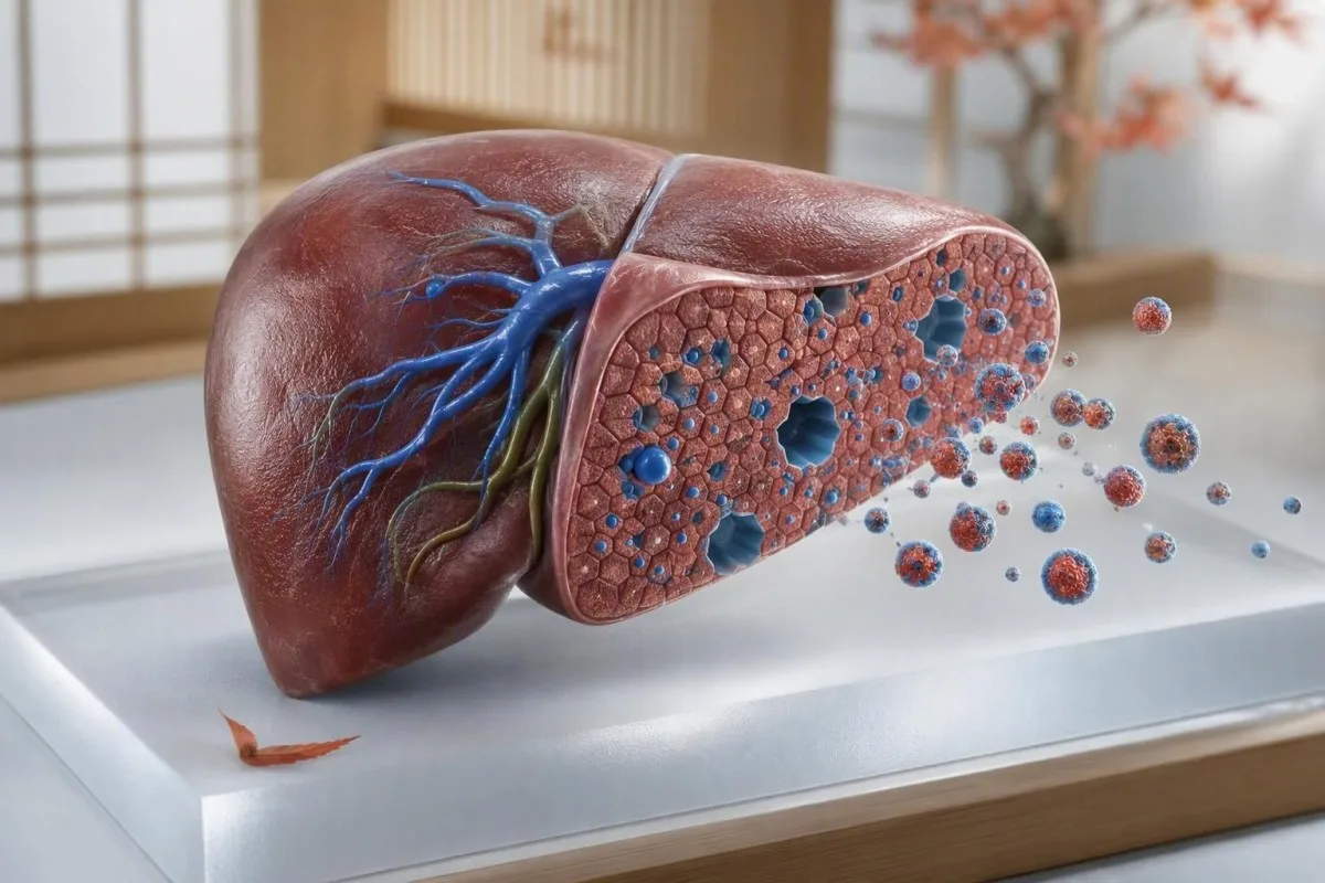
A nutritious diet can lower LDL cholesterol, but it cannot fully override inherited LDL clearance, thyroid dysfunction, certain medicines, or a misleading single test result. The useful next step is to identify which mechanism fits your own lipid pattern.


- LDL-C of 190 mg/dL (4.9 mmol/L) or higher warrants assessment for familial hypercholesterolaemia, even in a lean person who eats well.
- Familial hypercholesterolaemia affects roughly 1 in 250 people and usually reflects reduced LDL-receptor clearance from birth.
- TSH above 10 mIU/L with low free T4 can raise LDL substantially; thyroid testing is a sensible early check when LDL changes unexpectedly.
- ApoB above 130 mg/dL is an AHA/ACC risk-enhancing factor and can clarify particle burden when LDL-C and triglycerides disagree.
- Lipoprotein(a) of 50 mg/dL or 125 nmol/L or higher is a mostly inherited risk marker that diet usually does not lower meaningfully.
- Saturated fat response varies by genotype; coconut oil, butter, cheese, and high-fat low-carbohydrate diets can markedly raise LDL in susceptible people.
- Calculated LDL becomes less dependable when triglycerides exceed 400 mg/dL, making direct LDL-C, ApoB, or non-HDL-C more useful.
- Repeat lipids after 6 to 12 weeks of a stable diet, weight, medication schedule, and thyroid treatment plan before judging whether a change worked.
Why a healthy diet may not lower LDL cholesterol
High LDL cholesterol causes are often inherited or medical, not a failure of willpower. LDL can remain elevated despite a Mediterranean-style diet because the liver may remove LDL particles inefficiently, thyroid hormone may be low, a medicine may shift lipid handling, or the result may need confirmation under comparable conditions. As of August 2, 2026, I would not label diet ineffective until I have looked at the full panel, family pattern, thyroid status, and at least one well-timed repeat.
Most LDL particles are cleared by LDL receptors in the liver, not burned away by exercise. A person can eat legumes, vegetables, fish, and oats consistently yet still have LDL-C of 175 mg/dL (4.5 mmol/L) if receptor number or function is genetically reduced. Diet matters, but it typically changes LDL-C by a modest percentage rather than rewriting the inherited baseline.
In my clinical experience, the emotionally difficult part is that patients often assume a high result proves they have done something wrong. It does not. I am Dr. Thomas Klein, and when I review an LDL result, I first ask whether it is persistent, whether relatives had early heart disease, and whether ApoB, non-HDL-C, triglycerides, and Lp(a) point in the same direction; our guide to LDL targets by heart risk explains why one number rarely tells the whole story.
Kantesti is an AI blood test analyzer that reads a lipid panel alongside available thyroid, glucose, kidney, liver, and prior-result data rather than treating LDL-C as an isolated flag. That context can distinguish a longstanding inherited pattern from a new rise after a medication, menopause transition, illness, or dietary shift.
What LDL-C measures—and what it can miss
LDL-C estimates the cholesterol carried inside LDL particles, whereas ApoB approximates the number of atherogenic particles. LDL cholesterol is clinically useful, but two people with LDL-C of 150 mg/dL can have materially different particle counts and cardiovascular risk.
Every LDL particle contains one ApoB molecule, so ApoB is a practical proxy for the count of particles capable of entering the artery wall. Non-HDL-C equals total cholesterol minus HDL-C and captures LDL plus triglyceride-rich remnants; it is particularly informative when triglycerides are 150 mg/dL or higher. The difference between a lipid profile and lipid panel is usually naming rather than a different biological test.
A calculated LDL-C is commonly derived from total cholesterol, HDL-C, and triglycerides. The traditional Friedewald equation is not reported reliably when triglycerides are above 400 mg/dL (4.5 mmol/L), and it can underestimate LDL-C even at lower triglyceride values; direct LDL-C or ApoB is often cleaner in that setting.
Low HDL does not cancel a low LDL, and high HDL does not neutralise a high LDL particle burden. The reason clinicians care about LDL exposure is cumulative: particles retained in arterial walls can initiate plaque formation over decades, which is why a high result at age 32 can deserve more attention than the same number first seen at age 78.
When genetic high cholesterol is the main explanation
Genetic high cholesterol commonly reflects variants in LDLR, APOB, or PCSK9 that reduce hepatic LDL clearance. Heterozygous familial hypercholesterolaemia, or FH, affects about 1 in 250 people and can produce untreated LDL-C above 190 mg/dL (4.9 mmol/L) in adults.
FH is not diagnosed from an LDL number alone. Persistent LDL-C of 190 mg/dL or more, a parent or sibling with similar levels, tendon xanthomas, or coronary disease before age 55 in men or 65 in women strengthen the likelihood; the European Atherosclerosis Society consensus describes these as core clinical clues (Nordestgaard et al., 2013). Some families have polygenic high LDL rather than one identifiable variant, which can look similar in routine practice.
A normal body weight and excellent fitness do not rule FH out. I have seen endurance athletes with LDL-C around 230 mg/dL (6.0 mmol/L) whose triglycerides were 55 mg/dL and HbA1c was normal—the pattern was not metabolic syndrome, but a likely inherited clearance issue. A hereditary marker checklist can help organise the family details a clinician will need.
Kantesti is an AI-powered blood test analysis tool used across 127+ countries to identify repeated lipid patterns and prompt users to discuss possible inherited risk with a clinician. It cannot diagnose a genetic variant, but its 15,000+ biomarker guide helps place LDL-C, ApoB, Lp(a), thyroid tests, and metabolic markers in one review.
Familial hypercholesterolaemia signs that should prompt review
Familial hypercholesterolaemia signs include very high untreated LDL-C, premature coronary disease in relatives, and occasionally tendon xanthomas or a corneal arcus at a young age. Many people with FH have no visible signs at all, which is why a family history remains more useful than a mirror.
Tendon xanthomas are firm cholesterol deposits most often found around the Achilles tendon or extensor tendons of the hands. They are specific when present but absent in many people with heterozygous FH; relying on them alone misses cases. Corneal arcus is common with ageing, but a clear ring at the corneal edge before age 45 is more suggestive of inherited dyslipidaemia.
Ask relatives for actual numbers, not only whether they were told their cholesterol was high. A parent who started a statin at 42 after a heart attack, an aunt with LDL-C above 220 mg/dL, or a sibling with premature coronary calcium gives more diagnostic weight than a vague family story. For children, the age-based cholesterol screening guide is useful because FH can be detectable long before adulthood.
Genetic testing can confirm a causative variant, but a negative panel does not erase a convincing clinical pattern. Roughly 20% to 40% of clinically suspected FH cases may not have a single detectable variant on standard testing, depending on referral criteria and assay coverage. Treatment decisions should therefore reflect LDL burden and family risk, not genetic testing alone.
How thyroid function can raise LDL despite good nutrition
Overt hypothyroidism can raise LDL-C by reducing LDL-receptor activity and slowing cholesterol clearance. A high TSH with a low free T4 is the classic pattern, and treating confirmed hypothyroidism often improves LDL without any additional dietary restriction.
The thyroid-lipid link is strongest in overt disease. TSH above 10 mIU/L with free T4 below the laboratory range warrants a clinical discussion regardless of cholesterol, while mild subclinical hypothyroidism has a smaller and less predictable lipid effect. Clinicians usually repeat an unexpected TSH result unless symptoms, pregnancy, or a very abnormal value changes the urgency; see our practical thyroid test decoding guide.
Cold intolerance, constipation, dry skin, slowed thinking, heavier periods, and unexplained weight gain can support the possibility of hypothyroidism, but none is diagnostic. Conversely, people with autoimmune thyroid disease can have few symptoms. I routinely check whether a rising LDL-C coincided with TSH drift, because that sequence often changes the next step.
Kantesti is an AI lab test interpretation service that can flag the combination of LDL-C elevation, high TSH, and low or low-normal free T4 for clinician follow-up. It does not prescribe levothyroxine; it helps prevent the common mistake of treating a reversible endocrine signal as a diet failure.
Medicines and hormones that can push LDL upward
Several medicines can increase LDL-C or worsen the overall lipid pattern, including cyclosporine, some retinoids, corticosteroids, thiazide diuretics, certain antipsychotics, and some hormonal therapies. The magnitude varies widely, so stopping a prescribed medicine on your own is not a safe response.
Cyclosporine can cause a clinically meaningful LDL rise through effects on LDL receptor activity and bile-acid metabolism. Oral retinoids used for severe acne can elevate triglycerides more prominently, yet LDL-C may rise too; corticosteroids often shift triglycerides, glucose, weight, and LDL together. The pattern matters more than blaming every medication on a list.
Estrogen-containing contraception often lowers LDL-C slightly or has a neutral effect, while progestin type and dose can alter the response. Menopause itself may raise LDL-C as estrogen falls, even if meals and exercise are unchanged; our overview of lipids on birth control separates medication changes from age-related changes.
Bring the exact medicine names, doses, start dates, and supplements to a lipid appointment. Red yeast rice deserves special mention: its active compound can act like a variable-dose statin, and unregulated products may complicate both interpretation and safety. A prescriber can often adjust the treatment plan without sacrificing the drug's original benefit.
Why saturated fat affects some people far more than others
Saturated fat can raise LDL-C by suppressing hepatic LDL-receptor activity, but the size of the rise differs dramatically between individuals. Butter, ghee, coconut oil, fatty cheese, cream, and high-fat low-carbohydrate diets can produce an unexpectedly large LDL increase in a susceptible person.
A particularly striking pattern occurs in some lean people on ketogenic or carnivore-style diets: triglycerides fall below 70 mg/dL, HDL-C rises, and LDL-C climbs above 200 mg/dL. The proposed lean-mass hyper-responder phenotype is clinically interesting, but long-term outcome evidence remains incomplete; low triglycerides and high HDL should not be treated as proof that markedly elevated ApoB is harmless.
Replacing saturated fat with refined starch is not the answer. Replacing it with unsaturated fats—olive oil, rapeseed oil, nuts, seeds, avocado, and oily fish—and soluble fibre usually makes more physiological sense. The low-carbohydrate diet lipid guide explains why carbohydrate restriction can improve some markers while worsening LDL in others.
In a useful personal experiment, hold body weight and training steady, reduce saturated fat for 6 to 8 weeks, and repeat LDL-C, non-HDL-C, ApoB, and triglycerides. A fall of 30 to 60 mg/dL strongly suggests dietary responsiveness; little movement makes inherited or secondary causes more likely, though never proves them by itself.
When a high LDL result needs a careful retest
LDL-C should be retested when the result conflicts with prior values, follows major illness or weight change, or was calculated during high triglycerides. One measurement is a data point, not a diagnosis, and lab method, fasting status, menstrual timing, and recent dietary changes can all add noise.
LDL-C varies biologically from one draw to another, even when a person has done nothing wrong. For trend interpretation, compare the same laboratory where possible, note whether the sample was fasting, and avoid testing during an acute infection or immediately after a major lifestyle overhaul. Our article on meaningful changes between blood tests explains why small differences may be ordinary variation.
Non-fasting lipid panels are acceptable for routine cardiovascular risk assessment in many guidelines, but triglycerides rise after meals and can distort calculated LDL-C. If triglycerides are above 400 mg/dL, request a direct LDL measurement or use non-HDL-C and ApoB rather than over-interpreting a calculated value. Alcohol in the prior day can raise triglycerides disproportionately.
Kantesti AI compares serial laboratory values and highlights whether an apparent LDL rise travels with triglycerides, body weight, TSH, glucose, or medication timing. That longitudinal view is especially helpful when the difference is 10 to 15 mg/dL—an amount that may not represent a meaningful biological shift.
Which tests clarify persistent high LDL
ApoB, non-HDL-C, lipoprotein(a), TSH, HbA1c, kidney function, and liver tests can clarify why LDL stays high and how much risk it represents. These are not all needed for every person, but they are more informative than repeating total cholesterol alone.
ApoB above 130 mg/dL is considered a risk-enhancing factor in the 2018 AHA/ACC cholesterol guideline, especially when triglycerides are high or metabolic risk is present (Grundy et al., 2019). An ApoB result can also be useful when LDL-C is high after a low-carbohydrate diet, because it tests whether the particle count rose along with cholesterol content. Our explanation of high ApoB risk clues goes deeper into discordant results.
Lp(a) is largely inherited and generally stable after childhood. A value of 50 mg/dL or 125 nmol/L or higher is a guideline-recognised risk enhancer, although conversion between mg/dL and nmol/L is unreliable because particle size differs. Most adults should have Lp(a) measured at least once, particularly when a close relative had early coronary disease.
If LDL-C seems out of proportion to lifestyle, I also check TSH, creatinine/eGFR, urine albumin where indicated, HbA1c, and liver enzymes. Chronic kidney disease, nephrotic-range protein loss, cholestasis, diabetes, and hypothyroidism can each alter lipids through different mechanisms. A small-dense LDL overview may add context in selected insulin-resistant patterns, but it does not replace ApoB or clinical risk assessment.
LDL thresholds: when lifestyle alone is not enough
LDL-C of 190 mg/dL (4.9 mmol/L) or above is classified as severe primary hypercholesterolaemia in US guidance and generally merits prompt medical assessment without waiting for a risk calculator. Lower LDL targets depend on the person’s age, diabetes status, blood pressure, smoking, kidney disease, established cardiovascular disease, and inherited risk markers.
The 2019 ESC/EAS guideline uses risk-based LDL-C goals: below 116 mg/dL (3.0 mmol/L) for low risk, below 100 mg/dL (2.6 mmol/L) for moderate risk, below 70 mg/dL (1.8 mmol/L) for high risk, and below 55 mg/dL (1.4 mmol/L) for very high risk (Mach et al., 2020). These are treatment goals, not universal definitions of normality. A 145 mg/dL result means something different in a healthy 25-year-old and in a 58-year-old smoker with diabetes.
I, Dr. Thomas Klein, explain the decision this way: the concern is not an arbitrary bad number, but years of exposure to ApoB-containing particles. In the Cholesterol Treatment Trialists’ meta-analysis, each 1 mmol/L reduction in LDL-C was associated with about a 22% reduction in major vascular events over roughly five years (Baigent et al., 2010). Medication is therefore a risk-reduction tool, not evidence that someone has failed diet.
If medication is discussed, ask what baseline risk, absolute expected benefit, side effects, pregnancy plans, and follow-up tests apply to you. The clinicians on our Medical Advisory Board support a measured approach: start with an accurate diagnosis, then choose intensity proportional to lifetime risk.
Why relatives may need lipid testing too
First-degree relatives of a person with suspected familial hypercholesterolaemia should have a lipid panel, and sometimes genetic testing, because each child or sibling has a 50% chance of inheriting a causative variant. This process is called cascade screening and can find risk decades before a heart event.
A family conversation is often the most effective test in this situation. Tell relatives the actual untreated LDL-C value, the age of any cardiac events, and whether genetic testing confirmed a variant. Do not wait for symptoms: atherosclerosis develops silently, and cholesterol deposits do not cause chest pain until disease is advanced.
For a child with an affected parent, paediatric lipid testing is usually considered from age 2, with earlier targeted evaluation in unusual specialist circumstances. An LDL-C of 160 mg/dL (4.1 mmol/L) or more in a child with a positive family history raises concern for FH, though paediatric interpretation must be done with an experienced clinician. Our family history marker guide explains what records are most useful to save.
Family testing should be voluntary and handled with consent, particularly for adult relatives. A shared, dated list of results is often enough to make a primary-care appointment far more productive; our article on secure family lab sharing covers practical privacy considerations.
A practical 6-to-12-week plan when LDL is not lowering with diet
When LDL is not lowering with diet, use a structured 6-to-12-week review rather than adding random supplements. Confirm the result, identify reversible causes, measure the right additional markers, and make one or two meaningful dietary changes that can be evaluated.
Week 1 is for information: document untreated or pre-treatment lipid results, medication changes, supplements, menstrual or menopausal status, family cardiac history, smoking, blood pressure, and waist change. Ask about TSH and free T4, HbA1c, creatinine/eGFR, liver tests, ApoB, and one-time Lp(a) testing where appropriate. That list prevents a narrow focus on dietary cholesterol alone.
For the next 6 to 8 weeks, replace—not merely add—major saturated-fat sources with unsaturated fats, aim for 5 to 10 g of soluble fibre daily, and continue regular activity. A plant sterol intake of about 2 g per day can lower LDL-C by around 7% to 10% for some people, but it does not substitute for evaluation when LDL-C is 190 mg/dL or higher. Our blood-test-based nutrition planning guide shows how targeted changes can be linked to a retest.
Repeat the lipid panel under similar conditions, ideally at the same laboratory, and compare LDL-C, non-HDL-C, ApoB, triglycerides, and body weight together. Kantesti AI can organise that trend and its blood-test analysis technology guide describes how contextual pattern checks are designed. A 5 mg/dL change is usually less persuasive than a sustained 40 mg/dL movement accompanied by a consistent ApoB change.
When persistent high LDL needs medical review sooner
Arrange a medical review promptly for LDL-C of 190 mg/dL or higher, an abrupt unexplained increase, suspected hypothyroidism, kidney or liver symptoms, or a family history of early heart disease. High LDL itself rarely causes symptoms, so waiting for chest pain is the wrong safety strategy.
Seek emergency care for new chest pressure, breathlessness, fainting, sudden weakness on one side, or trouble speaking—those symptoms are not caused by the cholesterol number that morning, but they can indicate an acute cardiovascular event. For an isolated high LDL without symptoms, primary care, a lipid clinic, endocrinology, or cardiology review is normally the appropriate route.
Bring at least two lipid panels if possible, plus your medication list and family history. If total cholesterol rose but LDL-C did not, or triglycerides changed substantially, the explanation may be different; our guide to why cholesterol trends rise can help you prepare focused questions rather than arrive with a frightening but incomplete number.
Kantesti AI supports laboratory interpretation but does not replace diagnosis, examination, or prescribing. Our clinical review standards are described in the medical validation overview, and the safest bottom line is simple: healthy eating remains valuable, while persistent high LDL deserves an explanation and, for many people, more than diet alone.
Frequently Asked Questions
Why is my LDL high when I eat healthy and exercise?
LDL can remain high despite healthy eating and exercise because liver LDL clearance is strongly influenced by genetics, thyroid hormone, medication effects, and saturated-fat sensitivity. An untreated LDL-C of 190 mg/dL (4.9 mmol/L) or higher should prompt evaluation for familial hypercholesterolaemia, even in a lean and active person. Exercise improves blood pressure, insulin sensitivity, and triglycerides, but it usually lowers LDL-C less than inherited receptor function or medication treatment. A repeat lipid panel with ApoB, Lp(a), and TSH often gives a more useful explanation than stricter dieting alone.
Can familial hypercholesterolaemia be missed if I have no symptoms?
Yes, familial hypercholesterolaemia is commonly missed because most people have no symptoms until cardiovascular disease has developed. Heterozygous FH affects roughly 1 in 250 people and often causes untreated adult LDL-C above 190 mg/dL (4.9 mmol/L), although lower values can occur. Tendon xanthomas and early corneal arcus are helpful signs when present, but their absence does not rule FH out. A parent, sibling, or child with similar LDL levels or coronary disease before age 55 in men or 65 in women makes clinical review more urgent.
How much can hypothyroidism raise LDL cholesterol?
Overt hypothyroidism can raise LDL cholesterol substantially because low thyroid hormone reduces liver LDL-receptor activity. The most convincing laboratory pattern is a raised TSH with free T4 below the local reference range; TSH above 10 mIU/L generally deserves clinical assessment even if symptoms are mild. Mild subclinical hypothyroidism may have a smaller and variable LDL effect, so clinicians usually consider repeat testing, thyroid antibodies, symptoms, and cardiovascular risk together. LDL often improves after confirmed hypothyroidism is treated, but the timing and degree of change differ between patients.
What foods can raise LDL even on a healthy diet?
Foods high in saturated fat can raise LDL despite an otherwise nutritious diet, especially butter, ghee, coconut oil, cream, fatty cheese, pastries, and processed meats. Many people respond more strongly to saturated fat than expected, and some lean people on high-fat low-carbohydrate diets develop LDL-C above 200 mg/dL (5.2 mmol/L). Replacing saturated fat with olive or rapeseed oil, nuts, seeds, avocado, and oily fish is usually more effective than replacing it with refined carbohydrates. Adding 5 to 10 g of soluble fibre daily can lower LDL-C by roughly 5% to 10% in many people.
Should I repeat a high LDL cholesterol test?
A high LDL cholesterol result should usually be repeated when it is unexpected, differs from prior results, follows illness or weight change, or was calculated with high triglycerides. Triglycerides above 400 mg/dL (4.5 mmol/L) make calculated LDL-C unreliable, so direct LDL-C, non-HDL-C, or ApoB may be preferable. Retest after 6 to 12 weeks of stable diet, medication timing, body weight, and thyroid treatment, unless LDL-C is 190 mg/dL or higher or symptoms require earlier assessment. Use the same laboratory when possible and compare the entire lipid pattern rather than LDL-C alone.
Is ApoB better than LDL cholesterol?
ApoB is not universally better than LDL-C, but it can be more informative when cholesterol content and particle number do not match. Every atherogenic LDL, IDL, VLDL-remnant, and Lp(a) particle carries one ApoB molecule, so ApoB estimates total particle burden. ApoB above 130 mg/dL is an AHA/ACC risk-enhancing factor, particularly in people with elevated triglycerides or metabolic risk. LDL-C remains a validated treatment target, while ApoB is often the tie-breaker when the standard lipid panel leaves uncertainty.
Get AI-Powered Blood Test Analysis Today
Join over 2 million users worldwide who trust Kantesti for instant, accurate lab test analysis. Upload your blood test results and receive comprehensive interpretation of 15,000+ biomarkers in seconds.
📚 Referenced Research Publications
Klein, T., Mitchell, S., & Weber, H. (2026). B Negative Blood Type, LDH Blood Test & Reticulocyte Count Guide. Kantesti AI Medical Research.
Klein, T., Mitchell, S., & Weber, H. (2026). Diarrhea After Fasting, Black Specks in Stool & GI Guide 2026. Kantesti AI Medical Research.
📖 External Medical References
⚕️ Medical Disclaimer
This article is for educational purposes only and does not constitute medical advice. Always consult a qualified healthcare provider for diagnosis and treatment decisions.
E-E-A-T Trust Signals
Experience
Physician-led clinical review of lab interpretation workflows.
Expertise
Laboratory medicine focus on how biomarkers behave in clinical context.
Authoritativeness
Written by Dr. Thomas Klein with review by Dr. Sarah Mitchell and Prof. Dr. Hans Weber.
Trustworthiness
Evidence-based interpretation with clear follow-up pathways to reduce alarm.