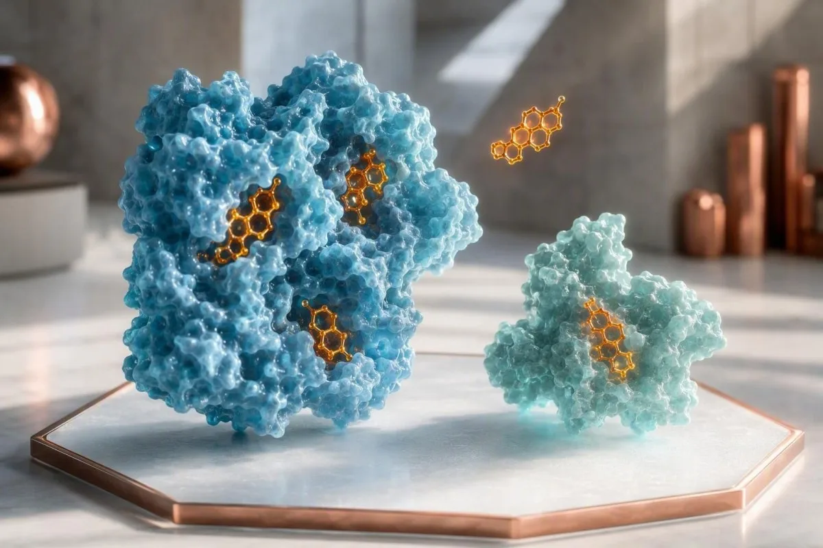
A low free testosterone result means the fraction of testosterone available to tissues may be reduced, even when total testosterone is normal. The result needs confirmation with the right method, morning timing, SHBG, albumin, symptoms, and medical context.


- Low free testosterone can reflect high SHBG rather than low testosterone production, especially when total testosterone remains normal.
- SHBG binds testosterone tightly; a higher SHBG concentration can lower calculated free testosterone without changing the total result much.
- Morning testing matters: testosterone should usually be measured between 7 and 10 am, ideally after a normal night's sleep and before food.
- Two results are usually needed before diagnosing male hypogonadism; one low hormone result after illness, poor sleep, or hard training is not enough.
- Direct analogue free-T assays are often unreliable; equilibrium dialysis or calculated free testosterone using total T, SHBG, and albumin is preferred.
- Male total testosterone below 264 ng/dL on repeat standardized morning testing supports biochemical deficiency in the Endocrine Society framework.
- Symptoms drive decisions: reduced spontaneous erections, sexual desire, anemia, loss of body hair, infertility, and low bone density carry more weight than nonspecific fatigue alone.
- Women need different interpretation: no laboratory cutoff defines androgen deficiency in women, and low free T alone does not establish a diagnosis.
What a Low Free Testosterone Result Actually Means
Low free testosterone means less unbound testosterone is estimated to be available to tissues, but it does not automatically mean your body is failing to make testosterone. A normal total testosterone result can coexist with low free testosterone when sex hormone-binding globulin, or SHBG, is elevated.
Only about 1% to 4% of circulating testosterone is truly unbound, while roughly 40% to 50% is loosely attached to albumin and much of the rest is tightly bound to SHBG. Free testosterone is therefore a small calculated or measured slice of a larger hormone pool, not a separate hormone your body produces.
In my clinical work, the classic mismatch is a 58-year-old man with total testosterone of 520 ng/dL and low calculated free testosterone because SHBG is 78 nmol/L. That pattern deserves a careful look at thyroid status, liver health, medications, weight change, and testing method before anyone talks about treatment.
Kantesti is an AI blood test analyzer that reads total testosterone, SHBG, albumin, thyroid markers, and liver results as one endocrine pattern rather than treating a low free-T flag as a diagnosis. For a plain-language comparison of methods, see our free testosterone calculator guide.
How SHBG and Albumin Change Free Testosterone Levels
SHBG is the main reason free testosterone levels can be low despite a normal total result. SHBG binds testosterone more tightly than albumin, so a rise in SHBG shifts a greater share of testosterone into the bound pool.
Albumin-bound testosterone is often called bioavailable because it separates from albumin relatively easily at tissues; SHBG-bound testosterone does not. A calculated free testosterone result uses total testosterone, SHBG, and albumin in a mass-action equation, most commonly the Vermeulen approach.
Albumin concentrations around 3.5 to 5.0 g/dL usually have a smaller effect than SHBG, but markedly low albumin from liver disease, kidney protein loss, or severe illness can distort calculated values. This is why an isolated free-T number without its companion measurements is incomplete.
I often ask patients to bring the original laboratory page because units vary: free testosterone may appear in pg/mL, ng/dL, or pmol/L, and those values cannot be compared casually. Our serum proteins reference guide explains why albumin is clinically useful beyond nutrition.
Why Total Testosterone Can Be Normal When Free T Is Low
A normal total testosterone result can mask a low free fraction when SHBG is high, but the reverse pattern is also common when SHBG is low. Total testosterone measures every circulating form together; it cannot reveal how much is tightly bound.
A total testosterone of 450 ng/dL may be fully reassuring in one man and less persuasive in another, depending on SHBG, albumin, age, symptoms, and assay quality. The Endocrine Society recommends checking free testosterone when total T is near the lower limit or when an SHBG abnormality is suspected (Bhasin et al., 2018).
High SHBG often produces normal total T with low calculated free T; obesity, insulin resistance, and hypothyroidism more often lower SHBG, making total T look low while free T is relatively preserved. That distinction helps prevent a surprisingly common error: labeling every low total result as testosterone deficiency.
Reference intervals for calculated free testosterone are method-specific and age-sensitive, with some adult male laboratories reporting approximately 47 to 244 pg/mL. The interval printed by the reporting laboratory is the one that applies to that sample; our sex-specific lab range explainer shows why borrowed internet ranges mislead.
Common Causes of High SHBG and Low Free Testosterone
High SHBG commonly results from ageing, hyperthyroidism, liver conditions, oral estrogen exposure, certain antiseizure medicines, and substantial weight loss. These causes can lower free testosterone without necessarily lowering total testosterone.
SHBG tends to rise gradually with age, particularly after 50 years, which is one reason total testosterone alone becomes less representative of tissue availability. Hyperthyroidism can increase SHBG within weeks, so a suppressed TSH and raised free T4 should be addressed before interpreting testosterone as a primary problem.
Liver-produced SHBG may rise with chronic liver disease, certain medications, and oral estrogen-containing medicines. A raised SHBG alongside elevated ALT, GGT, or bilirubin points toward a broader metabolic or hepatic review; start with our guide to what a liver panel includes.
Rapid calorie restriction is another under-recognized trigger. I have seen endurance athletes with SHBG above 90 nmol/L, reduced libido, and apparently normal total T after months of low energy availability; the useful next step was restoring training recovery and nutrition, not reflexively prescribing testosterone.
Which Free Testosterone Test Method Is Reliable?
Equilibrium dialysis is the reference method for free testosterone, while a calculated result using total T, SHBG, and albumin is often the most practical reliable option. Direct analogue immunoassays can give misleading free-T values, particularly when SHBG is abnormal.
Equilibrium dialysis physically separates the free hormone fraction and is analytically sound, but it is specialized, slower, and not available in every laboratory. Calculated free testosterone performs reasonably well when total testosterone is measured accurately and SHBG plus albumin are obtained from the same sample.
Direct analogue assays may report a precise-looking value such as 6.2 pg/mL, yet precision is not the same as accuracy. When I review discordant results, I first ask whether total testosterone came from LC-MS/MS or a quality immunoassay and whether the laboratory used dialysis, ultrafiltration, or calculation for free T.
Kantesti AI is an AI blood test interpretation platform that flags whether a reported free-T method is likely to need confirmation and compares it with the laboratory's own interval. Review our biomarker methods guide before comparing values from different laboratories.
Why Test Timing, Sleep, Food, and Illness Matter
Testosterone should usually be rechecked on two separate mornings, ideally between 7 and 10 am after normal sleep and before food. Late-day testing, acute illness, sleep restriction, and heavy exercise can create a temporary low result.
Testosterone follows a diurnal rhythm, with morning levels often 20% to 30% higher than afternoon values in younger men; the daily swing becomes smaller with age but does not disappear. A sample drawn at 4 pm should not usually be used to diagnose hypogonadism.
A 2011 sleep-restriction experiment by Leproult and Van Cauter found that one week of sleeping about 5 hours nightly reduced daytime testosterone by roughly 10% to 15% in healthy young men. Fever, hospitalization, under-fueling, and a punishing training block can have similar short-term effects, which is why waiting 2 to 4 weeks after recovery is often sensible.
Avoid trying to game a result with supplements or fasting extremes. Record sleep, illness, training, alcohol intake, and collection time alongside the number; our blood test fasting guide covers common pre-test distortions.
Low Free Testosterone Symptoms That Carry Most Weight
Low free testosterone symptoms are most meaningful when sexual, reproductive, blood, bone, or body-composition changes occur alongside repeatedly low results. Fatigue, low mood, and poor concentration alone are real symptoms but have many more common explanations.
In men, reduced morning erections, lower sexual desire, erectile difficulty, infertility, reduced shaving frequency, gynecomastia, anemia, and low-trauma fractures deserve attention. A hemoglobin drop below 13.0 g/dL in a man with confirmed low testosterone can strengthen the clinical picture, although iron deficiency and chronic disease remain more common causes.
Erectile dysfunction is not automatically hormonal; vascular disease, diabetes, depression, medication effects, and sleep apnea often contribute. Our erectile dysfunction blood test guide explains why glucose, lipids, prolactin, thyroid tests, and testosterone may all matter.
For women, low free testosterone does not define a recognized androgen-deficiency syndrome, and there is no validated diagnostic cutoff. The Global Consensus Position Statement supports testosterone therapy only for carefully assessed postmenopausal hypoactive sexual desire disorder, not for fatigue, low mood, muscle goals, or general wellbeing (Davis et al., 2019).
Low Free Testosterone Causes Beyond SHBG
Low free testosterone can reflect reduced hormone production, altered signaling from the pituitary, medication effects, obesity-related suppression, or chronic systemic illness. The pattern of LH, FSH, prolactin, and total testosterone helps separate these possibilities.
Primary testicular failure usually produces low testosterone with high LH and FSH, while secondary hypogonadism produces low testosterone with low or inappropriately normal LH and FSH. A single normal LH does not clear the pituitary; it must be read against the testosterone concentration and timing.
Obesity often lowers SHBG and total testosterone, but severe obesity, sleep apnea, type 2 diabetes, opioid use, glucocorticoids, and prolonged energy deficit can suppress the hormonal axis itself. A waist-focused metabolic review is often more useful than chasing a marginal free-T result; see testosterone in obesity.
Some causes are reversible. Opioid-associated suppression may improve after medication review, and testosterone can recover after resolution of severe illness, meaningful sleep-apnea treatment, or correction of major calorie deficit—though the timeline is variable and sometimes measured in months.
When a Low Free Testosterone Result Needs Prompt Medical Review
Low free testosterone warrants prompt medical review when it occurs with severe headache, visual change, breast discharge, infertility, unexplained anemia, fracture, or very low total testosterone. These features can point beyond ordinary ageing or SHBG variation.
A new persistent headache or loss of peripheral vision with low testosterone and elevated prolactin can indicate a pituitary mass and needs timely clinician assessment. Prolactin above the local reference interval should be repeated under calm conditions, because stress and some medicines can produce modest temporary elevations.
Testosterone below 150 ng/dL in an adult man, especially with low or normal LH, generally deserves endocrine evaluation and consideration of pituitary testing. The AUA guideline recommends measuring prolactin when low testosterone is paired with low or low-normal LH (Mulhall et al., 2018).
Low testosterone can reduce bone mineral density over time, but a fracture after a simple fall is the more urgent clue than a borderline laboratory value. Our review of high prolactin symptoms covers the headache, vision, and sexual-health pattern that should not wait.
A Practical Follow-Up Testing Plan
The best follow-up is two standardized morning hormone panels that include total testosterone, SHBG, albumin, and calculated free testosterone. If low values persist with compatible symptoms, LH, FSH, prolactin, CBC, thyroid testing, and targeted metabolic tests usually come next.
For most adult men, repeat total testosterone should be collected on separate mornings with the same laboratory where possible. The Endocrine Society's harmonized lower reference limit of 264 ng/dL applies to standardized assays in healthy non-obese men aged 19 to 39, not automatically to every laboratory or age group (Bhasin et al., 2018).
If repeated total T is near the lower boundary, add SHBG and albumin so free testosterone can be calculated; if total T is clearly low, measure LH and FSH to localize the problem. CBC can identify anemia or elevated hematocrit, while TSH, free T4, liver enzymes, A1c, and ferritin may reveal a correctable driver.
Dr. Thomas Klein's practical rule is simple: do not start a lifelong hormone decision from a one-off panel. Kantesti's clinical logic compares date, collection time, assay, symptoms, and related biomarkers; its approach is described in our medical validation overview.
How Clinicians Read Common Testosterone and SHBG Patterns
The combination of total testosterone, SHBG, calculated free testosterone, LH, and FSH is more informative than any single result. A pattern can suggest high binding, reduced production, functional suppression, or a result that simply needs repeating.
Normal total testosterone plus high SHBG and low calculated free T often indicates reduced free fraction rather than unequivocal gland failure. Normal total T plus normal SHBG and an isolated low direct-assay free T is more likely to be an assay problem, particularly if symptoms are absent.
Low total T with low SHBG and normal calculated free T frequently appears with obesity or insulin resistance; it may improve with treatment of the underlying metabolic condition. In contrast, low total T, low free T, and high LH is a more consistent primary hypogonadal pattern.
Patients receiving testosterone need a different interpretation because timing relative to the dose can dominate the result. Read our guidance on testosterone monitoring timing rather than comparing a post-dose peak with a pre-dose trough.
What to Do Before Considering Testosterone Treatment
Treating low free testosterone starts with confirming the diagnosis and addressing reversible causes, not buying testosterone boosters or beginning therapy from one borderline value. Testosterone therapy is appropriate only for selected people with persistent biochemical deficiency and relevant symptoms.
Improving sleep, resistance exercise, adequate protein and energy intake, alcohol moderation, diabetes management, and treatment of sleep apnea can improve testosterone physiology in some patients. These measures are worthwhile even when they do not move a result from 240 to 500 ng/dL, because sexual function and energy have multiple drivers.
Avoid over-the-counter products marketed as testosterone boosters. Some contain undisclosed hormones or liver-toxic ingredients, and high-dose zinc or herbal blends can create new abnormalities; our evidence review on foods and testosterone separates plausible nutrition from marketing.
Exogenous testosterone can suppress sperm production within months, so men pursuing fertility should speak with an endocrinologist or reproductive specialist before treatment. Monitoring typically includes testosterone timing, hematocrit, prostate-risk discussion where relevant, and symptom response—not simply chasing a high number.
Low Free Testosterone in Women: Why Interpretation Differs
A low free testosterone result in women is difficult to interpret and does not diagnose androgen deficiency by itself. Concentrations are much lower than in men, assays are less accurate at low levels, and oral estrogen can raise SHBG substantially.
Combined oral contraceptives and oral menopausal estrogen can raise SHBG and lower free testosterone without proving a hormone disorder. In women with irregular periods, acne, hair changes, infertility, or sexual symptoms, clinicians consider the menstrual cycle, medication exposure, thyroid function, prolactin, and adrenal context.
Kantesti is an AI-powered blood test analysis tool that places testosterone results beside SHBG, estradiol, DHEA-S, thyroid tests, and the collection date rather than applying male thresholds to female results. Our women's testosterone range guide explains why cycle phase and assay type matter.
The evidence for treating women is narrow. The 2019 Global Consensus group found benefit for postmenopausal hypoactive sexual desire disorder, while stating that a blood concentration should not be used alone to diagnose the condition (Davis et al., 2019).
How to Track Free Testosterone Results Without Overreacting
Free testosterone trends are useful only when collection time, laboratory method, SHBG, albumin, medications, and health status are comparable. A 15% change between two different laboratories may be analytical noise rather than a physiological shift.
Save the collection time, whether you were fasting, sleep duration, recent infection, training load, body weight, alcohol intake, and new medicines for every test. A meaningful personal baseline usually takes at least 2 to 3 comparable results, not one annual snapshot.
Kantesti AI interprets free testosterone trends by matching the reported assay, SHBG direction, albumin, and date-specific context across visits. The underlying workflow is explained in our AI technology guide, while our longitudinal results guide shows what to record.
As of August 6, 2026, no major guideline recommends routine free testosterone screening in asymptomatic adults. Testing is most helpful when symptoms, borderline total testosterone, altered SHBG, infertility evaluation, or a relevant medical condition gives the result a clinical question to answer.
Questions to Bring to Your Clinician After a Low Result
Ask whether your free testosterone was directly measured or calculated, whether the sample was collected early morning, and whether SHBG was abnormal. Those three questions often clarify whether a low result represents biology, temporary suppression, or a test limitation.
A useful appointment question is: “Do my symptoms and repeat results meet criteria for hypogonadism, or should we look first for thyroid, liver, sleep, medication, weight, or pituitary causes?” Ask for the actual values in ng/dL, nmol/L, g/dL, or pg/mL, not just “normal” or “low.”
Bring a list of prescription medicines, non-prescription products, fertility plans, and recent illnesses. Dr. Thomas Klein has found that this short history often changes the interpretation more than an extra one-off hormone test—especially with opioids, steroid tablets, antiseizure medicines, estrogen exposure, or major weight change.
Kantesti is an AI biomarker interpretation platform used to organize multi-marker laboratory context, but it does not replace diagnosis or treatment from a qualified clinician. Our physician standards and review process are available through the Medical Advisory Board; urgent neurologic symptoms, vision change, or severe systemic illness should be assessed promptly in person.
Frequently Asked Questions
Can free testosterone be low when total testosterone is normal?
Yes. Free testosterone can be low when total testosterone is normal because high SHBG binds a larger proportion of the circulating hormone, leaving a smaller unbound fraction. This pattern is particularly common with ageing, hyperthyroidism, liver conditions, oral estrogen exposure, and some antiseizure medicines. Confirmation should use a repeat 7 to 10 am sample with total testosterone, SHBG, albumin, and calculated free testosterone or equilibrium dialysis.
What is considered a low free testosterone level?
A low free testosterone level is a value below the reporting laboratory's method-specific reference interval, not one universal number. Adult male intervals may range roughly from 47 to 244 pg/mL depending on age, assay, and calculation method, so results from different laboratories should not be compared directly. In women, no free-testosterone threshold establishes androgen deficiency. A clinically meaningful result also requires compatible symptoms and repeat testing.
Should I worry about low free testosterone but normal total testosterone?
Low free testosterone with normal total testosterone is usually not an emergency, but it deserves follow-up if symptoms are present or SHBG is markedly high. Review thyroid tests, liver results, medications, oral estrogen exposure, recent weight loss, sleep, acute illness, and the laboratory method. Severe headache, visual symptoms, breast discharge, infertility, fracture, or total testosterone below 150 ng/dL should prompt faster medical assessment.
What time of day should free testosterone be tested?
Free testosterone should generally be tested between 7 and 10 am, preferably after a normal night's sleep and before food. Testosterone can be 20% to 30% higher in the morning than later in the day in younger men, making afternoon results less useful for diagnosis. Repeat testing on two separate mornings reduces the chance that sleep loss, illness, hard exercise, or normal day-to-day variation produces a false low result.
Is calculated free testosterone better than direct free testosterone?
Calculated free testosterone is often more useful than a direct analogue free-testosterone immunoassay when total testosterone, SHBG, and albumin are measured accurately from the same sample. Equilibrium dialysis remains the reference method because it physically measures the unbound fraction, although it is less widely available. Direct analogue assays can be misleading when SHBG is high or low. Ask the laboratory which method it used before acting on an isolated result.
Can low free testosterone cause fatigue?
Low free testosterone can contribute to fatigue, reduced exercise capacity, low mood, and poor concentration, but these symptoms are not specific enough to diagnose testosterone deficiency. Sleep apnea, anemia, iron deficiency, thyroid disease, depression, medication effects, diabetes, and inadequate calorie intake are frequent alternatives. Symptoms such as reduced spontaneous erections, lower libido, infertility, anemia below 13.0 g/dL in men, or low bone density provide more specific clinical context.
Get AI-Powered Blood Test Analysis Today
Join over 2 million users worldwide who trust Kantesti for instant, accurate lab test analysis. Upload your blood test results and receive comprehensive interpretation of 15,000+ biomarkers in seconds.
📚 Referenced Research Publications
Klein, T., Mitchell, S., & Weber, H. (2026). aPTT Normal Range: D-Dimer, Protein C Blood Clotting Guide. Kantesti AI Medical Research.
Klein, T., Mitchell, S., & Weber, H. (2026). Serum Proteins Guide: Globulins, Albumin & A/G Ratio Blood Test. Kantesti AI Medical Research.
📖 External Medical References
⚕️ Medical Disclaimer
This article is for educational purposes only and does not constitute medical advice. Always consult a qualified healthcare provider for diagnosis and treatment decisions.
E-E-A-T Trust Signals
Experience
Physician-led clinical review of lab interpretation workflows.
Expertise
Laboratory medicine focus on how biomarkers behave in clinical context.
Authoritativeness
Written by Dr. Thomas Klein with review by Dr. Sarah Mitchell and Prof. Dr. Hans Weber.
Trustworthiness
Evidence-based interpretation with clear follow-up pathways to reduce alarm.