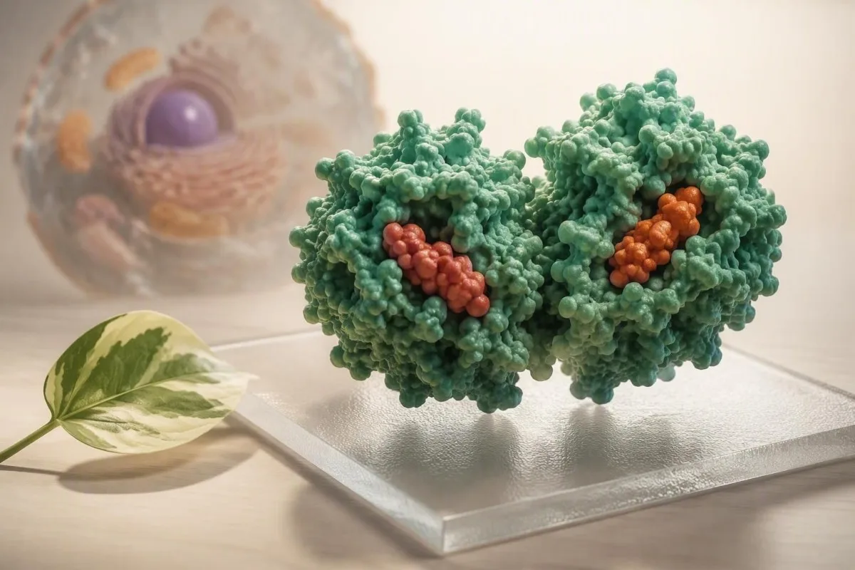
A high SHBG result usually reflects thyroid status, estrogen exposure, liver signaling, energy deficit, aging, or medicines—not a diagnosis on its own. The practical issue is whether it makes your available testosterone or estradiol lower than the total-hormone result suggests.


- High SHBG is most often caused by estrogen exposure, hyperthyroidism, chronic liver disease, calorie deficit, older age, or certain medicines.
- Reference intervals vary by assay; many adult laboratories use roughly 18-54 nmol/L for men and 24-122 nmol/L for premenopausal women.
- Free testosterone can be low despite a normal total testosterone when SHBG is high, especially in men over 50 and people using oral estrogen.
- Thyroid testing matters because suppressed TSH with high free T4 can raise SHBG before obvious hyperthyroid symptoms appear.
- Oral estrogen usually raises SHBG more than transdermal estrogen because first-pass liver exposure changes hepatic protein production.
- Energy availability below roughly 30 kcal/kg fat-free mass/day can suppress reproductive signaling and raise SHBG in endurance athletes.
- Liver context matters: SHBG may rise in hepatitis, cirrhosis, or cholestatic disease even when ALT is only mildly abnormal.
- Do not treat SHBG alone; repeat a morning hormone panel with albumin and interpret calculated free testosterone or free androgen index.
What a High SHBG Result Actually Means
High sex hormone-binding globulin means the liver is producing more of a carrier protein that binds testosterone and estradiol tightly. The result can lower the fraction of hormone available to tissues even when total testosterone or total estradiol sits inside the laboratory range.
SHBG is a glycoprotein made mainly by hepatocytes, and each dimer has high-affinity steroid-binding sites. In practical terms, testosterone bound to SHBG is not necessarily absent from the body, but it is less readily available than albumin-bound or free testosterone. A value of 82 nmol/L in a man with total testosterone of 520 ng/dL can produce a very different clinical picture from the same total testosterone with SHBG of 22 nmol/L.
When I review this pattern, I do not diagnose “high SHBG disease.” I ask what is driving liver production and whether the calculated free testosterone agrees with symptoms, examination, and repeat testing. Dr. Thomas Klein’s rule in clinic is simple: a binding protein result is a clue, not a treatment target.
Kantesti is an AI blood test analyzer that reads SHBG alongside total testosterone, albumin, estradiol, thyroid tests, liver markers, and prior results rather than treating one flagged number as a diagnosis. For a primer on the protein itself, see our SHBG test guide.
Why total hormone can mislead
Total testosterone includes SHBG-bound, albumin-bound, and free hormone. In men, only about 1-4% circulates as free testosterone, so a change in SHBG can materially alter the biologically available fraction without moving total testosterone very much.
Reference Ranges, Assays and When a Number Is Truly High
Adult SHBG reference ranges are assay-, sex-, and age-dependent, so the laboratory interval printed beside your result outranks any universal internet cutoff. Many laboratories report approximately 18-54 nmol/L for adult men and 24-122 nmol/L for premenopausal women.
A result above 100 nmol/L deserves context, but it is not automatically dangerous. In a 30-year-old woman taking a combined oral contraceptive, 120 nmol/L may be medication-related; in a 35-year-old man with weight loss, tremor, and TSH below 0.01 mIU/L, the same result pushes thyroid disease much higher on the list.
SHBG is commonly measured by immunoassay, while free testosterone may be directly measured, calculated from total testosterone-SHBG-albumin, or measured by equilibrium dialysis. Direct analogue free-testosterone assays are often unreliable at low concentrations; the Endocrine Society advises equilibrium dialysis or a validated calculation when SHBG is altered (Bhasin et al., 2018).
Repeat an unexpected result after 6-12 weeks under comparable conditions, particularly after acute illness, major diet change, or a medicine switch. A blood-test change guide explains why a small numerical difference can be normal biological variation.
Hyperthyroidism Is a Commonly Missed Cause of High SHBG
Hyperthyroidism raises SHBG because thyroid hormone stimulates hepatic SHBG gene expression. A suppressed TSH below 0.1 mIU/L with raised free T4 or free T3 makes thyroid excess a more likely explanation than an isolated hormone problem.
I have seen men referred for “low testosterone symptoms” whose total testosterone was respectable at 18 nmol/L, yet SHBG was 96 nmol/L and TSH was undetectable. Their palpitations, heat intolerance, sleep disruption, and falling weight were thyroid clues, not evidence that testosterone therapy was the answer.
Free T4 above the assay upper limit plus low TSH should prompt a clinician-led hyperthyroidism evaluation, especially with resting pulse persistently above 100 beats/minute, new atrial fibrillation, tremor, or eye changes. Biotin can distort some thyroid immunoassays, so disclose supplements and stop high-dose biotin only if the ordering clinician advises it.
A normal TSH makes overt thyroid-driven SHBG elevation less likely, though not impossible in central thyroid disorders or during treatment changes. Review thyroid retest timing rather than changing thyroid medication from one panel alone.
Thyroid treatment changes the interpretation
SHBG can take weeks to months to settle after thyroid levels normalize. A repeat hormone panel 8-12 weeks after stable thyroid treatment is usually more interpretable than a test drawn during active dose adjustment.
Estrogen Exposure and High SHBG in Women
Estrogen exposure is one of the most frequent high SHBG causes in women, particularly combined oral contraception, pregnancy, and oral menopausal hormone therapy. Oral estrogen has a stronger effect than transdermal estrogen because it reaches the liver first at a high concentration.
High SHBG in women can lower the free androgen index, calculated as total testosterone divided by SHBG times 100 when both values use nmol/L. A low index on combined contraception does not diagnose androgen deficiency, because the pill both raises SHBG and suppresses ovarian androgen production; it is a pharmacologic pattern.
Pregnancy also raises SHBG substantially, often into the 200 nmol/L range or higher by late gestation, so non-pregnant reference ranges should not be applied. In patients evaluating acne, hirsutism, irregular cycles, or possible PCOS, hormone testing while using hormonal contraception may not represent untreated physiology.
Our birth-control hormone testing article explains which values change on treatment. Kantesti is an AI blood test interpretation platform that flags estrogen exposure as a possible confounder when interpreting SHBG and calculated androgen measures.
Liver Health, Alcohol and SHBG Production
The liver produces SHBG, so hepatitis, cirrhosis, cholestasis, and some alcohol-related liver changes can raise it. High SHBG with bilirubin elevation, low albumin, prolonged INR, or low platelets needs more attention than high SHBG with otherwise normal liver markers.
The confusing point is that metabolic insulin resistance often lowers SHBG, while advanced or hormonally active liver disease may raise it. Therefore, SHBG cannot diagnose liver disease by itself. ALT above 55 IU/L, AST above 40 IU/L, alkaline phosphatase above the local upper limit, or rising bilirubin changes the next step far more than SHBG alone.
Alcohol deserves a careful, nonjudgmental history because intake can influence liver enzymes, nutrition, sleep, and gonadal hormone metabolism simultaneously. A normal ALT does not exclude significant fibrosis, particularly in people with diabetes, obesity, viral hepatitis risk, or sustained heavy alcohol exposure.
Use a full panel—ALT, AST, ALP, GGT, bilirubin, albumin, platelet count, and sometimes FIB-4—rather than chasing one liver enzyme. Our MASLD testing guide and bilirubin pattern guide help distinguish common patterns.
When liver symptoms need urgent assessment
Jaundice, dark urine, pale stool, confusion, abdominal swelling, vomiting blood, or severe right-upper abdominal pain should prompt urgent medical assessment. Those symptoms are never explained away by a high SHBG result.
Calorie Restriction, Weight Loss and Endurance Training
Sustained low energy availability can raise SHBG and reduce reproductive hormone signaling, particularly in endurance athletes and people losing weight rapidly. The concern is not a single low-calorie day; it is a prolonged mismatch between training demand and energy intake.
In sports medicine, energy availability below about 30 kcal/kg fat-free mass/day is associated with endocrine disruption, though an exact individual threshold is not universal. SHBG may rise alongside low-normal free T3, declining libido, missed periods, recurrent bone stress injury, or unexpectedly poor recovery.
A 52-year-old marathon runner with SHBG of 88 nmol/L, low calculated free testosterone, ferritin of 18 ng/mL, and an escalating training load needs a broader conversation than “eat more protein.” I would look at total energy intake, carbohydrate timing, sleep, iron status, thyroid tests, and fracture history; each changes risk differently.
Rapid weight loss can also improve insulin resistance and thereby release a previously low SHBG toward normal or high-normal values. For athletes, our RED-S laboratory patterns offer a more useful checklist than SHBG in isolation.
Aging, Body Composition and Insulin Signaling
SHBG tends to rise with age in men, while obesity and hyperinsulinemia often lower it. The combination of aging and a normal total testosterone can therefore conceal a clinically meaningful fall in calculated free testosterone.
In men, total testosterone often declines gradually with age while SHBG rises, creating a double effect on free testosterone. A total testosterone of 420 ng/dL may be reassuring in a 65-year-old only after SHBG, albumin, symptoms, timing, and repeat morning testing are considered.
Conversely, low SHBG frequently accompanies central adiposity, fatty liver, high triglycerides, and insulin resistance. That does not make low SHBG protective; it means total testosterone can look deceptively low when free testosterone is less reduced than expected. The free testosterone calculation guide covers why methods disagree.
Kantesti AI interprets SHBG results against age, sex, albumin, total hormone values, and longitudinal trend data; a single reference flag is rarely enough. As of September 19, 2026, no major guideline recommends treatment solely to force SHBG into a reference interval.
Medicines and Supplements That Can Raise SHBG
Combined estrogen-containing contraceptives, oral estrogen therapy, thyroid hormone excess, some anticonvulsants, and certain antiretroviral regimens can increase SHBG. The medication list is often the fastest route to an explanation for an unexpected result.
Enzyme-inducing anticonvulsants such as carbamazepine and phenytoin can alter hepatic hormone metabolism and SHBG. Levothyroxine is appropriate when hypothyroidism is real, but an excessive dose can reproduce the SHBG pattern of hyperthyroidism; dose changes should be guided by TSH and free T4 rather than symptoms alone.
Do not stop contraception, epilepsy treatment, HIV treatment, or hormone therapy just to normalize a laboratory number. The safe question is whether the medicine is providing more benefit than risk and whether a different formulation would change the interpretation—particularly oral versus transdermal estrogen.
Bring every prescription, injection, patch, inhaler, supplement, and recent dose change to the appointment. The medication safety trend guide is useful when several results shifted after the same treatment change.
How High SHBG Changes Free Testosterone Interpretation
High SHBG can make total testosterone appear normal while calculated free testosterone is low. For men with symptoms and borderline total testosterone, a morning repeat test with SHBG and albumin is the usual next step.
The Endocrine Society recommends diagnosing male hypogonadism only when symptoms are present and testosterone is unequivocally and consistently low, confirmed with repeat morning fasting total testosterone; free testosterone is particularly helpful when SHBG is altered (Bhasin et al., 2018). One result after a poor night’s sleep, heavy exercise, or acute illness is a weak basis for diagnosis.
Calculated free testosterone commonly uses the Vermeulen equation with total testosterone, SHBG, and albumin. It is not a direct tissue measurement, but in my experience it is far more clinically useful than interpreting total testosterone alone when SHBG is 70-100 nmol/L.
Low libido, fewer spontaneous erections, low energy, depressed mood, anemia, and reduced bone density can overlap with low androgen availability, but none is specific. Read our low free testosterone overview before assuming one symptom identifies the cause.
High SHBG in Women: Symptoms and Hormone Clues
High SHBG in women may reduce the free fraction of testosterone, but symptoms such as low libido, fatigue, vaginal dryness, or mood change are not specific enough to diagnose androgen deficiency. Menopause stage, contraception, estradiol exposure, thyroid disease, iron status, sleep, and relationship factors often matter more.
A high SHBG result is common after starting a combined oral contraceptive and does not mean the medicine is harming you. Yet if new symptoms began after a specific formulation, the timing is clinically useful and worth discussing with the prescriber; switching formulation is a medical decision, not a DIY experiment.
Postmenopausal hormone results fluctuate and may not correlate tightly with symptoms. Total testosterone in women is often near assay limits, so liquid chromatography-tandem mass spectrometry is preferable when an accurate low-level testosterone result will change a clinical decision.
Cycle timing remains relevant when periods occur. Compare results with our testosterone ranges for women and menopause estradiol guide rather than borrowing male cutoffs.
A Practical Follow-Up Panel for Unexpectedly High SHBG
An unexpectedly high SHBG result is best repeated with a targeted panel: total testosterone, albumin, calculated free testosterone, TSH, free T4, liver tests, and medication review. Add estradiol, LH, FSH, prolactin, pregnancy testing, or nutritional markers only when the history makes them relevant.
For men, collect testosterone between 7 and 10 am after usual sleep, ideally before intense training and not during a febrile illness. For women, the best timing depends on the clinical question: cycle day, contraceptive use, pregnancy, and menopausal status should all be recorded beside the result.
Useful liver context includes ALT, AST, ALP, GGT, bilirubin, albumin, platelet count, and hepatitis testing when indicated by risk. If restrictive eating, heavy training, or fatigue is present, a CBC, ferritin, iron studies, vitamin B12, vitamin D, and phosphate can uncover parallel problems that SHBG does not explain.
Kantesti is an AI-powered blood test analysis tool used across 127+ countries to organize these related results into a clinician-ready trend view. Our biomarker reference guide can help you verify units before an appointment.
What not to do before the repeat test
Do not fast for 24 hours, add testosterone boosters, start iodine, or alter prescribed thyroid medication to improve the number. Those changes can create a misleading panel and sometimes cause real harm.
High SHBG Symptoms: What You Feel and What Needs Urgent Care
High SHBG itself usually causes no direct symptoms; symptoms come from the underlying condition or from lower free hormone availability. Urgent assessment is warranted for jaundice, confusion, chest pain, fainting, severe shortness of breath, or a rapid resting heartbeat with thyroid symptoms.
Possible low-free-testosterone symptoms include reduced sexual desire, erectile changes, reduced morning erections, lower exercise tolerance, and low mood in men. In women, libido changes and fatigue can occur, but symptoms overlap heavily with iron deficiency, depression, sleep disorders, menopause, medicines, and chronic illness.
A high SHBG result should not be used to explain away palpitations, unintentional weight loss, tremor, new diarrhea, or heat intolerance. Those features call for timely thyroid testing; see our palpitations blood-test guide for the broader differential.
Dr. Thomas Klein advises patients to seek same-day care for yellow eyes with dark urine, severe confusion, or a pulse persistently above 120 beats/minute at rest. Laboratory interpretation is helpful, but it is not a substitute for triage when symptoms are escalating.
Common Misconceptions About Lowering SHBG
There is no evidence-based supplement regimen designed to lower SHBG safely in everyone. Treating an underlying thyroid, liver, nutritional, or medication issue is more rational than trying to suppress a protective carrier protein.
Boron, nettle-root products, and “testosterone boosters” are often marketed around SHBG, but clinical evidence for meaningful durable benefit is limited and product quality varies. More concerningly, androgenic supplements can suppress natural hormone production, affect lipids, and sometimes contain undeclared substances.
People also assume high SHBG means “too much estrogen.” That is incomplete: high SHBG can occur with low estradiol, normal estradiol, pregnancy, hyperthyroidism, older age, and liver disease. The pattern of total and free hormones—not SHBG alone—determines whether estrogen exposure is even plausible.
If nutrition is part of the story, aim for adequate total energy, carbohydrate around training, dietary fat, protein, calcium, and iron rather than a single magic food. Our intermittent fasting lab guide explains why fasting routines can alter interpretation.
Questions to Take to Your Clinician After a High SHBG Result
The most useful questions are: Is my free testosterone or free androgen index actually low, could thyroid or liver disease explain this, and should we repeat testing under standardized conditions? A good consultation connects the number to symptoms, medicines, and trends.
Bring the original report rather than a cropped screenshot, because units and reference intervals matter. Ask whether albumin was measured, which free-testosterone method was used, and whether the sample was drawn during illness, pregnancy, estrogen use, or a major training block.
If there are liver abnormalities, ask whether FIB-4, ultrasound, viral hepatitis testing, alcohol review, or medication changes are appropriate. If thyroid symptoms are present, ask whether TSH, free T4, free T3, thyroid antibodies, and repeat timing fit the clinical picture rather than requesting every test at once.
Our medical advisory board oversees clinician-facing safety standards, and medical validation information explains how Kantesti AI supports—not replaces—medical judgment. The aim is a better conversation and a sensible next test, not self-diagnosis.
The Bottom Line on High SHBG Causes
High SHBG is most commonly explained by estrogen exposure, hyperthyroidism, liver signaling, prolonged calorie restriction, aging, or medicines. The next decision should rest on calculated free hormone values and the cause of the rise, not on SHBG alone.
A stable, mildly elevated SHBG with normal thyroid and liver tests, no concerning symptoms, and a normal calculated free hormone level is often simply a personal physiological pattern. In contrast, rising SHBG plus suppressed TSH, abnormal bilirubin, low albumin, marked weight loss, or new symptoms warrants timely clinical review.
Rosner and colleagues cautioned that testosterone testing has substantial measurement and interpretation pitfalls, especially when binding proteins are abnormal (Rosner et al., 2007). That remains true in 2026: the best answer usually comes from a repeated, well-timed panel and a good history, not from one dramatic-looking flag.
If you are comparing multiple reports, the Kantesti AI technology guide describes the contextual checks our system uses for longitudinal lab interpretation. Keep the result, note medicines and training changes, and take the whole pattern to your clinician.
Frequently Asked Questions
What is the most common cause of high SHBG?
Estrogen exposure is among the most common causes of high SHBG, especially combined oral contraceptives, pregnancy, and oral menopausal hormone therapy. Hyperthyroidism, liver disease, older age, sustained calorie restriction, and some anticonvulsants are also frequent explanations. In adult men, an SHBG result above roughly 70 nmol/L should usually prompt review of total testosterone, albumin, calculated free testosterone, TSH, liver tests, and medicines. The laboratory reference interval remains the correct first benchmark because assays differ.
Can high SHBG cause low testosterone symptoms?
High SHBG can contribute to low testosterone symptoms by lowering calculated free testosterone even when total testosterone is normal. Symptoms may include lower libido, fewer spontaneous erections, lower energy, depressed mood, reduced exercise tolerance, or reduced bone density, but none is specific to testosterone. The Endocrine Society recommends repeat morning total testosterone testing and assessment of free testosterone when SHBG is abnormal. A single result, particularly after poor sleep, acute illness, or hard training, should not establish hypogonadism.
What level of SHBG is considered too high?
There is no universal dangerous SHBG cutoff because reference ranges vary by sex, age, laboratory, and assay. Many laboratories use about 18-54 nmol/L for adult men and 24-122 nmol/L for premenopausal women, while pregnancy can produce values above 200 nmol/L. In men, values above 70-80 nmol/L often make free-testosterone calculation particularly useful. A level above 100 nmol/L is not an emergency by itself, but it warrants review for thyroid disease, estrogen exposure, liver abnormalities, nutritional deficit, and medicines.
Does high SHBG mean too much estrogen?
High SHBG does not automatically mean too much estrogen. Oral estrogen and pregnancy can raise SHBG, but hyperthyroidism, liver disease, aging, low energy availability, and medicines can do the same with normal or low estradiol. Estradiol interpretation depends on sex, menstrual-cycle phase, pregnancy, menopausal stage, and treatment use. A clinician should interpret estradiol and SHBG together rather than treating either number as proof of hormone excess.
Can fasting or weight loss increase SHBG?
Prolonged calorie restriction and rapid weight loss can increase SHBG, particularly when energy intake remains low relative to endurance training volume. In athletes, energy availability below approximately 30 kcal/kg fat-free mass/day is associated with endocrine disruption, although individual thresholds vary. Weight loss can also improve insulin sensitivity, which may raise SHBG from a previously low level. Persistent fatigue, missed periods, low libido, stress injuries, or low ferritin make a fuller nutritional and endocrine assessment sensible.
How can I lower high SHBG naturally?
There is no proven universal natural treatment for high SHBG because SHBG is a marker of several different physiological states. Addressing hyperthyroidism, correcting sustained under-fuelling, reviewing oral estrogen or interacting medicines, and assessing liver health may normalize SHBG when one of those causes is present. Supplements marketed to lower SHBG have limited high-quality evidence and may create medication interactions or misleading hormone results. The safer goal is normal free-hormone function and treatment of the underlying cause, not a specific SHBG number.
Get AI-Powered Blood Test Analysis Today
Join over 2 million users worldwide who trust Kantesti for instant, accurate lab test analysis. Upload your blood test results and receive comprehensive interpretation of 15,000+ biomarkers in seconds.
📚 Referenced Research Publications
Klein, T., Mitchell, S., & Weber, H. (2026). AI Blood Test Analyzer: 2.5M Tests Analyzed | Global Health Report 2026. Kantesti AI Medical Research.
Klein, T., Mitchell, S., & Weber, H. (2026). RDW Blood Test: Complete Guide to RDW-CV, MCV & MCHC. Kantesti AI Medical Research.
📖 External Medical References
⚕️ Medical Disclaimer
This article is for educational purposes only and does not constitute medical advice. Always consult a qualified healthcare provider for diagnosis and treatment decisions.
E-E-A-T Trust Signals
Experience
Physician-led clinical review of lab interpretation workflows.
Expertise
Laboratory medicine focus on how biomarkers behave in clinical context.
Authoritativeness
Written by Dr. Thomas Klein with review by Dr. Sarah Mitchell and Prof. Dr. Hans Weber.
Trustworthiness
Evidence-based interpretation with clear follow-up pathways to reduce alarm.