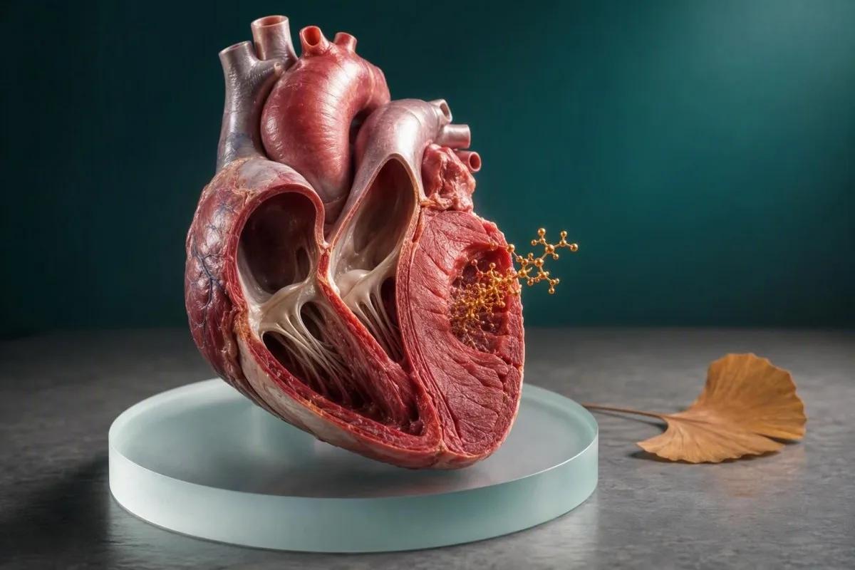
CK-MB er en eldre, men fortsatt nyttig markør for muskelskade som kan involvere hjertet. Verdien avhenger av tidspunkt, symptomer, EKG-funn, total CK og – vanligvis – troponin.


- CK-MB betydning: CK-MB står for kreatinkinase myokardialt bånd, en isoenzym som hovedsakelig finnes i hjertemuskelen, men også i skjelettmuskelen.
- Normale verdier: Mange CK-MB masseanalyser bruker en øvre referansegrense nær 3–5 ng/mL, men det laboratoriespesifikke området har alltid prioritet.
- Tidspunkt: CK-MB stiger vanligvis 3–6 timer etter myokardskade, topper seg ved 12–24 timer, og returnerer ofte mot baseline innen 48–72 timer.
- Troponin først: Høy-sensitivitets kardial troponin er den foretrukne blodmarkøren for mistenkt hjerteinfarkt i moderne akuttmedisin.
- Ingen enkeltstående testdiagnose: En forhøyet CK-MB alene kan ikke diagnostisere et hjerteinfarkt, fordi hard trening, muskelskader, kirurgi og muskelsykdommer kan øke den.
- Relativ indeks: En CK-MB-indeks over ca. 51 % kan støtte en kardiell årsak, men den blir upålitelig når total CK er markert forhøyet.
- Urgent symptoms: Ny brysttrykk, pustevansker, besvimelse, svetting eller smerter som sprer seg til kjeven eller armen krever akutt vurdering uavhengig av et tidligere CK-MB-resultat.
- Trenden betyr noe: En økning og nedgang på gjentatte prøver er mer informativ enn ett isolert tall.
CK-MB er myokardial-bandformen av kreatinkinase
CK-MB står for kreatinkinase myokardbånd. Det er en enzymform som måles i en CK-MB-blodprøve når klinikere undersøker mulig skade på hjertemuskelen, selv om en unormal verdi i seg selv ikke beviser et hjerteinfarkt.
Kreatinkinase hjelper celler med å raskt regenerere ATP, den umiddelbare energikilden som brukes under muskelarbeid. CK-MB er konsentrert i hjertemuskelen, mens CK-MM dominerer i skjelettmuskulaturen og CK-BB finnes mer utbredt i hjerne- og glatte vev. For en grunnleggende forståelse av morsenzymet, se vår veiledning til kreatinkinase.
Et CK-MB-resultat rapporteres vanligvis som masse-konsentrasjon i ng/mL eller, sjeldnere, enzymaktivitet i U/L. Mange nåværende masseanalyser setter en øvre grense rundt 3–5 ng/mL, men kjønnsspesifikke grenser og analysekalibrering varierer såpass at et tall fra ett laboratorium ikke kan overføres til et annet.
Kantesti er en AI-blodprøveanalysator som viser CK-MB sammen med analysens referanseintervall, total CK, troponin og tidspunkt for prøvetaking. Dette har betydning, fordi en verdi på 6 ng/mL etter 2 timer med symptomer gir et helt annet spørsmål enn 6 ng/mL etter et maraton.
Suffikset kan virke definitivt, og pasienter hører forståelig nok “myokardiell” som “hjerteinfarkt”. I reell klinisk praksis betyr det at laboratoriet har målt ett spor om skade på muskelceller – ikke årsaken, lokaliseringen eller alvorlighetsgraden av den skaden.
Hvorfor navnet fortsatt vises på rapporter
Noen sykehus beholder CK-MB i bestillingssett for akuttmottak eller sykehusinnlagte pasienter, spesielt når de ønsker en kortvarig markør for å hjelpe til med å vurdere mulig tilbakevendende skade. I mange samfunnsbaserte settinger har det imidlertid i stor grad blitt erstattet av høy-sensitivitet troponin for innledende vurdering av hjerteinfarkt.
CK-MB og total CK måler relaterte, men forskjellige ting
Total CK måler enzym frigjort fra alle muskelrike vev, mens CK-MB måler én CK-fraksjon. Total CK kan stige til tusenvis av U/L etter anstrengende aktivitet eller muskelskade uten å indikere et hjerteproblem.
En total CK på 1500 U/L etter uvant trening kan gjenspeile stress i skjelettmuskulaturen, dehydrering, medikamenteffekter eller rabdomyolyse; det identifiserer ikke kilden alene. CK-MB kan også øke i den settingen fordi skjelettmuskulatur inneholder små mengder av MB-fraksjonen.
Den gamle CK-MB relative indeks beregnes som CK-MB delt på total CK, deretter multiplisert med 100. En indeks under 3% lener tradisjonelt mot skjelettmuskulatur, mens en indeks over 5% kan støtte en kardial kilde – men disse grenseverdiene er støttende bevis, ikke en dom.
Når total CK er ekstremt høy, kan indeksen villede fordi både telleren og nevneren endres av forskjellige biologiske årsaker. Dette er en grunn til at klinikere nå legger større vekt på serielle høyfølsomhetstroponiner og EKG fremfor å prøve å matematisk redde et tvetydig CK-MB-resultat.
Et praktisk eksempel: en styrkeutøver med total CK 4 000 U/L og CK-MB 12 ng/mL kan ha en lav relativ andel og ingen iskemiske symptomer. Samme CK-MB-verdi hos en stillesittende person med ny, knusende brystsmerte fortjener en helt annen respons.
Hvorfor total CK kan være høy
Trening, fall, langvarig immobilitet, anfall, intramuskulære injeksjoner, hypotyreose, statiner og inflammatoriske muskelsykdommer kan øke total CK. Vår guide til farlig høy CK forklarer når mørk urin, alvorlig svakhet eller redusert urinproduksjon endrer alvoret.
Hvorfor en kliniker fortsatt kan bestille en CK-MB blodprøve
Klinikere kan bestille CK-MB ved evaluering av brystsymptomer, mulig tilbakevendende myokardskade eller en uklar økning i andre muskelenzymer. Det bestilles vanligvis sammen med EKG-testing og troponin, snarere enn som en frittstående screening.
I de første 24–72 timene etter en bekreftet kardial hendelse kan CK-MBs relativt raske nedgang noen ganger hjelpe med å identifisere en mulig ny skade. Troponin forblir ofte forhøyet lenger, så en ny CK-MB-økning kan gi kontekst, selv om lokale protokoller varierer og bevisene ikke er helt ensartede.
En kliniker kan også bruke CK-MB når et eldre laboratorieinformasjonssystem fortsatt inkluderer det sammen med total CK. Etter min erfaring blir pasienter ofte skremt av en automatisk generert CK-MB-flagg som deres behandlende team aldri anså som bevis på infarkt, fordi troponin og EKG var betryggende.
CK-MB er ikke en screeningtest for stille koronar plakk, fremtidig hjertrisiko eller generell kondisjon. Blodtrykk, diabetesstatus, LDL-kolesterol, ApoB, røyking, nyrefunksjon og familiehistorie besvarer mer nyttige forebyggende spørsmål; vår ApoB og ApoA1-ratio guide forklarer et av disse risikoplanene.
Hvis du er symptomfri og ser CK-MB på en rapport fra en yrkesmessig, idretts- eller innlagte panel, spør hvorfor den ble bestilt og hva legen din sammenlignet den med. Ordringskonteksten forteller oss ofte mer enn et mildt unormalt resultat.
En test er ikke en diagnose
Hjerteinfarkt er kliniske diagnoser basert på symptomer, undersøkelse, EKG- eller bildediagnostiske bevis, og biomarkørdynamikk. Laboratoriemarkører identifiserer myokardskade; de etablerer ikke uavhengig om en blokkert kransarterie forårsaket den.
CK-MB-tidsforløpet forklarer hvorfor et enkelt resultat kan villede
CK-MB begynner vanligvis å stige omtrent 3–6 timer etter akutt myokardskade, topper seg rundt 12–24 timer, og faller ofte mot baseline innen 48–72 timer. En normal tidlig prøve kan derfor være for tidlig til å berolige.
Tidsberegning er en av de minst verdsatte delene av kardial blodtesting. En person som ankommer 45 minutter etter plutselige symptomer, kan ha en CK-MB innenfor normalområdet til tross for reell skade, mens en person testet 2 dager etter en hendelse kan ha en fallende, men fortsatt unormal verdi.
Dr. Thomas Klein har sett dette oftest etter forsinket presentasjon: en pasient føler seg uvel over natten, venter til neste morgen, og lurer deretter på hvorfor klinikere gjentar en test som allerede er høy. Gjentakelsesprøven ser etter en meningsfull endring, ikke bare bekrefter at et tall krysset en grense.
Høyfølsom troponin kan stige tidligere enn CK-MB, ofte innen 1–3 timer, avhengig av analysen og tidspunktet for skaden. Vanlige protokoller for serietesting bruker prøver ved presentasjon og 1–3 timer senere; din akuttavdeling følger sin validerte lokale vei.
Hard trening skaper en annen tidsfelle. CK og CK-MB kan øke 24–72 timer etter eksentrisk trening, spesielt nedoverbakkeløping eller uvant styrketrening. Vår maratonløper-labguide forklarer hvorfor timingen av treningsøkten hører hjemme i den kliniske historien.
Den relevante klokken starter ved symptomdebut.
Laboratorier registrerer tidspunktet for prøvetaking, men klinikere trenger også det beste estimatet på når brysttrykk, tungpust, kollaps eller et annet bekymringsfullt symptom startet. Hvis symptomer kommer og går, dokumenter den første og mest alvorlige episoden i stedet for å gjette senere.
CK-MB referanseområder og resultatutvikling
De fleste CK-MB masseanalyser definerer normal som omtrent 0–3 eller 0–5 ng/mL, men laboratoriets øvre referansegrense er den eneste passende grensen for rapporten din. En liten forhøyelse krever klinisk korrelasjon; det finnes ikke noe universelt CK-MB-tall som tilsvarer et hjerteinfarkt.
Laboratorier etablerer referanseområder ved hjelp av sin egen analyse, kalibrering og referansepopulasjon. Noen europeiske laboratorier rapporterer CK-MB-aktivitet i U/L i stedet for masse i ng/mL, noe som gjør direkte internettsammenligninger spesielt lite nyttige.
En CK-MB-verdi på 7 ng/mL når den øvre grensen er 5 ng/mL, er en 1,4-ganger forhøyelse, ikke en diagnostisk merkelapp. Et resultat 10 ganger den øvre grensen er mer bekymringsfullt for betydelig muskelskade, men selv da må klinikere identifisere om kilden er kardial eller skjelettmuskulær.
AST kan stige etter både kardial og skjelettmuskulær stress, noe som noen ganger kompliserer et gammeldags enzympanel. Gjennomgang av høye AST-mønstre kan bidra til å forklare hvorfor AST, CK og leverprøver noen ganger beveger seg sammen etter trening.
Den praktiske regelen er enkel: behold de eksakte enhetene, referanseområdet, tidspunktet for prøvetaking, symptomer og nylig aktivitet når du diskuterer CK-MB. Et beskåret skjermbilde som bare viser den røde varsellampen, er sjelden nok til å tolkes trygt.
Hvorfor en rød varsellampe ikke automatisk er farlig
Referanseintervaller beskriver hvor resultater faller i en valgt populasjon; de sorterer ikke folk i trygge og utrygge grupper. For en bredere forklaring av flagg og laboratorieformatering, les hva utenfor referanseområdet betyr.
CK-MB versus troponin: hvorfor troponin vanligvis leder
Høy-sensitivitets kardial troponin er mer kardial-spesifikk og mer sensitiv enn CK-MB for diagnostisering av akutt myokardskade. CK-MB kan legge til valgt informasjon, men det bør ikke overstyre en validert troponin-vei.
Troponin I og troponin T er proteiner som er integrert i den kardiale kontraktile apparatet. CK-MB er en enzymisoform som hovedsakelig, men ikke utelukkende, finnes i hjertemuskelen; denne biologiske forskjellen er grunnen til at CK-MB vs troponin ikke er en jevn kamp for initial diagnose.
ESCs retningslinje for akutt koronarsyndrom fra 2021 anbefaler validerte algoritmer for høyfølsomt kardialt troponin som bruker assayspesifikke 99-persentils øvre referansegrense og serielle endringer (Collet et al., 2021). Et troponin over denne grensen indikerer myokardskade, men det betyr fortsatt ikke automatisk at en koronarblokkering forårsaket den.
Troponin kan forbli forhøyet i 7–14 dager etter et stort hjerteinfarkt, mens CK-MB ofte normaliseres i løpet av 2–3 dager. Det kortere vinduet er den historiske begrunnelsen for CK-MB ved mistanke om reinfarkt, selv om mange sentre nå bruker kliniske, EKG-, bildediagnostiske og troponin-endringsbevis i stedet.
Troponinresultater er også assayspesifikke: hs-cTnI og hs-cTnT-verdier kan ikke konverteres direkte, og et “normalt” tall har ulik betydning før og etter 8 timer fra symptomdebut. Sammenlign assaysene i vår troponin I versus T forklarer.
Ingen av markørene identifiserer årsaken alene
Sepsis, rask arytmi, lungeemboli, myokarditt, alvorlig anemi, nyresykdom og hjertesvikt kan øke troponin uten et klassisk type 1 hjerteinfarkt. CK-MB har sine egne ikke-kardiale årsaker, så hele det kliniske bildet er fortsatt ikke-forhandlingsbart.
Hvordan klinikere diagnostiserer et hjerteinfarkt utover CK-MB
Et hjerteinfarkt krever akutt myokardskade pluss bevis på iskemi, ikke bare et forhøyet CK-MB eller troponin. Bevis kan inkludere iskemiske symptomer, nye EKG-endringer, bildediagnostiske funn eller en koronar blodpropp.
Den fjerde universelle definisjonen av myokardinfarkt definerer akutt myokardskade som en kardial troponinverdi over assays 99-persentil med en økning og/eller fall; myokardinfarkt krever også kliniske bevis på iskemi (Thygesen et al., 2019). CK-MB erstatter ikke dette rammeverket.
Kantesti er en AI-blodprøvetolkningsplattform som kan organisere rapporterte verdier og trender, men den kan ikke diagnostisere et hjerteinfarkt fra en PDF eller et bilde. Akutte symptomer krever sanntidsvurdering, EKG-tolkning, vitale tegn og klinisk bedømmelse – ikke en app-basert trygghetsløkke.
Vurder en 52 år gammel løper med CK-MB 6 ng/mL etter et 42 km-løp: et normalt EKG, lavt stabilt troponin, og såre leggmuskler peker i én retning. En 52 år gammel person med samme CK-MB pluss ny sentral trykkfølelse, svetting og en dynamisk troponinøkning hører hjemme i akuttmottaket.
For pasienter med tung pust snarere enn åpenbar smerte, kan risikoen fortsatt være betydelig, spesielt hos eldre, personer med diabetes og kvinner. Vår veiledning for blodprøver ved tungpustethet dekker den bredere laboratoriekonteksten.
Type 1 og type 2 myokardinfarkt er forskjellige
Type 1 myokardinfarkt involverer vanligvis akutt koronar plakkruptur og trombose. Type 2 myokardinfarkt reflekterer ubalanse mellom oksygentilbud og etterspørsel, som alvorlig anemi eller vedvarende rask arytmi, og håndteringen retter seg mot utløseren så vel som koronarrisikoen.
Ikke-kardiale årsaker til forhøyet CK-MB
Skjelettmuskelskader, betydelig trening, traumer, kirurgi, inflammatorisk myopati og nyresvikt kan gi et forhøyet CK-MB. Disse årsakene er grunnen til at klinikere aldri tolker CK-MB isolert.
Skjelettmuskulatur inneholder en beskjeden CK-MB-fraksjon, så skadet eller regenererende muskler kan frigjøre nok til å utløse en unormal analyse. Nylige fall, knusningsskader, langvarig immobilisering, anfall, muskelinjeksjoner og ortopedisk kirurgi bør alle opplyses om når et resultat diskuteres.
Kroniske muskelsykdommer kan gi vedvarende unormale total CK og CK-MB-mønstre som ikke har noe med akutt koronarsykdom å gjøre. I sjeldne tilfeller skaper makro-CK-komplekser eller analyseinterferens forvirrende resultater, og laboratoriet kan undersøke med isoenzymseparasjon eller en alternativ metode.
En diskrepans fortjener oppmerksomhet: markert forhøyet CK-MB med normal serielt troponin og ingen symptomer bør føre til en gjennomgang av muskelhistorikk og analytiske problemer, heller enn en reflekserende kardiell bildebehandling. Hvis prøven ble skadet under innsamling, kan en veileder for ny prøvetaking ved hemolyse være relevant.
Nyresvikt kan komplisere tolkningen av flere biomarkører, spesielt troponin, gjennom kronisk myokardskade og effekter relatert til redusert clearance. Det gir ikke tillatelse til å avvise et stigende resultat; det gjør trender og symptomer enda mer verdifulle.
Indeksen har begrensninger ved muskelsykdom
CK-MB-indeksen ble utviklet for en snevrere klinisk setting enn mange pasienter antar. Ved omfattende muskelskade eller kronisk myopati kan en indeks over 5% fortsatt være misvisende, så moderne klinikere prioriterer troponinbaserte algoritmer.
Trening kan øke CK-MB uten hjerteinfarkt
Langdistanseløping, CrossFit-lignende treningsøkter, tung eksentrisk løfting og utholdenhetsarrangementer kan øke total CK og noen ganger CK-MB i 24–72 timer. Trenerelaterte forhøyninger er mer sannsynlig når symptomer mangler og troponin og EKG ikke viser et iskemisk mønster.
Poenget er at muskler ikke vet at din årlige blodprøve er i morgen. Nedoverbakkeløping og motstandstrening med høyt volum skaper mikroskopisk muskelskade, og CK kan toppe seg senere enn stølhet – ofte 24–48 timer etter økten.
Jeg råder pasienter til ikke å prøve å “bestå” en test ved å trene annerledes uten å informere legen sin. Registrer treningstype, varighet, muskelsmerter, hydrering, kosttilskudd, feber og medisiner; denne informasjonen kan forhindre unødvendig alarm og beskytter fortsatt mot å overse ekte sykdom.
Mørk cola-farget urin, alvorlig hevelse, dyp svakhet, forvirring eller fallende urinmengde etter anstrengelse er forskjellig fra vanlig forsinket-innsettende stølhet og krever akutt vurdering. Risikoen der er rabdomyolyse og nyreskade, ikke bare en upraktisk høy CK.
Hvis brystubehag, uvanlig tungpustethet, kollaps eller hjertebank oppstod under trening, ikke tilskriv en unormal CK-MB til formen. Se vår blodprøveguide for hjertebank mens du ordner rask klinisk vurdering.
Hvor lenge man skal hvile før en rutinemessig prøvetaking
For en ikke-akutt basismåling av CK, er det en rimelig praktisk tilnærming å unngå ukjent høyintensiv trening i omtrent 48–72 timer, med mindre legen din råder annerledes. Ikke utsett akutt testing for brystsymptomer for å “hvile” først.
Hvordan laboratoriet måler CK-MB og hvor feil oppstår
De fleste moderne CK-MB-tester bruker immuntester som kvantifiserer CK-MB-masse i ng/mL, mens eldre metoder målte enzymaktivitet eller skilte ut isoenzymer. Ulike metoder kan gi forskjellige tall og referanseintervaller.
Masseanalyser identifiserer CK-MB-protein med antistoffer og er generelt mer spesifikke enn eldre aktivtetsanalyser. Likevel kan heterofile antistoffer, makro-CK, problemer med prøvekvalitet og svært høy total CK av og til skape et resultat som ikke passer til det kliniske bildet.
Et laboratorium kan bruke deltachekker – sammenligning med tidligere resultater – for å markere et biologisk usannsynlig hopp. Plutselige endringer er ikke alltid feil, men det er fornuftig å sammenligne innsamlingsforhold, medisinendringer og nylig aktivitet; se vår forklaring av lab-delta-sjekk.
Faste er vanligvis ikke nødvendig for CK-MB, og det er fornuftig å drikke vann med mindre du har væskerestriksjoner. Biotin påvirker hovedsakelig visse immuntester heller enn CK-MB universelt, så oppgi høydose kosttilskudd, vanligvis 5–10 mg/dag eller mer, heller enn å stoppe foreskrevet behandling på egen hånd.
Fra og med 25. august 2026 bør en god laboratorierapport identifisere analytten, enheter, lokalt intervall, innsamlingstidspunkt og flaggstatus. Mangler noen av disse elementene, svekkes sikker fjern tolkning betydelig.
Spør hvilken analyse som ble brukt
Hvis en CK-MB-verdi står i skarp kontrast til dine symptomer eller troponinresultater, spør om laboratoriet målte CK-MB-masse eller aktivitet, og om interferens ble vurdert. Det fokuserte spørsmålet er mer nyttig enn å lete etter en universell “normal CK-MB”-verdi.
Hvordan lese en CK-MB-rapport uten å overreagere
Les CK-MB med enhetene, laboratoriets øvre grense, total CK, troponiner, EKG-resultat og tidslinje for symptomer. Et enkelt høyt utslag, spesielt etter trening eller skade, har lav diagnostisk spesifisitet.
Start med det eksakte resultatet og den lokale øvre referansegrensen. Spør deretter om prøven ble tatt før symptomer, innen 3 timer, 6–12 timer, eller dager senere; samme konsentrasjon har ulik betydning på hvert punkt i denne kurven.
Se deretter etter et mønster. En stigende troponin med iskemiske symptomer og utviklende EKG-forandringer veier langt tyngre enn CK-MB alene, mens forhøyet CK etter ben-tung trening med stabil troponin ofte peker mot skjelettmuskulatur.
[brukere] sin trendanalyse hjelper brukere med å bevare tidligere resultater og innsamlingskontekst, i stedet for å behandle hver røde varsellampe som en ny krise. En tidligere CK, nylig trening og gjentatt verdi svarer ofte på spørsmål som en isolert rapport ikke kan.
Ikke sammenlign CK-MB fra forskjellige laboratorier som om de var identiske, spesielt hvis ett bruker U/L og et annet ng/mL. Å beholde en blodprøvetidslinje gjør oppfølgingsavtaler mer produktive.
Spørsmål som er verdt å ta med til en time
Spør: Var dette CK-MB masse eller aktivitet? Hva var troponin-trenden min? Viste EKG-en min iskemi? Kan nylig muskelskade forklare det? Trenger jeg en ny prøve, ekkokardiografi, eller ingen videre kardiologisk testing?
Symptomer som krever øyeblikkelig hjelp uavhengig av CK-MB
Ring nødnummeret nå ved nytt brysttrykk som varer mer enn 5–10 minutter, alvorlig tungpustethet, besvimelse, kaldsvetting eller smerte som sprer seg til kjeven, armen, ryggen eller øvre del av magen. Ikke vent på et CK-MB-resultat eller prøv å tolke det hjemme.
Symptomer på hjerteinfarkt er ikke alltid dramatiske. Kvinner, eldre og personer med diabetes kan ha kvalme, uvanlig tretthet, tungpustethet, ubehag i ryggen, eller en følelse av at noe er alvorlig galt, i stedet for klassiske brystsmerter.
Dr. Thomas Kleins regel i klinisk praksis er direkte: hvis symptomene er aktuelle og bekymringsfulle, er tallene du så i går sekundære. Akuttpersonell kan ta EKG raskt, gjenta høy-sensitiv troponin med riktig intervall, og vurdere blodtrykk, rytme, oksygenering og alternative diagnoser.
En høy troponin er i seg selv ikke synonymt med en koronar blokkering, men den bør aldri selv-triage fra en portal-resultat. Les høy troponin akutte tegn for situasjoner der øyeblikkelig hjelp er det tryggere valget.
Hvis symptomene har gitt seg, men var nye, anstrengelsesutløste eller tilbakevendende, søk legehjelp samme dag. Stabile symptomer fortjener fortsatt en leges vurdering, spesielt ved kjent koronarsykdom, røyking, diabetes, kronisk nyresykdom eller en sterk familiehistorie.
Ikke kjør selv hvis symptomene er alvorlige
Nødetattransport tillater overvåking og tidlig behandling hvis en farlig rytme utvikler seg. Tygging eller inntak av medisin bør følge råd fra nødoperatør eller lege, spesielt hvis du bruker antikoagulantia, har allergier eller har fått beskjed om ikke å ta aspirin.
Hvem trenger oppfølging etter et unormalt CK-MB-resultat
Et unormalt CK-MB-resultat krever oppfølging når det er nytt, stigende, uforklart, kombinert med symptomer, eller ledsaget av unormal troponin eller EKG-funn. Neste steg kan være gjentatt testing, ikke automatisk en skanning eller prosedyre.
For en person med lav risiko og uten symptomer etter åpenbar anstrengende trening, kan en lege gjenta CK og CK-MB etter hvile og hydrering. For noen med aktive brystsymptomer eller en unormal troponin, er akutt vurdering mer passende enn poliklinisk retesting.
Oppfølging avhenger også av baseline kardiovaskulær risiko. Diabetes, tidligere koronarsykdom, røyking, høy LDL, høy lipoprotein(a), hypertensjon og nyresykdom kan senke terskelen for rask undersøkelse; vår Veiledning for hjertetest hos kvinner dekker vanlige oversette risikofaktorer.
Kantesti er en Analyseverktøy for blodprøver drevet av KI som kan gi kombinasjoner for diskusjon, inkludert CK-MB, CK, kreatinin, AST og serielle datoer. Det bør støtte – ikke erstatte – legen som kjenner dine symptomer, undersøkelse, EKG og medisiner.
Gjentatte tester bør ideelt sett bruke samme laboratorium og samles inn med et klinisk meningsfullt intervall. Å gjenta en CK-MB-prøve 15 minutter senere kan gi lite, mens en troponinprotokoll på 1–3 timer kan være svært informativ når den er validert av sykehuset.
Hva du skal ta med til oppfølging
Ta med hele rapporten, tidligere verdier, medisin- og tilskuddsliste, en redegjørelse for nylig trening eller skade, og en klar symptomtidslinje. Disse detaljene kan spare deg for både falsk trygghet og unødvendig testing.
Sikker bruk av AI-tolkning for CK-MB-resultater
KI kan forklare CK-MB-terminologi og organisere trender, men den kan ikke avgjøre om brystsmerter er en nødsituasjon. Sikkerhet avhenger av å bevare den originale rapporten, anerkjenne usikkerhet og eskalere akutte symptomer til personlig omsorg.
Kantesti KI tolker CK-MB-resultater ved å vurdere enheter, referanseintervaller, relaterte biomarkører og tidligere resultater, heller enn å erklære en diagnose basert på én markering. Vår biomarkørreferanseguide forklarer hvorfor laboratorieintervaller og målemetoder betyr noe.
For personvern, fjern eller beskytt unødvendig identifiserbar informasjon før du deler en rapport, og bruk tjenester med klare standarder for datahåndtering. Kantesti betjener brukere i mer enn 127 land og støtter flerspråklig tolkning av rapporter, men lokale nødetater og ditt kliniske team er fortsatt den riktige veien for akutte symptomer.
Våre klinikere gjennomgår medisinsk innhold mot kliniske standarder, og vår technology guide beskriver grensene for KI-assistert laboratorietolkning. En forklaring er mest nyttig når den forteller deg hvilken informasjon som mangler og hvilket spørsmål du skal stille videre.
Bruk aldri en KI-tolkning til å utsette behandling for nye trykkende brystsmerter, kollaps eller alvorlig tung pust. Et sikkert verktøy bør gjøre den grensen veldig tydelig.
Hva en nyttig KI-forklaring inkluderer
En klinisk ansvarlig forklaring identifiserer enheten, laboratorieintervallet, mulige ikke-kardiale årsaker, ledsagende tester, tidsbegrensninger og akutte symptomer. Den lover ikke at et unormalt eller normalt CK-MB alene utelukker eller bekrefter et hjerteinfarkt.
Den praktiske bunnlinjen for CK-MB-resultater
CK-MB er en hjerterelatert kreatinkinasisfraksjon, men høyfølsom troponin, EKG-funn, symptomer og gjentatt testing driver nå de fleste beslutninger om hjerteinfarkt. Et CK-MB-resultat behandles best som ett bevisstykke, aldri en diagnose i seg selv.
Hvis CK-MB-verdien din er litt høy etter tung trening, skade, kirurgi eller muskelsmerter, ikke få panikk – men gi legen din konteksten. Hvis den er høy med brystsmerter, tung pust, svetting, besvimelse eller unormal troponin, behandle det som et tidssensitivt klinisk spørsmål.
Kantesti kan hjelpe deg med å forberede en kortfattet spørsmålsliste og sammenligne tidligere rapporter, mens vår kliniske valideringsstandarder forklarer hvordan vi nærmer oss sikker tolkning av resultater. Den kan ikke bekrefte eller utelukke akutt koronarsyndrom hjemmefra.
Dr. Thomas Klein anbefaler å beholde den originale rapporten i stedet for å bare kopiere den unormale linjen. Den nøyaktige prøvetiden, enheter, lokalt referanseintervall, total CK, troponinverdier, EKG-uttalelse, medisiner og nylig trening er dataene som gjør CK-MB tolkningsbart.
For lege-gjennomgått helseutdanning og menneskene bak våre kliniske standarder, bli kjent med Kantesti medisinsk rådgivende styre. Det beste neste steget er det som samsvarer med symptomene og risikoen din – ikke det skumleste tallet på siden.
En siste sikkerhetssjekk
Nye eller pågående mulige hjerte-symptomer har alltid prioritet over et beroligende resultat fra tidligere. Søk øyeblikkelig hjelp først; gjennomgå laboratoriedetaljene senere, når du er trygg.
Ofte stilte spørsmål
Hva står CK-MB for i en blodprøve?
CK-MB står for kreatinkinase-myokardband, en form av kreatinkinaseenzymet som hovedsakelig finnes i hjertemuskelen, men også i mindre mengder i skjelettmuskulaturen. Mange laboratorier rapporterer CK-MB-masse i ng/mL, med øvre referansegrenser vanligvis nær 3–5 ng/mL. Et høyt resultat kan indikere skade på muskelceller, men CK-MB alene kan ikke diagnostisere et hjerteinfarkt. Klinikere tolker det sammen med symptomer, EKG-funn, serielle troponiner, total CK og testtidspunkt.
Hva er et normalt CK-MB-nivå?
Et normalt CK-MB-masseresultat er ofte under ca. 3–5 ng/mL, selv om det korrekte normalområdet er intervallet som er angitt av laboratoriet som utførte analysen. Noen laboratorier rapporterer CK-MB-aktivitet i U/L i stedet, så verdier kan ikke sammenlignes direkte mellom metoder. En verdi like over øvre grense kan skyldes nylig trening eller skade på skjelettmuskulatur. Det er ingen enkelt CK-MB-grenseverdi som bekrefter eller utelukker hjerteinfarkt.
Er CK-MB eller troponin bedre for et hjerteinfarkt?
High-sensitivity cardiac troponin er generelt bedre enn CK-MB for å oppdage akutt myokardskade fordi den er mer sensitiv og mer spesifikk for hjertemuskelen. ESC-retningslinjene fra 2021 bruker assayspesifikke, serielle algoritmer for high-sensitivity troponin, ofte med prøver ved presentasjon og 1–3 timer senere. CK-MB kan noen ganger gi supplerende informasjon fordi den ofte returnerer mot baseline innen 48–72 timer. Ingen av biomarkørene alene etablerer et hjerteinfarkt uten bevis på iskemi, som symptomer, EKG-endringer, billeddiagnostiske funn eller en koronar blodpropp.
Kan trening øke CK-MB?
Ja, anstrengende trening kan øke CK-MB fordi skjelettmuskulatur inneholder en liten CK-MB-fraksjon. Utholdenhetsarrangementer, uvant tung løfting, løping nedoverbakke og høyintensiv intervalltrening kan øke total CK og noen ganger CK-MB i omtrent 24–72 timer. Trening gjør ikke brysttrykk, besvimelse, alvorlig tungpustethet eller svetting trygt å ignorere. Disse symptomene trenger fortsatt akutt klinisk vurdering med EKG og serielle troponinmålinger.
Hva betyr en CK-MB-indeks over 51 %?
En CK-MB relativ indeks over ca. 5% har tradisjonelt antydet at en større andel av målt kreatinkinase kan komme fra hjertemuskelen snarere enn skjelettmuskulaturen. Indeksen beregnes som CK-MB delt på total CK, multiplisert med 100, og verdier under 3% har historisk favoriserte skjelettmuskulaturen. Denne beregningen blir upålitelig etter alvorlig muskelskade, med svært høy total CK, eller ved kronisk muskelsykdom. Moderne omsorg baserer seg derfor tyngre på serielle høysensitivitet troponin og EKG-funn.
Hvor raskt stiger CK-MB etter et hjerteinfarkt?
CK-MB begynner vanligvis å stige omtrent 3–6 timer etter akutt myokardskade, når toppen rundt 12–24 timer, og går ofte tilbake mot grunnlinjen innen 48–72 timer. Et normalt CK-MB-resultat oppnådd innen de første 1–2 timene etter symptomdebut kan være for tidlig til å utelukke skade. Akuttmottak bruker gjentatte prøver og validerte høyfølsomhetstroponinprotokoller i stedet for å stole på én CK-MB-måling. Tidspunktet for symptomdebut bør deles så nøyaktig som mulig.
Få AI-drevet analyse av blodprøver i dag
Bli med over 2 millioner brukere over hele verden som stoler på Kantesti for øyeblikkelig, nøyaktig analyse av laboratorietester. Last opp blodprøveresultatene dine og få omfattende tolkning av 15,000+-biomarkører på sekunder.
📚 Refererte forskningspublikasjoner
Klein, T., Mitchell, S., & Weber, H. (2026). Nipah-virus blodprøve: Veiledning for tidlig oppdagelse og diagnostikk 2026. Kantesti KI medisinsk forskning.
Klein, T., Mitchell, S., & Weber, H. (2026). B-negativ blodtype, LDH-blodprøve og veiledning for retikulocyttelling. Kantesti KI medisinsk forskning.
📖 Eksterne medisinske referanser
⚕️ Medisinsk ansvarsfraskrivelse
Denne artikkelen er kun for informasjonsformål og utgjør ikke medisinsk rådgivning. Rådfør deg alltid med en kvalifisert helsepersonell for beslutninger om diagnose og behandling.
E-E-A-T tillitssignaler
Experience
Klinisk gjennomgang ledet av lege av arbeidsflyter for laboratorietolkning.
Expertise
Fagområde laboratoriemedisin med fokus på hvordan biomarkører oppfører seg i klinisk kontekst.
Authoritativeness
Skrevet av Dr. Thomas Klein med gjennomgang av Dr. Sarah Mitchell og Prof. Dr. Hans Weber.
Trustworthiness
Evidensbasert tolkning med tydelige oppfølgingsveier for å redusere uro.