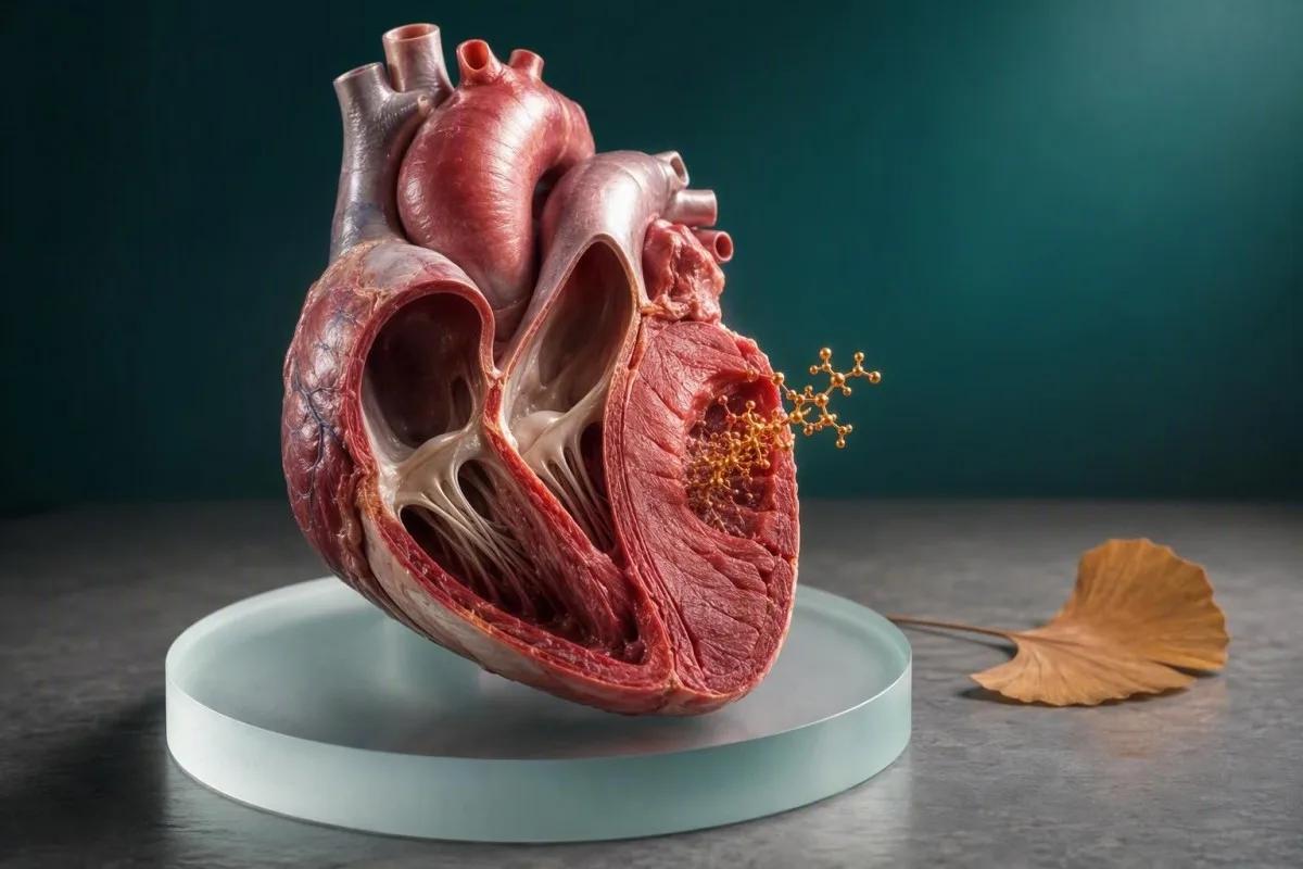
CK-MB on vanhempi, mutta edelleen hyödyllinen sydänlihaksen vaurioitumisen merkkiaine. Sen arvo riippuu ajoituksesta, oireista, EKG-löydöksistä, kokonais-CK:sta ja yleensä troponiinista.


- CK-MB:n merkitys: CK-MB tulee sanoista kreatiinikinaasi sydänlihasosa (creatine kinase myocardial band), joka on entsyymi-isomuoto, jota esiintyy pääasiassa sydänlihaksessa, mutta myös luustolihaksessa.
- Normaaliarvot: Monet CK-MB-massamääritykset käyttävät ylärajaa lähellä 3–5 ng/mL, mutta laboratorion oma viiteväli on aina ensisijainen.
- Ajoitus: CK-MB yleensä nousee 3–6 tuntia sydänlihasvaurion jälkeen, saavuttaa huippunsa 12–24 tunnissa ja palaa usein takaisin perustasolle 48–72 tunnin kuluessa.
- Troponiini ensin: Herkät sydäntroponiinit ovat ensisijainen verestä mitattava merkkiaine epäillyssä sydänkohtauksessa nykyaikaisessa ensihoidossa.
- Ei yksittäisellä testillä diagnoosia: Kohonnut CK-MB ei yksinään voi diagnosoida sydänkohtausta, koska kova liikunta, lihasvamma, leikkaus ja lihassairaus voivat nostaa sitä.
- Suhteellinen indeksi: CK-MB-indeksi yli noin 5% voi tukea sydänperäistä syytä, mutta se muuttuu epäluotettavaksi, kun kokonais-CK on merkittävästi koholla.
- Kiireelliset oireet: Uusi rintakipu, hengenahdistus, pyörtyminen, hikoilu tai leukaan tai käsivarteen säteilevä kipu vaatii hätäarviointia riippumatta aiemmasta CK-MB-tuloksesta.
- Trendi ratkaisee: Nousu ja lasku uusintanäytteissä on informatiivisempaa kuin yksi erillinen numero.
CK-MB on kreatiinikinaasin sydänlihasmuoto.
CK-MB tulee sanoista kreatiinikinaasi, sydänlihasosa. Se on entsyymi, jota mitataan CK-MB-verikokeella, kun lääkärit tutkivat mahdollisia sydänlihasvaurioita, vaikka poikkeava arvo ei yksin todista sydänkohtausta.
Kreatiinikinaasi auttaa soluja nopeasti regeneroimaan ATP:tä, välitöntä polttoainetta, jota käytetään lihastyön aikana. CK-MB on keskittynyt sydänlihakseen, kun taas CK-MM hallitsee luustolihaksissa ja CK-BB:tä löytyy laajemmalti aivo- ja sileistä kudoksista. Perusteet emäentsyymistä löytyvät artikkelistamme kreatiinikinaasioppaamme.
CK-MB-tulosta raportoidaan yleensä massakonsentraationa ng/mL tai harvemmin entsyymiaktiivisuutena U/L. Monet nykyiset massa-analyysit asettavat ylärajan noin 3–5 ng/mL, mutta sukupuolikohtaiset rajat ja analyysien kalibrointi eroavat niin paljon, että yhden laboratorion numeroa ei voi siirtää toiseen.
Kantesti on Tekoälyinen verikoeanalysaattori jossa näkyy CK-MB yhdessä analyysin viitevälin, kokonais-CK:n, troponiinin ja näytteenottoajan kanssa. Se on tärkeää, koska 6 ng/mL kahden tunnin kuluttua oireista herättää aivan erilaisen kysymyksen kuin 6 ng/mL maratonin jälkeen.
Pääte voi kuulostaa lopulliselta, ja potilaat kuulevat ymmärrettävästi “myocardial” eli “sydänkohtaus”. Varsinaisessa kliinisessä työssä se tarkoittaa, että laboratorio on mitannut yhden vihjeen lihassolujen vauriosta – ei vaurion syytä, sijaintia tai vakavuutta.
Miksi nimi esiintyy edelleen raporteissa
Jotkut sairaalat säilyttävät CK-MB:n ensiapu- tai sairaalahoito-ohjelmissa, erityisesti silloin, kun he haluavat lyhytkestoisen merkkiaineen mahdollisen toistuvan vamman arvioimiseksi. Monissa kunnallisissa terveydenhuollon yksiköissä se on kuitenkin suurelta osin korvattu korkeamman herkkyyden troponiinilla sydänkohtauksen alustavassa arvioinnissa.
CK-MB ja kokonais-CK mittaavat toisiinsa liittyviä, mutta eri asioita.
Kokonais-CK mittaa kaikkien lihasrikkaiden kudosten vapauttamaa entsyymiä, kun taas CK-MB mittaa yhtä CK-fraktiota. Kokonais-CK voi nousta tuhansiin U/L:iin rankan liikunnan tai lihasvamman jälkeen ilman, että se osoittaa sydänongelmaa.
1 500 U/L:n kokonais-CK epätavallisen harjoituksen jälkeen voi heijastaa luustolihasten stressiä, dehydraatiota, lääkevaikutuksia tai rabdomyolyysiä; se ei yksinään tunnista syytä. CK-MB voi myös nousta tässä tilanteessa, koska luustolihakset sisältävät pieniä määriä MB-fraktiota.
Vanha CK-MB suhteellinen indeksi lasketaan jakamalla CK-MB:n kokonais-CK:lla ja kertomalla sitten 100:lla. Indeksi alle 3% viittaa perinteisesti luustolihakseen, kun taas indeksi yli 5% voi tukea sydänperäistä syytä – mutta nämä rajat ovat tukevia todisteita, eivät ratkaisu.
Kun kokonais-CK on erittäin korkea, indeksi voi johtaa harhaan, koska sekä osoittaja että nimittäjä muuttuvat eri biologisista syistä. Tämä on yksi syy siihen, miksi lääkärit nyt painottavat enemmän sarjamittauksisia korkean herkkyyden troponiinia ja EKG:tä sen sijaan, että yrittäisivät matemaattisesti pelastaa epäselvän CK-MB-tuloksen.
Käytännön esimerkki: voimaurheilijalla, jonka kokonais-CK on 4 000 U/l ja CK-MB 12 ng/ml, voi olla alhainen suhteellinen osuus eikä iskeemisiä oireita. Sama CK-MB-arvo istumatyötä tekevällä henkilöllä, jolla on uutta puristavaa rintakipua, vaatii täysin erilaista reagointia.
Miksi kokonais-CK voi olla korkea
Liikunta, kaatumiset, pitkäaikainen liikkumattomuus, kouristukset, lihakseen annetut pistokset, hypotyreoosi, statiinit ja tulehdukselliset lihassairaudet voivat nostaa kokonais-CK:ta. Oppaamme vaarallisesti korkeaan CK:hon selittää, milloin tumma virtsa, vakava heikkous tai vähentynyt virtsaneritys muuttavat kiireellisyyttä.
Miksi lääkäri saattaa edelleen määrätä CK-MB-veritestiä
Lääkärit voivat määrätä CK-MB:tä arvioidessaan rintaoireita, mahdollista toistuvaa sydänlihasvauriota tai muiden lihasentsyymien epäselvää nousua. Se määrätään yleensä EKG-testauksen ja troponiinin kanssa sen sijaan, että se olisi erillinen seulontatesti.
Ensimmäisten 24–72 tunnin aikana vahvistetun sydäntapahtuman jälkeen CK-MB:n suhteellisen nopea lasku voi joskus auttaa tunnistamaan mahdollisen toisen vamman. Troponiini pysyy koholla pidempään, joten uusi CK-MB-nousu voi lisätä kontekstia, vaikka paikalliset protokollat vaihtelevat ja todisteet eivät ole täysin yhtenäisiä.
Lääkäri voi käyttää CK-MB:tä myös silloin, kun vanhempi laboratoriotietojärjestelmä sisällyttää sen edelleen kokonais-CK:hon. Kokemukseni mukaan potilaat säikähtävät usein automaattisesti generoituvaa CK-MB-merkintää, jota hoitava tiimi ei koskaan pitänyt infarktin todisteena, koska troponiini ja EKG olivat rauhoittavia.
CK-MB ei ole seulontatesti hiljaiselle sepelvaltimoplakille, tulevalle sydänriskille tai yleiskunnon arviointiin. Verenpaine, diabetesstatus, LDL-kolesteroli, ApoB, tupakointi, munuaisten toiminta ja sukuhistoria vastaavat hyödyllisempiin ehkäisyyn liittyviin kysymyksiin; meidän ApoB ja ApoA1 -suhdeopas selittää yhden näistä riskikuvioista.
Jos olet oireeton ja näet CK-MB:n työterveys-, urheilu- tai sairaalapaneelin raportissa, kysy miksi se tilattiin ja mihin sitä verrattiin. Tilauksen konteksti kertoo usein enemmän kuin lievästi epänormaali tulos.
Tutkimus ei ole diagnoosi
Sydänkohtaukset ovat kliinisiä diagnooseja, jotka perustuvat oireisiin, tutkimukseen, EKG- tai kuvantamistutkimuksiin ja biomarkkeridynamiikkaan. Laboratoriotutkimukset tunnistavat sydänlihasvaurion; ne eivät itsenäisesti vahvista, aiheuttiko tukkeutunut sepelvaltimo sen.
CK-MB:n aikataulu selittää, miksi yksittäinen tulos voi johtaa harhaan.
CK-MB alkaa yleensä nousta noin 3–6 tunnin kuluttua akuutista sydänlihasvauriosta, saavuttaa huippunsa noin 12–24 tunnin kuluttua ja laskee usein perustasolle 48–72 tunnin kuluessa. Normaali varhainen näyte voi siksi olla liian aikaisin rauhoittumiseen.
Ajoitus on yksi vähiten arvostetuista osista sydänveritutkimuksessa. Henkilö, joka saapuu 45 minuuttia äkillisten oireiden jälkeen, voi saada CK-MB:n normaalialueella todellisen vamman tapauksessa, kun taas henkilö, jolta otetaan näyte 2 päivää tapahtuman jälkeen, voi saada laskevan mutta silti epänormaalin arvon.
Dr. Thomas Klein on nähnyt tämän useimmiten viivästyneen esiintymisen jälkeen: potilas tuntee olonsa huonoksi yön aikana, odottaa seuraavaan aamuun ja ihmettelee sitten, miksi lääkärit toistavat testin, joka on jo korkea. Toistuva näyte etsii merkityksellistä muutosta, ei pelkästään vahvista, että yksi numero ylitti rajan.
Korkean herkkyyden troponiini voi nousta aikaisemmin kuin CK-MB, usein 1–3 tunnin kuluessa, riippuen määrityksestä ja vamman ajoituksesta. Sarjatestausprotokollat käyttävät yleensä näytteitä esiintymishetkellä ja 1–3 tuntia myöhemmin; ensiapuosastonne noudattaa validoitua paikallista käytäntöään.
Kova harjoittelu luo toisen ajoitusansan. CK ja CK-MB voivat nousta 24–72 tuntia eksentrisen harjoituksen, erityisesti alamäkilaskun tai epätavallisen vastustusharjoituksen jälkeen. Meidän maratonjuoksijan laboratoriopainopisteoppaan selittää, miksi harjoituksen ajoitus kuuluu kliiniseen anamneesiin.
Asiaankuuluva kello alkaa oireiden alkamisesta
Laboratoriot kirjaavat näytteenottopäivämäärän, mutta kliinikot tarvitsevat myös parhaan arvion siitä, milloin rintakipu, hengenahdistus, romahtaminen tai muu huolestuttava oire alkoi. Jos oireet tulevat ja menevät, dokumentoi ensimmäinen ja vakavin jakso myöhemmin arvaamisen sijaan.
CK-MB:n viitearvot ja tuloskuviot
Useimmat CK-MB-massamääritykset määrittelevät normaalin alueeksi noin 0–3 tai 0–5 ng/mL, mutta laboratoriosi yläviitearvo on ainoa asianmukainen raja raportissasi. Pieni kohotus vaatii kliinistä korrelaatiota; ei ole olemassa yleismaailmallista CK-MB-lukua, joka vastaisi sydänkohtausta.
Laboratoriot perustavat alueet oman määrityksensä, kalibrointinsa ja viiteväestönsä perusteella. Jotkut eurooppalaiset laboratoriot raportoivat CK-MB-aktiivisuuden U/L:na massan ng/mL sijaan, mikä tekee suorista internetvertailuista erityisen hyödyttömiä.
CK-MB-arvo 7 ng/mL, kun yläraja on 5 ng/mL, on 1,4-kertainen kohotus, ei diagnoosi. Tulos, joka on 10 kertaa ylärajan yläpuolella, on huolestuttavampi merkittävästä lihasvauriosta, mutta silloinkin kliinikoiden on selvitettävä, onko lähde sydämellinen vai luustolihas.
AST voi kohota sekä sydän- että luustolihasten rasituksen jälkeen, mikä satunnaisesti monimutkaistaa vanhanaikaista entsyymipaneelia. Tarkastelu korkeista AST-malleista voi auttaa selittämään, miksi AST, CK ja maksatestit joskus liikkuvat yhdessä harjoituksen jälkeen.
Käytännön sääntö on yksinkertainen: säilytä tarkat yksiköt, viitealue, näytteenottopäivämäärä, oireet ja viimeaikainen toiminta, kun keskustelet CK-MB:stä. Rajattu kuvakaappaus, joka näyttää vain varoituslipun, on harvoin riittävä turvalliseen tulkintaan.
Miksi varoituslippu ei ole automaattisesti vaarallinen
Viitevälimäärät kuvaavat, missä tulokset sijoittuvat valittuun populaatioon; ne eivät jaa ihmisiä turvallisiin ja turvattomiin ryhmiin. Laajemman selityksen lipuista ja laboratorion muotoilusta lue mitä ulkona oleva tarkoittaa.
CK-MB vastaan troponiini: Miksi troponiini yleensä johtaa
Herkkyystroponiini on sydänspesifisempi ja herkempi kuin CK-MB akuutin sydänlihasvaurion diagnosoinnissa. CK-MB voi lisätä valittua tietoa, mutta se ei saa ohittaa validoitua troponiinipolkua.
Troponiini I ja troponiini T ovat sydämen supistuslaitteen rakenteellisia proteiineja. CK-MB on entsyymi-isoformi, jota esiintyy pääasiassa, muttei yksinomaan, sydänlihaksessa; tämä biologinen ero selittää, miksi CK-MB vs troponiini ei ole tasainen vastus ensidiagnostiikassa.
Eurooppalaisen kardiologisen seuran (ESC) akuutin sepelvaltimotautioireyhtymän vuoden 2021 ohjeisto suosittelee validoituja korkean herkkyyden sydäntroponiinialgoritmeja käyttäen määrityskohtaista 99. persentiilin ylärajaa ja sarjamuutosta (Collet et al., 2021). Troponiinitaso tuon rajan yläpuolella viittaa sydänlihasvaurioon, mutta se ei silti automaattisesti tarkoita, että sepelvaltimon tukos olisi sen aiheuttanut.
Troponiini voi kohota 7–14 päivää suuren infarktin jälkeen, kun taas CK-MB normalisoituu usein 2–3 päivässä. Tämä lyhyempi ikkuna on historiallinen peruste CK-MB:n käytölle epäiltäessä uudelleeninfarktia, vaikka monet keskukset käyttävät nykyään kliinistä, EKG:tä, kuvantamista ja troponiinimuutoksiin perustuvaa näyttöä sen sijaan.
Troponiinitulokset ovat myös määritysriippuvaisia: hs-cTnI- ja hs-cTnT-arvoja ei voi muuntaa suoraan, ja “normaalilla” lukemalla on eri merkitys ennen oireiden alkua verrattuna 8 tuntiin oireiden alkamisen jälkeen. Vertaile määrityksiä selityksessämme troponiini I vs T -selittäjässä.
Kumpikaan merkkiaine ei yksinään tunnista syytä
Sepsis, nopea rytmihäiriö, keuhkoembolia, myokardiitti, vaikea anemia, munuaissairaus ja sydämen vajaatoiminta voivat nostaa troponiinia ilman klassista tyypin 1 sydänkohtausta. CK-MB:llä on omat ei-sydänperäiset syynsä, joten koko kliininen kuva pysyy neuvoteltavissa olevana.
Kuinka lääkärit diagnosoivat sydänkohtauksen CK-MB:n lisäksi
Sydänkohtaus vaatii akuutin sydänlihasvaurion sekä iskemian osoitusta, ei pelkästään kohonnutta CK-MB:tä tai troponiinia. Näyttö voi sisältää iskeemisiä oireita, uusia EKG-muutoksia, kuvantamislöydöksiä tai sepelvaltimotukoksen.
Myocardial Infarctionin neljäs universaali määritelmä määrittelee akuutin sydänlihasvaurion sydäntroponiiniarvona, joka ylittää määrityksen 99. persentiilin nousulla ja/tai laskulla; sydäninfarkti vaatii myös iskemian kliinistä näyttöä (Thygesen et al., 2019). CK-MB ei korvaa tätä viitekehystä.
Kantesti on AI verikoe-tulkinta-alusta jotka voivat järjestää raportoidut arvot ja trendit, mutta eivät voi diagnosoida sydänkohtausta PDF-tiedostosta tai valokuvasta. Akuutit oireet vaativat reaaliaikaista arviointia, EKG-tulkintaa, vitaalielintoimintoja ja lääkärin harkintaa – ei sovelluspohjaista rauhoittelukierrettä.
Harkitse 52-vuotiasta juoksijaa, jolla on CK-MB 6 ng/mL 42 km:n kilpailun jälkeen: normaali EKG, matala vakaa troponiini ja kipeät jalkalihakset viittaavat yhteen suuntaan. 52-vuotias, jolla on sama CK-MB sekä uusi keskivartalon paine, hikoilu ja dynaaminen troponiinin nousu, kuuluu ensiapuun.
Potilailla, joilla on hengenahdistusta eikä ilmeistä kipua, riski voi silti olla merkittävä, erityisesti vanhemmilla aikuisilla, diabeetikoilla ja naisilla. Selityksemme hengenahdistuksen verikoeg opas kattaa laajemman laboratoriotarkastelun.
Tyypin 1 ja tyypin 2 sydäninfarkti eroavat toisistaan
Tyypin 1 sydäninfarkti liittyy yleensä sepelvaltimon plakin akuuttiin häiriöön ja tromboosiin. Tyypin 2 sydäninfarkti heijastaa hapen saannin ja kulutuksen epätasapainoa, kuten vaikeaa anemiaa tai pitkittynyttä nopeaa rytmihäiriötä, ja hoito kohdistuu laukaisijaan sekä sepelvaltimoriskiin.
Ei-sydänperäiset syyt CK-MB:n kohonneisuudelle
Luustolihasvaurio, raskas liikunta, trauma, leikkaus, tulehduksellinen myopatia ja munuaisten vajaatoiminta voivat tuottaa kohonnutta CK-MB:tä. Nämä syyt selittävät, miksi lääkärit eivät koskaan tulkitse CK-MB:tä erikseen.
Luustolihas sisältää kohtuullisen osuuden CK-MB:tä, joten vaurioitunut tai uusiutuva lihas voi vapauttaa sitä riittävästi epätavallisen määrityksen käynnistämiseksi. Tuoreet kaatumiset, murskavammat, pitkittynyt liikkumattomuus, kouristukset, lihasinjektiot ja ortopediset leikkaukset tulisi kaikki ilmoittaa tulosta keskusteltaessa.
Krooniset lihassairaudet voivat tuottaa pysyvästi epätavallisia kokonais-CK- ja CK-MB-kuvioita, joilla ei ole mitään tekemistä akuutin sepelvaltimotaudin kanssa. Harvinaisissa tapauksissa makro-CK-kompleksit tai määrityksen häiriöt luovat hämmentäviä tuloksia, ja laboratorio voi tutkia niitä isoentsyymien erottelulla tai vaihtoehtoisella menetelmällä.
Huomiotta jäävä epäselvyys: huomattavasti kohonnut CK-MB, normaali sarjamittainen troponiini ja oireettomuus tulisi ohjata lihashistorian ja analyyttisten ongelmien tarkasteluun pikaisen sydämen kuvantamisen sijaan. Jos näyte vaurioitui keräyksen aikana, hemolyysin uusintanäytteen ohje voi olla merkityksellinen.
Munuaisten vajaatoiminta voi vaikeuttaa useiden biomarkkereiden, erityisesti troponiinin, tulkintaa kroonisen sydänlihasvaurion ja alentuneen eliminaation vuoksi. Se ei anna lupaa sivuuttaa nousevaa tulosta; se tekee trendeistä ja oireista entistäkin arvokkaampia.
Indeksillä on rajoituksia lihassairauksissa
CK-MB-indeksi kehitettiin kapeampaan kliiniseen tilanteeseen kuin monet potilaat olettavat. Laajassa lihasvauriossa tai kroonisessa myopatiassa yli 5%:n indeksi voi silti olla harhaanjohtava, joten nykyajan kliinikot priorisoivat troponiinipohjaisia algoritmeja.
Liikunta voi nostaa CK-MB:tä ilman sydänkohtausta
Pitkän matkan juoksu, CrossFit-tyyliset harjoitukset, raskas eksentrinen nostaminen ja kestävyyskilpailut voivat nostaa kokonais-CK:ta ja joskus CK-MB:tä 24–72 tunnin ajaksi. Liikuntaan liittyvä kohotus on todennäköisempi, kun oireita ei ole ja troponiini ja EKG eivät osoita iskemiaa.
Asia on niin, että lihakset eivät tiedä, että vuosittainen verikokeesi on huomenna. Alamäkeen juoksu ja suuren volyymin vastusharjoittelu aiheuttavat mikroskooppista lihasvauriota, ja CK voi huipentua myöhemmin kuin lihaskipu – usein 24–48 tuntia harjoituksen jälkeen.
Kehotan potilaita olemaan yrittämättä “läpäistä” testiä harjoittelemalla eri tavalla kertomatta siitä lääkärilleen. Kirjaa harjoituksen tyyppi, kesto, lihaskipu, nesteytys, lisäravinteet, kuume ja lääkkeet; tämä tieto voi estää tarpeettoman hälytyksen ja silti suojata todellisen sairauden havaitsematta jättämiseltä.
Tumman kolaa muistuttava virtsa, voimakas turvotus, syvä heikkous, sekavuus tai virtsanerityksen lasku rasituksen jälkeen eroaa tavallisesta viivästyneestä lihaskivusta ja vaatii kiireellistä arviointia. Riski on silloin rabdomyolyysi ja munuaisvaurio, ei pelkästään hankalasti korkea CK.
Jos rintakipua, epätavallista hengenahdistusta, lyyhistymistä tai sydämentykytystä esiintyi liikunnan aikana, älä selitä epänormaalia CK-MB:tä kuntoilulla. Tutustu oppaaseemme sydämentykytys verikoe opas samalla kun järjestät nopean kliinisen arvioinnin.
Kuinka kauan levätä ennen rutiininäytteen ottoa
Ei-kiireellistä perus-CK-mittausta varten on kohtuullinen käytännön lähestymistapa välttää tuntematonta korkeatehoista liikuntaa noin 48–72 tuntia, ellei lääkäri toisin neuvo. Älä viivytä rintakipuoireiden vuoksi tehtävää hätätestausta “lepäämisen” vuoksi.
Kuinka laboratorio mittaa CK-MB:tä ja missä virheitä ilmenee
Useimmat nykyaikaiset CK-MB-testit käyttävät immunomäärityksiä, jotka kvantifioivat CK-MB-massan ng/mL, kun taas vanhemmat menetelmät mittasivat entsyymiaktiivisuutta tai erottivat isoentsyymejä. Eri menetelmät voivat tuottaa erilaisia tuloksia ja viitevälejä.
Massamääritykset tunnistavat CK-MB-proteiinin vasta-aineilla ja ovat yleensä spesifisempiä kuin vanhemmat aktiivisuusmääritykset. Kuitenkin heterofiiliset vasta-aineet, makro-CK, näytteen laatuongelmat ja erittäin korkea kokonais-CK voivat ajoittain aiheuttaa tuloksen, joka ei sovi kliiniseen kuvaan.
Laboratorio voi käyttää delta-tarkistuksia – vertailua aikaisempiin tuloksiin – merkitsemään biologisesti epäuskottavan hypyn. Äkilliset muutokset eivät aina ole virheitä, mutta näytteenotto-olosuhteiden, lääkemuutosten ja äskettäisen toiminnan vertailu on järkevää; katso oppaamme labradelta-tarkistuksen selittäjä.
Paasto ei yleensä ole tarpeen CK-MB:lle, ja veden juominen on järkevää, ellei nesteen saantia ole rajoitettu. Biotiini vaikuttaa pääasiassa tiettyihin immunomäärityksiin eikä universaalisti CK-MB:hen, joten ilmoita suuriannoksista lisäravinteista, yleensä 5–10 mg/päivä tai enemmän, sen sijaan, että lopettaisit määrätyn hoidon omin päin.
Elokuusta 25. päivästä 2026 lähtien hyvän laboratorioraportin tulisi tunnistaa analyytti, yksiköt, paikallinen väli, näytteenottoaika ja liputustila. Jonkin näistä elementeistä puuttuminen heikentää merkittävästi turvallista etätulkintaa.
Kysy, mitä määritystä käytettiin
Jos CK-MB-arvo on jyrkässä ristiriidassa oireidesi tai troponiinitulostesi kanssa, kysy, mittasiko laboratorio CK-MB-massaa vai aktiivisuutta ja harkittiinko häiriöitä. Tämä kohdennettu kysymys on hyödyllisempi kuin yleisen “normaalin CK-MB”-arvon etsiminen.
Kuinka lukea CK-MB-raportti yli-reagoimatta
Lue CK-MB yksikköineen, laboratorion yläraja, kokonais-CK, troponiinisarja, EKG-löydös ja oireiden aikajana. Yksittäinen korkea hälytys, erityisesti rasituksen tai vamman jälkeen, on vähän diagnostisesti spesifi.
Aloita tarkasta tuloksesta ja sen paikallisesta yläviitearvosta. Kysy sitten, otettiinko näyte ennen oireita, 3 tunnin kuluessa, 6–12 tuntia myöhemmin vai päiviä myöhemmin; samalla pitoisuudella on eri merkitys kussakin kohdassa kyseistä käyrää.
Seuraavaksi etsi kuvio. Nouseva troponiini iskeemisten oireiden ja kehittyvien EKG-muutosten kanssa on paljon painavampi kuin pelkkä CK-MB, kun taas kohonnut CK jalkapainotteisen harjoittelun jälkeen vakaalla troponiinilla viittaa yleensä luurankolihakseen.
Kantesti:n trendianalyysi auttaa käyttäjiä säilyttämään aiemmat tulokset ja keräyskontekstin sen sijaan, että jokaista punaista lippua käsiteltäisiin uutena kriisinä. Aiempi CK, äskettäinen treeni ja toistuva arvo vastaavat usein kysymyksiin, joihin yksittäinen raportti ei pysty.
Älä vertaa eri laboratorioiden CK-MB-tuloksia ikään kuin ne olisivat identtisiä, etenkään jos toinen käyttää yksikköä U/L ja toinen ng/mL. Seuraamisen säilyttäminen verikokeen aikajanalle tekee seurantakäynneistä tuottavampia.
Kysymyksiä, joita kannattaa ottaa mukaan vastaanotolle
Kysy: Oliko tämä CK-MB-massaa vai aktiivisuutta? Mikä oli troponiinitrendini? Osoittiko EKG iskemiaa? Voisiko äskettäinen lihasvamma selittää sen? Tarvitsenko uusintanäytteen, sydämen ultraäänitutkimuksen vai muuta sydäntutkimusta?
Oireet, jotka vaativat kiireellistä hoitoa riippumatta CK-MB:stä
Soita hätäpalveluun nyt, jos uusi rintapaine kestää yli 5–10 minuuttia, vakava hengenahdistus, pyörtyminen, kylmänhikisyys tai kipu leviää leukaan, käsivarteen, selkään tai ylävatsaan. Älä odota CK-MB-tulosta tai yritä tulkita sitä kotona.
Sydänkohtauksen oireet eivät aina ole dramaattisia. Naiset, vanhukset ja diabeetikot voivat oireilla pahoinvoinnilla, epätavallisella väsymyksellä, hengenahdistuksella, selkäkivulla tai tunteella, että jokin on pahasti vialla, klassisen rintakivun sijaan.
Tohtori Thomas Kleinin kliininen sääntö on suora: jos oireet ovat ajankohtaisia ja huolestuttavia, eilen nähdyt numerot ovat toissijaisia. Hätäryhmät voivat ottaa EKG:n nopeasti, toistaa korkeapitoisen troponiinin oikealla aikavälillä ja arvioida verenpainetta, rytmiä, happeutumista ja vaihtoehtoisia diagnooseja.
Korkea troponiini itsessään ei ole synonyymi sepelvaltimon tukkeutumalle, mutta sitä ei koskaan saa itse triage-arvioida portaalin tuloksesta. Lue korkea troponiini kiireelliset merkit tilanteisiin, joissa välitön hoito on turvallisempi valinta.
Jos oireet ovat hävinneet, mutta olivat uusia, rasitukseen liittyviä tai toistuvia, hakeudu saman päivän lääkäriapuun. Vakaat oireet ansaitsevat silti lääkärin arvioinnin, erityisesti tunnetun sepelvaltimotaudin, tupakoinnin, diabeteksen, kroonisen munuaissairauden tai vahvan sukuhistorian tapauksessa.
Älä aja itse, jos oireet ovat vakavia
Hätäkuljetus mahdollistaa seurannan ja varhaisen hoidon, jos kehittyy vaarallinen rytmihäiriö. Lääkkeiden pureskelu tai ottaminen tulee tehdä hätäkeskuksen tai lääkärin ohjeiden mukaisesti, etenkin jos käytät antikoagulantteja, sinulla on allergioita tai sinulle on kerrottu, ettet saa ottaa aspiriinia.
Kuka tarvitsee seurantaa epänormaalin CK-MB-tuloksen jälkeen
Epänormaali CK-MB-tulos vaatii seurantaa, kun se on uusi, nouseva, selittämätön, yhdistetty oireisiin tai siihen liittyy epänormaali troponiini- tai EKG-löydös. Seuraava vaihe voi olla uusintatestaus, ei automaattisesti kuvantaminen tai toimenpide.
Matalan riskin, oireettoman henkilön kohdalla selvän raskaan liikunnan jälkeen lääkäri voi uusia CK:n ja CK-MB:n levon ja nesteytyksen jälkeen. Aktiivisten rintaoireiden tai epänormaalitroponiinin tapauksessa hätäarviointi on tarkoituksenmukaisempaa kuin avohoidon uusintatestaus.
Seuranta riippuu myös perussydän- ja verisuonitautien riskistä. Diabetes, aiempi sepelvaltimotauti, tupakointi, korkea LDL, korkea lipoproteiini(a), verenpainetauti ja munuaissairaus voivat alentaa kynnystä nopealle tutkimukselle; meidän naisten sydäntestiopas kattaa yleisesti huomiotta jäävät riskimarkkerit.
Kantesti on AI-pohjainen verikokeiden analyysityökalu jotka voivat tuoda esiin keskusteltavia yhdistelmiä, mukaan lukien CK-MB, CK, kreatiniini, AST ja sarjapäivämäärät. Sen tulisi tukea – ei korvata – lääkäriä, joka tuntee oireesi, tutkimuksesi, EKG:si ja lääkityksesi.
Toistettujen testien tulisi ihanteellisesti käyttää samaa laboratoriota ja ne tulisi kerätä kliinisesti merkityksellisellä aikavälillä. CK-MB-näytteen toistaminen 15 minuutin kuluttua voi lisätä vähän, kun taas 1–3 tunnin troponiiniprotokolla voi olla erittäin informatiivinen, kun se on validoitu kyseisessä sairaalassa.
Mitä ottaa mukaan seurantaan
Ota mukaan koko raportti, aiemmat arvot, lääke- ja lisäravinneluettelo, selvitys viimeaikaisesta liikunnasta tai vammasta ja selkeä oireiden aikajana. Nämä yksityiskohdat voivat säästää sinut sekä virheelliseltä varmuudelta että tarpeettomalta testaukselta.
CE
Tekoäly voi selittää CK-MB-terminologiaa ja järjestää trendejä, mutta se ei voi määrittää, onko rintakipu hätätilanne. Turvallisuus riippuu alkuperäisen raportin säilyttämisestä, epävarmuuden tunnustamisesta ja kiireellisten oireiden eskaloimisesta henkilökohtaiseen hoitoon.
Kantesti AI tulkitsee CK-MB-tuloksia tarkastelemalla yksiköitä, viitevälejä, siihen liittyviä biomarkkereita ja aiempia tuloksia sen sijaan, että se julistaisi diagnoosin yhdestä lipusta. Meidän biomarkkerien viiteopas selittää, miksi laboratoriovälit ja mittausmenetelmät ovat tärkeitä.
Yksityisyyden suojaamiseksi poista tai suojaa tarpeeton tunnistamistieto ennen raportin jakamista ja käytä palveluja, joilla on selkeät tiedonkäsittelystandardit. Kantesti palvelee käyttäjiä yli 127 maassa ja tukee monikielistä raporttitulkintaa, mutta paikalliset hätäpalvelut ja kliininen tiimisi ovat edelleen oikea reitti akuuttien oireiden tapauksessa.
Kliinikkamme tarkistavat lääketieteellisen sisällön kliinisten standardien mukaisesti, ja meidän teknologiaopas kuvaa tekoälyavusteisen laboratoriotulkinnan rajoituksia. Selitys on hyödyllisin, kun se kertoo, mitä tietoa puuttuu ja mitä kysymystä kysyä seuraavaksi.
Älä koskaan käytä tekoälyn tulkintaa viivästyttääksesi hoitoa uuden paineentuntuisen rintakivun, romahtamisen tai vakavan hengenahdistuksen vuoksi. Turvallisen työkalun tulisi tehdä tämä raja hyvin selväksi.
Mitä hyödyllinen tekoälyselitys sisältää
Kliinisesti vastuullinen selitys tunnistaa yksikön, laboratoriovälin, mahdolliset ei-sydämperäiset syyt, liitännäistestit, aikataulurajoitukset ja kiireelliset oireet. Se ei lupaa, että poikkeava tai normaali CK-MB yksinään sulkee sydänkohtauksen pois tai vahvistaa sen.
Käytännön ydinasia CK-MB-tuloksista
CK-MB on sydämeen liittyvä kreatiinikinaasin fraktio, mutta korkean herkkyyden troponiini, EKG-löydökset, oireet ja toistotestaus ohjaavat nyt useimpia sydänkohtausten päätöksiä. CK-MB-tulosta on parasta pitää yhtenä todisteena, ei koskaan diagnoosina itsessään.
Jos CK-MB on hieman koholla raskaan liikunnan, vamman, leikkauksen tai lihaskivun jälkeen, älä panikoi – mutta anna kliinikollesi konteksti. Jos se on koholla rintakipujen, hengenahdistuksen, hikoilun, pyörtymisen tai poikkeavan troponiinin kanssa, kohtele sitä aikasidonnaisena kliinisenä kysymyksenä.
Kantesti voi auttaa sinua laatimaan tiiviin kysymyslistan ja vertailemaan aiempia raportteja, kun taas meidän kliinisen validoinnin standardiemme selittää, miten lähestymme turvallista tulosten tulkintaa. Se ei voi vahvistaa tai poissulkea akuuttia sepelvaltimotautia kotoa käsin.
Dr. Thomas Klein suosittelee alkuperäisen raportin säilyttämistä sen sijaan, että kopioitaisiin vain poikkeava rivi. Tarkka näyteaika, yksiköt, paikallinen viiteväli, kokonais-CK-, troponiiniarvot, EKG-lausunto, lääkitykset ja viimeaikainen liikunta ovat dataa, jotka tekevät CK-MB:stä tulkittavan.
Lääkärien tarkistamaa terveyskoulutusta ja kliinisten standardiemme takana olevia ihmisiä varten, tapaa Kantesti lääketieteellinen neuvottelukunta. Paras seuraava askel on se, joka vastaa oireitasi ja riskiäsi – ei sivun pelottavimmalta näyttävä luku.
Viimeinen turvallisuustarkistus
Uudet tai jatkuvat mahdolliset sydänoireet ovat aina tärkeämpiä kuin rauhoittavan näköinen aiempitulos. Hakeudu ensin ensiapuun; tarkista laboratoriotiedot myöhemmin, kun olet turvassa.
Usein kysytyt kysymykset
Mitä CK-MB tarkoittaa verikokeessa?
CK-MB tulee sanoista kreatiinikinaasi-myokardiaalinen bandaasi, joka on kreatiinikinaasientsyymin muoto. Sitä esiintyy pääasiassa sydänlihaksessa, mutta pienempiä määriä myös luustolihaksessa. Monet laboratoriot raportoivat CK-MB-massan ng/mL, ja ylärajat ovat yleensä noin 3–5 ng/mL. Korkea tulos voi viitata lihassolujen vaurioitumiseen, mutta CK-MB ei yksinään voi diagnosoida sydänkohtausta. Kliinikot tulkitsevat sen yhdessä oireiden, EKG-löydösten, sarjahoitoisten troponiinien, kokonais-CK:n ja testien ajoituksen kanssa.
Mikä on normaali CK-MB-pitoisuus?
Normaalilla CK-MB-massapitoisuudella tarkoitetaan usein alle noin 3–5 ng/mL arvoa, vaikka oikea normaaliarvoalue onkin analyysin suorittaneen laboratorion ilmoittama väli. Jotkin laboratoriot ilmoittavat CK-MB-aktiivisuuden U/L yksiköissä, joten arvoja ei voi verrata suoraan eri menetelmien välillä. Raja-arvoa hieman korkeampi arvo voi johtua lähiaikaisesta rasituksesta tai luustolihasvammasta. Yksittäistä CK-MB-vertailuarvoa, joka varmistaisi tai sulkisi pois sydäninfarktin, ei ole.
Kumpi on parempi sydänkohtauksen diagnosoinnissa, CK-MB vai troponiini?
Suuren herkkyyden sydäntroponiini on yleisesti ottaen parempi kuin CK-MB akuutin sydänlihasvaurion havaitsemisessa, koska se on herkempi ja spesifisempi sydänlihakselle. Vuoden 2021 ESC-ohjeistuksessa käytetään analyysikohtaisia sarjamuotoisia suuren herkkyyden troponiinin algoritmeja, usein näytteillä esittelyssä ja 1–3 tuntia myöhemmin. CK-MB voi joskus tarjota lisätietoa, koska se palaa usein perustasolle 48–72 tunnin kuluessa. Kumpikaan biomarkkeri yksinään ei vahvista sydänkohtausta ilman iskemiaan viittaavaa näyttöä, kuten oireita, EKG-muutoksia, kuvantamislöydöksiä tai sepelvaltimotukosta.
Voiko liikunta nostaa CK-MB:tä?
Kyllä, raskas liikunta voi nostaa CK-MB:tä, koska luustolihaksissa on pieni CK-MB-osuus. Kestävyyssuoritukset, totuteltu raskas nostaminen, alamäkijuoksu ja korkeatehoinen intervalliharjoittelu voivat nostaa kokonais-CK:ta ja joskus CK-MB:tä noin 24–72 tunnin ajaksi. Liikunta ei tee turvalliseksi jättää huomiotta rintakipua, pyörtymistä, vaikeaa hengenahdistusta tai hikoilua. Nämä oireet vaativat edelleen kiireellistä kliinistä arviointia EKG:llä ja sarjamääräisillä troponiinitesteillä.
Mitä CK-MB-indeksi yli 51 % tarkoittaa?
Suhteellinen CK-MB-indeksi, joka on noin 51 TP54T:n yläpuolella, on perinteisesti viitannut siihen, että suurempi osa mitatusta kreatiinikinaasista voi tulla sydänlihaksesta eikä luustolihaksesta. Indeksi lasketaan jakamalla CK-MB kokonais-CK:lla ja kertomalla 100:lla. Arvot, jotka ovat alle 31 TP54T:n, ovat historiallisesti viitanneet luustolihakseen. Tämä laskelma muuttuu epäluotettavaksi merkittävän lihasvaurion, erittäin korkean kokonais-CK:n tai kroonisen lihassairauden jälkeen. Nykyaikainen hoito perustuu siksi vahvemmin sarjamittauksina tehtyihin korkeaherkkyyksisen troponiinin mittauksiin ja EKG-löydöksiin.
Kuinka nopeasti CK-MB nousee sydänkohtauksen jälkeen?
CK-MB alkaa yleensä nousta noin 3–6 tunnin kuluttua akuutista sydänlihasvauriosta, saavuttaa huippunsa noin 12–24 tunnissa ja palaa usein takaisin perustasolle 48–72 tunnin kuluessa. Normaali CK-MB-tulos, joka saadaan 1–2 tunnin kuluessa oireiden alkamisesta, voi olla liian aikainen vamman poissulkemiseksi. Päivystyspoliklinikat käyttävät uusintanäytteitä ja validoituja korkean herkkyyden troponiiniprotokollia sen sijaan, että luottaisivat yhteen CK-MB-mittaukseen. Oireiden alkamisaika tulisi jakaa mahdollisimman tarkasti.
Hanki tekoälypohjainen verikoeanalyysi tänään
Liity yli 2 miljoonan käyttäjän joukkoon maailmanlaajuisesti, jotka luottavat Kantesti:hen saadakseen välittömän ja tarkan laboratoriotestianalyysin. Lataa verikoetuloksesi ja saat kattavan tulkinnan 15,000+-biomarkkereista sekunneissa.
📚 Viitatut tutkimusjulkaisut
Klein, T., Mitchell, S., & Weber, H. (2026). Nipah-viruksen verikoe: Varhaisen havaitsemisen ja diagnoosin opas 2026. Kantesti AI Medical Research.
Klein, T., Mitchell, S., & Weber, H. (2026). B-negatiivinen veriryhmä, LDH-verikoe ja retikulosyyttimäärän opas. Kantesti AI Medical Research.
📖 Ulkoiset lääketieteelliset lähteet
⚕️ Lääketieteellinen vastuuvapauslauseke
Tämä artikkeli on tarkoitettu vain koulutustarkoituksiin eikä se muodosta lääketieteellistä neuvontaa. Ota aina yhteyttä pätevään terveydenhuollon ammattilaiseen diagnoosi- ja hoitopäätöksiä varten.
E-E-A-T-luottamussignaalit
Kokea
Lääkärin johtama kliininen arviointi laboratoriotulkinnan työnkuluista.
Asiantuntemus
Laboratoriolääketiede keskittyy siihen, miten biomarkkerit käyttäytyvät kliinisessä kontekstissa.
Auktoriteetti
Kirjoittanut tohtori Thomas Klein, tarkistanut tohtori Sarah Mitchell ja professori tohtori Hans Weber.
Luotettavuus
Näyttöön perustuva tulkinta selkeillä jatkopoluilla, jotka vähentävät hälytystä.