川崎病を確定できる単独の血液検査はありません。診断では、発熱の経過、診察所見、炎症マーカー、補助的な検査所見、心エコー検査を組み合わせます。小児の発熱が続く場合は、典型的な症状がすべて現れるまで待たず、速やかに評価を受ける必要があります。.

要点
- 診断 臨床的な評価が必要です。川崎病を確実に確定または除外できる単独の血液検査はありません。.
- 発熱が持続 5日間続く場合は、川崎病の評価を速やかに受ける必要があります。ただし、子どもの具合が非常に悪そうな場合は、さらに早く受診してください。.
- 炎症マーカー CRP ≥30 mg/LまたはESR ≥40 mm/hourは、米国心臓協会の不全型川崎病の診断経路において、さらなる評価を支持します。.
- 不全型川崎病 は、典型的な特徴が2~3つしか認められない場合にも起こり得ます。乳児では、目に見える徴候がごくわずかでも、発熱が長引くことがあります。.
- 血小板 発症7日目以降の血小板数 ≥450 × 10⁹/Lは診断を支持する所見です。第1週に血小板数が正常でも、川崎病は否定できません。.
- 診断を支持する所見 には、アルブミン ≤30 g/L、年齢相応の貧血、ALT上昇、WBC ≥15 × 10⁹/L、尿中白血球が視野あたり10個以上であることが含まれます。.
- 心エコー検査 冠動脈と心機能を評価します。早期の検査が正常でも、川崎病は否定できません。.
- 治療のタイミング 重要です。病院での治療には通常、静注用免疫グロブリンが含まれ、理想的には発症から10日以内に、すべての症状が現れるのを待たずに投与します。.
川崎病の血液検査で診断を確定できますか?
川崎病の診断を確定できる単一の血液検査はありません。. 血液検査では炎症や臓器の反応を測定し、医療従事者は発熱、診察所見、心臓画像検査からこの病気を特定します。すべての症状がそろっていなくても、発熱が5日続く小児には速やかな評価が必要です。.
川崎病は冠動脈に影響を及ぼすことがある小児期の血管炎です。. 2017年の米国心臓協会の声明では、未治療例の約25%に冠動脈瘤が生じると推定されています。この数値が示すのは未治療の病気についてであり、発熱が長引く小児やCRP値が高い小児すべてについてではありません(McCrindle et al., 2017)。罹患する小児の大半は5歳未満ですが、年長児も発症することがあります。.
炎症マーカーの異常は免疫反応を示すものであり、特定の病気を示すものではありません。. CRPが80 mg/Lでも、川崎病、細菌感染症、その他の炎症性疾患で見られることがあるため、その結果だけでは診断を確定できません。同様に、異常値が1つしかない検査報告でも、採血が早い時期に行われた場合や成人の基準範囲と照らして解釈された場合には、懸念の程度を過小評価している可能性があります。.
Kantestiは、川崎病を診断することなく、CRP、CBC、肝機能検査の用語を説明できるAI血液検査分析ツールです。. 私たちの 小児向けの解釈における安全対策 3歳児の検査結果を成人の前提で解釈できない理由を説明します。情報 当組織について この教育サービスの提供者を明らかにします。発熱が続く場合は、引き続き医療従事者による評価が必要です。.
小児の発熱が続く場合、いつ評価を受けるべきですか?
発熱が5日続く小児には、川崎病の検討を含む速やかな臨床評価が必要です。. 重い病気、懸念される症状、または乳児がいる場合は、より早く受診してください。診断上の発熱期間は、5日目まで自宅で待つよう指示するものではありません。.

生後3か月未満の乳児で38°C以上の発熱がある場合は、緊急の医学的評価が必要です。. 年齢を問わず、呼吸困難、異常な眠気、反応の鈍さ、圧迫しても消えない発疹、著しい脱水、または循環不全の徴候がある場合は、救急医療を受けてください。これらの所見は川崎病に特有のものではありません。敗血症など、迅速な対応を要する他の病態も考慮する必要があります。.
パラセタモールで一時的に熱が下がっても、川崎病は否定できません。. 子どもは熱が上がる合間に一時的に楽そうに見えることがありますが、基礎にある病気は続いている可能性があり、症状が日を追って現れることもあります。体温計で一度正常だったかどうかだけでなく、発熱が始まった時期、実際の体温、薬、尿量、目、唇、手、足の変化を記録してください。.
生後6か月未満の乳児では、典型的な発疹や目の充血がなく、発熱が長引くことがあります。. 原因不明の発熱が7日以上続くことは、AHAの診療経路において不全型の評価を検討する契機として認められています。当社の 検体準備に関するガイダンス は予定された検査には役立ちますが、具合の悪い子どもを、絶食、都合のよい予約日時、または症状がすべてそろうまで決して待たせてはいけません。.
川崎病は臨床徴候からどのように診断されますか?
典型的な川崎病は通常、5日以上続く発熱に加えて、主要な臨床的特徴5項目のうち少なくとも4項目がある場合に診断されます。. Clinicians can diagnose some strongly characteristic cases earlier; neither the fever count nor the symptom count replaces clinical judgment or urgent evaluation.

The 5 principal features are bilateral non-purulent conjunctival redness, mouth changes, a polymorphous rash, extremity changes, and cervical lymphadenopathy. Mouth changes include red or cracked lips and a strawberry tongue; hand and foot changes include redness or swelling. The lymph-node criterion is usually a cervical node at least 1.5 cm across, often on one side.
The clinical features do not have to occur simultaneously. A photograph of a rash from illness day 3 may remain diagnostically useful when the skin looks different on day 6, and a previous episode of swollen hands should not disappear from the history because swelling has improved. Peeling around the fingertips often occurs during weeks 2–3, too late to use as a reason to delay assessment.
Certain findings point toward alternative explanations rather than strengthening the classic Kawasaki pattern. Purulent eye discharge, prominent exudative tonsillitis, or discrete mouth ulcers deserve separate consideration, although no single finding resolves every case. Our laboratory marker reference guide explains test terminology; it cannot substitute for examining the 5 clinical feature groups or arranging appropriate imaging.
不全型川崎病の診断に、検査値はどのように役立ちますか?
Incomplete Kawasaki disease means a child has fewer classic features but enough clinical, laboratory, or echocardiographic evidence to warrant diagnosis and treatment. The AHA evaluation pathway considers children with fever for at least 5 days and 2–3 features, and certain infants with unexplained fever for at least 7 days.
CRP ≥30 mg/L or ESR ≥40 mm/hour supports proceeding through the AHA incomplete-disease evaluation pathway. When inflammation reaches either threshold, clinicians assess echocardiography and supplementary laboratory criteria rather than waiting for another visible symptom. The thresholds are decision supports for a compatible illness—not proof that Kawasaki disease is the cause (McCrindle et al., 2017).
The 6 supplementary laboratory criteria are albumin ≤30 g/L, anemia for age, elevated ALT, WBC ≥15 × 10⁹/L, platelets ≥450 × 10⁹/L after day 7, and urine ≥10 white cells per high-power field. In the appropriate clinical setting, at least 3 supplementary criteria or a positive echocardiogram can support treatment through the pathway. These are not independent points to count without reviewing competing diagnoses.
Fewer than 3 supplementary criteria does not create a permanent rule-out result. Persistent fever may require repeated examination, repeat laboratory testing, and imaging, particularly if the first evaluation occurred early. Systemic juvenile idiopathic arthritis is one possible alternative; our childhood arthritis laboratory guide explains why shared inflammatory findings do not make these conditions interchangeable.
川崎病におけるCRPとESRの結果は何を意味しますか?
CRP and ESR support Kawasaki evaluation by showing systemic inflammation, but neither identifies its cause. In the AHA incomplete-disease pathway, CRP ≥30 mg/L or ESR ≥40 mm/hour is a threshold for further assessment, not a severity scale or a stand-alone indication for treatment.
CRP of 3 mg/dL equals 30 mg/L. This 10-fold unit difference can make identical results appear very different when families compare reports from different laboratories, while common CRP reference limits are around 5 mg/L but vary by method. Our WBCとCRPの比較 explains why a high CRP and a less impressive white-cell count can coexist.
ESR is influenced by factors beyond the immediate inflammatory response, including anemia and circulating proteins. A result of 45 mm/hour is not directly comparable to CRP of 45 mg/L; the tests measure different processes and have different kinetics. When both markers are below the pathway thresholds, Kawasaki disease becomes less likely, but an early sample or a concerning clinical course still requires judgment.
High-sensitivity CRP measures the same protein as conventional CRP, but adult cardiovascular-risk categories do not apply to a febrile child. A value of 30 mg/L during illness should not be interpreted using an adult 1–3 mg/L cardiovascular-risk framework. Our hs-CRP interpretation guide explains that distinction; for acute pediatric illness, the clinical question is inflammation and its cause.
| レベル | 範囲 | 意味 |
|---|---|---|
| 一般的なCRP基準値上限 | Often <5 mg/L; laboratory-specific | A reference interval, not a Kawasaki exclusion threshold; early disease may require reassessment. |
| Incomplete-disease CRP trigger | ≥30 mg/L, equivalent to ≥3 mg/dL | Supports further AHA pathway evaluation when fever and clinical history are compatible. |
| Common ESR reference interval | Often approximately 0–20 mm/hour; age- and method-dependent | Use the reporting laboratory's pediatric interval; ESR is affected by anemia and proteins. |
| Incomplete-disease ESR trigger | ≥40 mm/hour | Supports further evaluation; neither confirms Kawasaki disease nor defines an emergency by itself. |
川崎病を支持するCBC所見は何ですか?
Kawasaki disease lab findings on a CBC can include neutrophil-predominant leukocytosis, anemia for age, and a later rise in platelets. WBC ≥15 × 10⁹/L and platelets ≥450 × 10⁹/L after day 7 are supplementary incomplete-disease criteria, but the illness day changes their meaning.
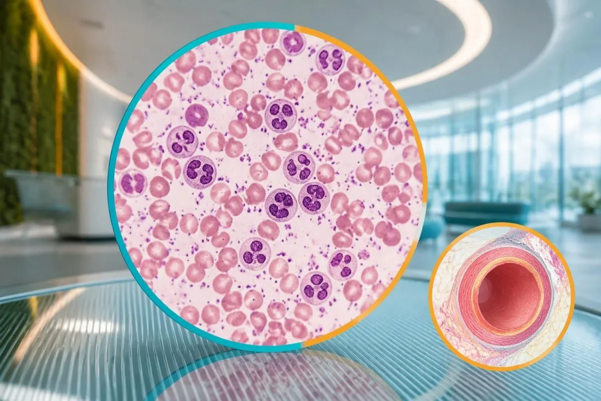
Platelets often rise during the second week rather than at the start of fever. A platelet count of 280 × 10⁹/L on illness day 4 therefore does not exclude Kawasaki disease, and waiting for thrombocytosis can delay care. Conversely, a count of 520 × 10⁹/L after day 7 supports the pattern but can also occur during recovery from other inflammatory illnesses.
Anemia in the incomplete-disease criteria is defined against the child's age, not a universal hemoglobin cutoff. For a hypothetical 3-year-old, hemoglobin of 9.8 g/dL would generally be low, but the cause could include inflammation, pre-existing iron deficiency, or both. WBC also needs age-specific context; our 好中球基準範囲ガイド explains why pediatric differential counts differ from adult ones.
Low platelets can occur in severe Kawasaki presentations and should not be treated as reassuring. A febrile child with platelets of 90 × 10⁹/L, poor circulation, or worsening organ function needs urgent assessment for shock, sepsis, consumption, or macrophage activation. Platelet size indices and neutrophil-to-lymphocyte ratios remain contextual findings; they are not substitutes for the 6 established supplementary criteria.
アルブミン値や肝機能検査値が変化するのはなぜですか?
Low albumin and elevated ALT can support incomplete Kawasaki disease because systemic inflammation affects protein distribution and liver function. Albumin ≤30 g/L is a defined supplementary criterion, while ALT is judged against the laboratory's pediatric upper reference limit rather than one universal Kawasaki cutoff.
Albumin of 3.0 g/dL equals 30 g/L. During acute illness, a low result can reflect reduced synthesis, increased vascular permeability, dilution, or protein loss—not simply poor dietary protein intake. Our 血清タンパク質解釈ガイド explains these mechanisms; nutritional advice alone would miss the clinical significance of low albumin combined with persistent fever.
An ALT result above the pediatric upper limit counts as supporting evidence, but its magnitude does not diagnose Kawasaki disease. For example, a hypothetical child with albumin 28 g/L and ALT 72 IU/L may meet 2 supplementary criteria if that ALT exceeds the local limit. The same combination can arise from other illnesses or medication effects, as our ALT用語ガイド が説明しています。.
Kantesti is an AI blood test interpretation platform that can explain why albumin 28 g/L and ALT 72 IU/L require clinical context. 私たちの AI技術の説明 describes the interpretation workflow, not a validated Kawasaki diagnostic test. Growing children can also have relatively high alkaline phosphatase from bone activity; that finding is not interchangeable with the ALT criterion.
尿検査やナトリウム値は評価の助けになりますか?
Urine white cells can support incomplete Kawasaki evaluation, while electrolyte changes help assess illness severity and alternative causes. Urine ≥10 white cells per high-power field is a supplementary criterion; low sodium is a possible associated finding but is not one of the 6 AHA supplementary laboratory criteria.
Sterile pyuria means urine white cells are present without a demonstrated bacterial cause on an appropriately collected culture. Kawasaki disease can produce this pattern, but a child with urine white cells still needs assessment for urinary infection. A urine dipstick alone cannot establish sterile pyuria; our 尿沈渣の解釈ガイド distinguishes microscopy findings from dipstick screening results.
A report of 10 white cells per high-power field cannot be directly equated with an automated result of 10 cells/µL. Instruments, specimen preparation, and counting methods differ, so clinicians may need the laboratory's conversion or interpretive guidance. Collection quality also matters: contamination can introduce cells, whereas prior antibiotics can reduce culture yield without explaining the entire fever pattern.
Serum sodium below 135 mmol/L is generally classified as hyponatremia, but its cause must be assessed separately. Acute inflammation, fluid intake, gastrointestinal losses, and altered water regulation can all contribute; the number is not a Kawasaki confirmation test. Our 電解質パターンガイド explains why sodium 130 mmol/L with neurological symptoms is a different problem from an isolated mildly low value.
心エコー検査は診断に何を加えますか?
Echocardiography assesses coronary artery dimensions, heart function, and other cardiac findings that blood tests cannot show. A coronary artery Z-score ≥2.5 can support Kawasaki diagnosis in the incomplete-disease pathway, but a normal early echocardiogram does not exclude the illness or justify delaying indicated treatment.
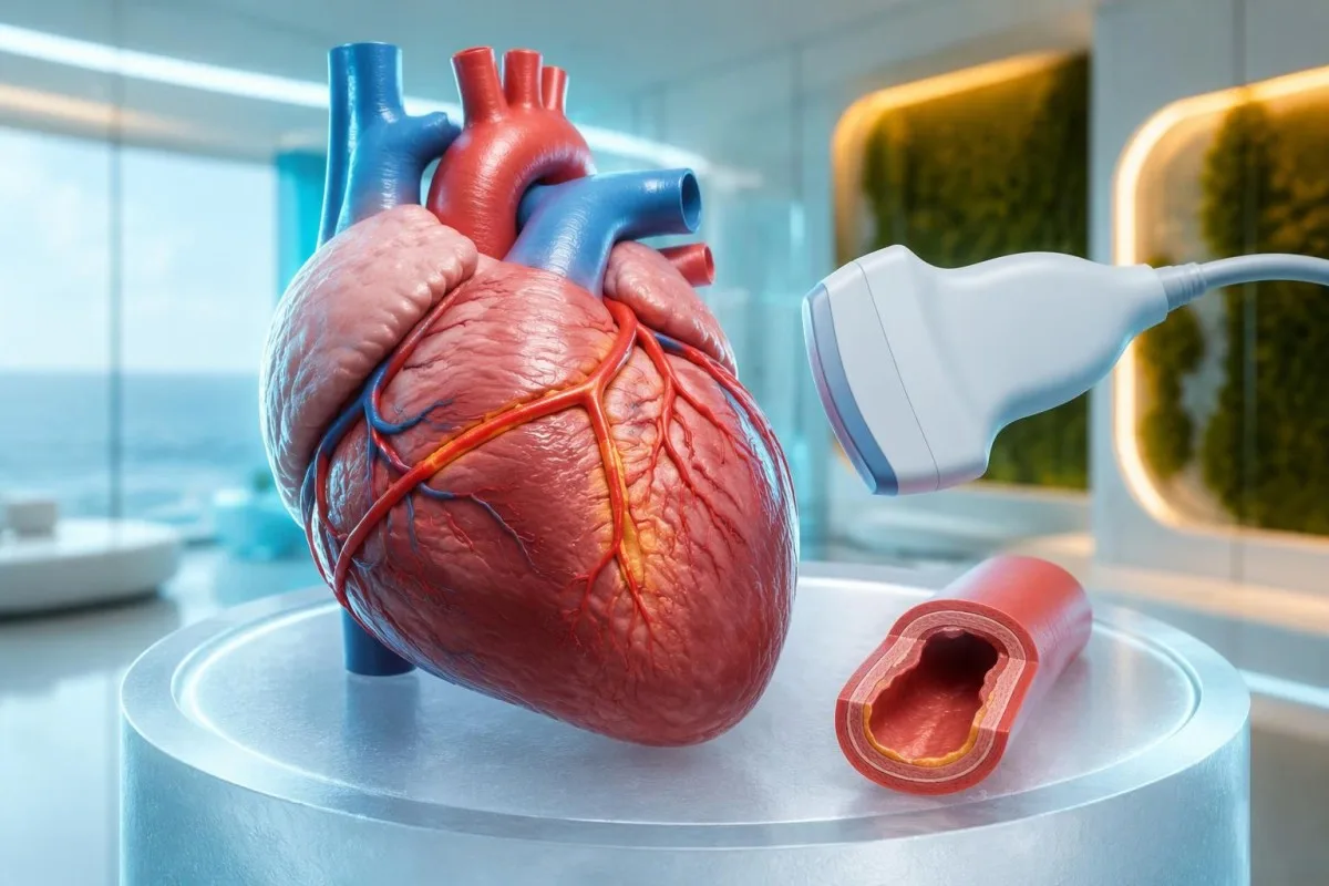
A coronary Z-score compares artery size with the expected size for a child's body surface area. A raw measurement such as 3 mm therefore has different implications in a small infant and a much larger child. The 2024 AHA statement emphasizes coronary assessment and risk-based management; the number should be interpreted with the measurement method and clinical setting (Jone et al., 2024).
A positive echocardiogram in the AHA incomplete-disease pathway can include a coronary aneurysm or a qualifying coronary Z-score. It can also involve at least 3 suggestive findings: decreased left-ventricular function, mitral regurgitation, pericardial effusion, or left anterior descending/right coronary Z-scores of 2 to less than 2.5. This is specialist image interpretation, not a checklist parents should apply to a brief scan summary.
Troponin and BNP or NT-proBNP may help evaluate cardiac involvement but do not replace echocardiography. A normal troponin on 1 sample cannot show that the coronary arteries are unaffected, and an elevated BNP can have several explanations. Our myocarditis laboratory guide explains the distinction between evidence of heart-muscle involvement and the separate question of coronary artery changes.
臨床医は川崎病と類似疾患をどのように区別しますか?
Clinicians distinguish Kawasaki disease from other causes of fever by combining the illness timeline, examination, targeted infection testing, laboratory patterns, and heart imaging. MIS-C, bacterial infections, adenovirus, scarlet fever, and systemic inflammatory disorders can overlap; a CRP result of 80 mg/L cannot separate them reliably.
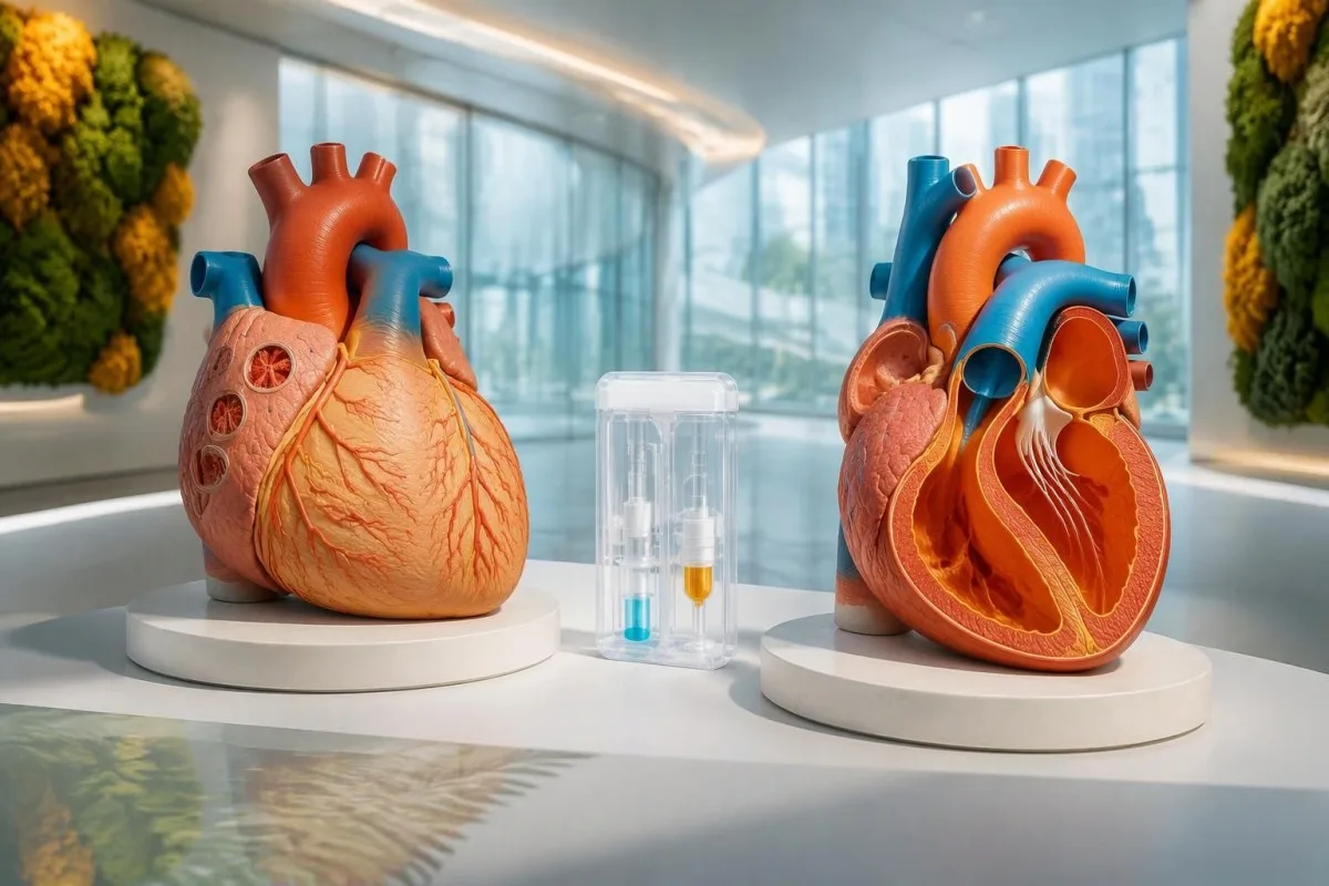
MIS-C often follows SARS-CoV-2 infection by approximately 2–6 weeks and frequently affects older children than classic Kawasaki disease. Prominent gastrointestinal symptoms, shock, lymphopenia, relatively low platelets, and ventricular dysfunction may favor MIS-C, but overlap is substantial. A positive SARS-CoV-2 antibody result alone does not settle the distinction because previous infection or vaccination can affect interpretation.
Procalcitonin can rise in Kawasaki disease and should not automatically be treated as proof of a bacterial cause. Clinicians assess cultures, examination, organ function, and the broader trajectory before deciding what an elevated result means. Our プロカルシトニンの解釈ガイドでは、 explains why 1 biomarker cannot safely decide between sepsis, Kawasaki disease, or another inflammatory syndrome.
Finding a respiratory virus does not necessarily exclude coexisting Kawasaki disease. A hypothetical child with 6 days of fever, characteristic mouth changes, swollen feet, and an adenovirus-positive swab still needs a complete clinical assessment rather than automatic dismissal of the other findings. Travel, medication exposure, urinary symptoms, and recurrent fever patterns guide additional testing; there is no universal panel that rules out every mimic.
発症日や再検査によって状況が変わるのはなぜですか?
Kawasaki laboratory findings evolve, so the illness day and treatment history are part of every result's meaning. A normal platelet count early in the first week can become elevated later, while intravenous immunoglobulin can raise ESR independently of ongoing inflammation and make post-treatment ESR misleading.
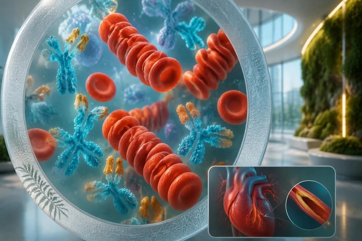
A changing CBC can strengthen suspicion without making the first sample wrong. For example, a hypothetical child might have WBC 13 × 10⁹/L and platelets 280 × 10⁹/L on day 4, then platelets 480 × 10⁹/L and albumin 29 g/L on day 7. Clinicians should not postpone a concerning assessment until those later findings appear; our 検査室タイムラインガイド explains evolving results.
ESR should not be used as the main measure of response after intravenous immunoglobulin. The infused proteins can increase sedimentation, so an ESR of 70 mm/hour after treatment does not necessarily mean treatment failure. Fever, clinical recovery, CRP trends, and cardiac assessment are more useful in context; the 2017 AHA statement specifically describes this post-infusion limitation (McCrindle et al., 2017).
Kantesti can help explain serial reports, but a trend graph cannot determine whether a child needs retreatment. Record the sample date, illness day, infusion completion time, and laboratory units alongside each value; comparing 2 results without that information can produce a false impression of deterioration. Immunoglobulin can also complicate some antibody tests through passive antibody transfer, so the treating team needs the infusion history.
すべての症状が現れるまで治療を待つべきでないのはなぜですか?
Treatment should begin promptly when Kawasaki disease is diagnosed, including incomplete disease, rather than waiting for every classic feature. Standard initial hospital treatment includes intravenous immunoglobulin at 2 g/kg, generally within the first 10 illness days; later treatment may still be indicated when fever or inflammation persists.
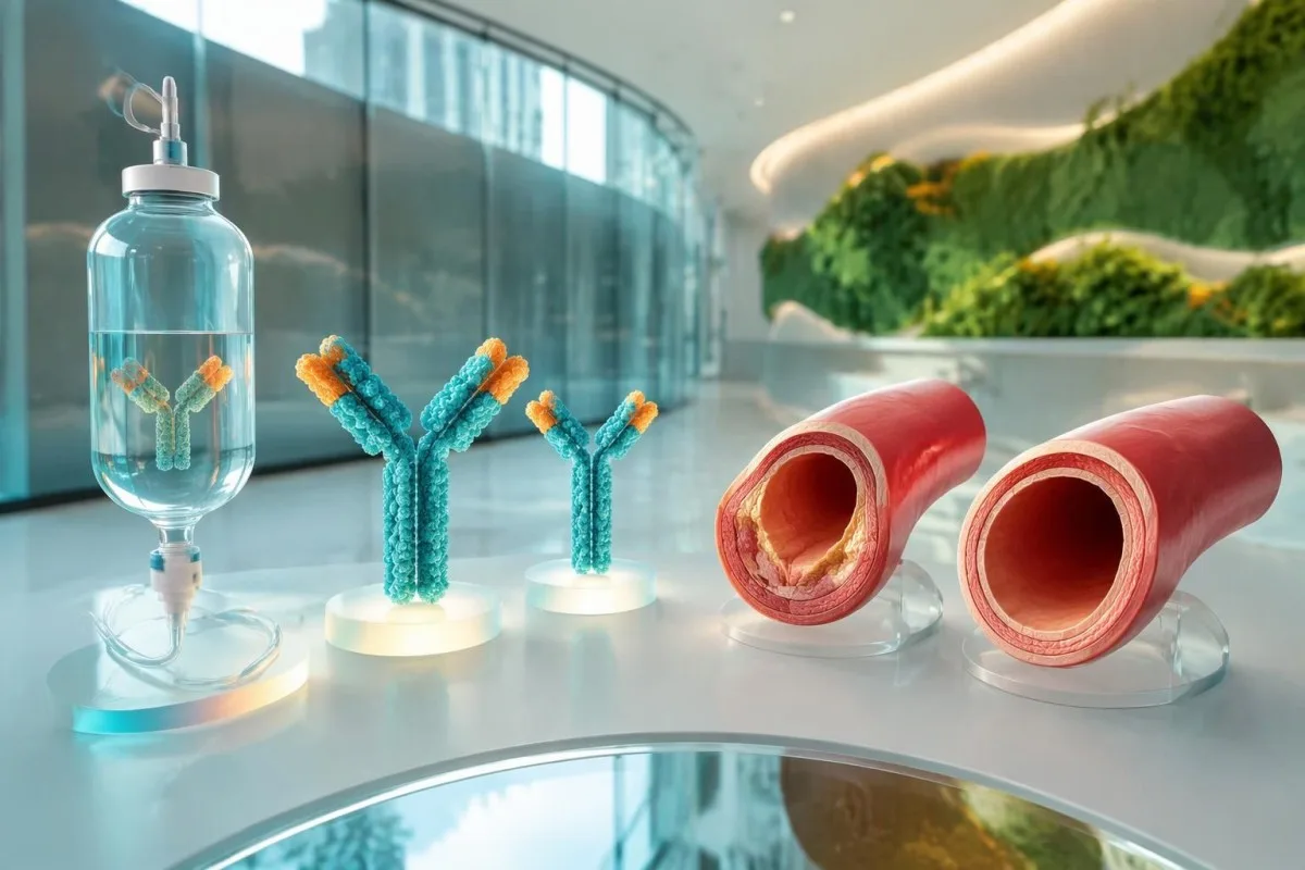
The 10-day treatment window is a target for timely care, not a reason to delay until day 10. The ACR/Vasculitis Foundation guideline recommends prompt treatment of incomplete Kawasaki disease once diagnosed, rather than waiting for the child to satisfy classic criteria (Gorelik et al., 2022). For a hypothetical 12-kg child, 2 g/kg represents 24 g of immunoglobulin administered under hospital supervision.
Aspirin treatment in Kawasaki disease requires a pediatric specialist's prescription and monitoring. Acute regimens vary internationally, including 30–50 or 80–100 mg/kg/day, followed in many cases by antiplatelet dosing of 3–5 mg/kg/day; families should not start, stop, or calculate treatment themselves. Our aspirin toxicity guidance explains why a prescribed Kawasaki regimen is not ordinary home fever treatment.
Approximately 10–20% of patients have persistent or recurrent fever after initial immunoglobulin treatment. Fever at least 36 hours after completion of the infusion is a commonly used threshold for IVIG resistance, after other causes are considered. Additional treatment may include corticosteroids or other specialist therapies; infants younger than 6 months and children with coronary Z-scores ≥2.5 can need particular attention, but risk scores do not perform equally across populations.
血液検査値が改善した場合、どのような経過観察が必要ですか?
Improving CRP and CBC results do not prove that coronary artery risk has resolved. Follow-up is based on clinical recovery and echocardiographic findings, including the largest coronary Z-score recorded; children with coronary aneurysms need a different surveillance plan from children whose coronary studies remain normal.
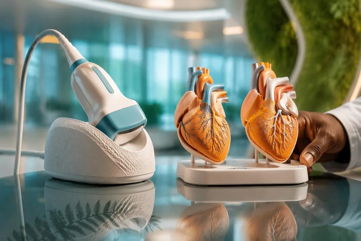
Uncomplicated follow-up often includes echocardiography around 1–2 weeks and 4–6 weeks after treatment, but the schedule is individualized. The 2024 AHA statement supports risk-based assessment, and normal early studies can affect whether further routine imaging adds value (Jone et al., 2024). Persistent coronary changes, poor visualization, recurrent symptoms, or an abnormal initial study can require a different timetable.
A platelet count that remains elevated during recovery is not, by itself, proof of treatment failure. A hypothetical count of 600 × 10⁹/L in week 2 may fit the expected illness phase even when fever has stopped and CRP is falling, but symptoms and cardiac findings still govern management. Our 入院後の検査室変化」 guide explains why normalization is not simultaneous across all markers.
High-dose immunoglobulin can affect the timing of certain live vaccines. After IVIG 2 g/kg, measles- and varicella-containing vaccines are commonly deferred for 11 months under immunization guidance, with exceptions assessed by the child's team and local schedule. Ask for a written plan covering vaccines, medicines, activity, repeat imaging, and which symptoms require reassessment; coronary follow-up should not be replaced by an apparently reassuring laboratory report.
評価中、AIによる説明を安全に活用するにはどうすればよいですか?
AI explanations can help families understand laboratory language, but they cannot confirm Kawasaki disease, assess a child's circulation, or interpret coronary imaging independently. With fever lasting 5 days—or concerning symptoms sooner—clinical assessment takes priority over uploading reports, comparing reference ranges, or obtaining another automated interpretation.

Kantesti is an AI lab test interpretation service that can explain what CRP 30 mg/L or platelets 450 × 10⁹/L mean in a report. Those numbers are not sufficient to decide whether a child has Kawasaki disease, and an explanation cannot replace documenting illness day, examination findings, or an echocardiogram. Our 責任あるAIの原則 describe why interpretation must remain separate from urgent pediatric decision-making.
A useful review packet combines at least 3 types of information: the fever timeline, complete laboratory reports, and the treating team's assessment. Preserve units, pediatric reference intervals, medication dates, and imaging conclusions rather than sharing only red flags or screenshots of isolated values. Our medical validation documentation describes evaluation standards and limitations; technical performance should not be read as condition-specific proof of diagnostic safety.
Kantesti's research publications listed below describe platform evaluation and aggregate report analysis, not a Kawasaki diagnostic assay. The Clinical Validation Framework v2.0 and Global Health Report 2026 are hosted on Zenodo, and their DOI identifiers establish persistent records rather than peer-review status. Neither publication should be interpreted as establishing sensitivity or specificity for incomplete Kawasaki disease; clinical recommendations here rely on the 2017 and 2024 AHA statements and the ACR/Vasculitis Foundation guideline.
よくある質問
川崎病を確定できる血液検査はありますか?
川崎病を確定する単一の血液検査はありません。臨床医は、発熱の経過、診察所見、炎症マーカー、補助的な検査結果、心エコー検査を総合して判断します。CRP ≥30 mg/LまたはESR ≥40 mm/hourは、AHAの不全型川崎病の診断経路において、さらなる評価を支持しますが、炎症の原因を特定するものではありません。特に発症早期には、個々の検査結果が正常であっても、この疾患を安全に否定することはできません。.
すべての症状がなくても、子どもが川崎病にかかることはありますか?
小児は、5つの主要な臨床所見がすべてそろっていなくても、不全型川崎病を発症することがあります。AHAの診療アルゴリズムでは、発熱が5日以上続き、2~3つの所見がある小児に加え、原因不明の発熱が7日以上続く特定の乳児も対象としています。診断と治療の根拠となる検査値の異常や冠動脈所見が認められることがあります。家族は、皮膚の落屑やほかの症状が現れるのを待たずに、速やかに診察を受けてください。.
川崎病でよく見られる血液検査結果は何ですか?
川崎病でよくみられる検査所見には、CRPまたはESRの上昇、白血球増加、年齢に比して貧血、アルブミン低値、ALT上昇、およびその後の血小板増加が含まれます。不全型の補助的な診断基準値には、アルブミン ≤30 g/L、WBC ≥15 × 10⁹/L、および発症7日目以降の血小板 ≥450 × 10⁹/Lが含まれます。これらの所見は川崎病に特異的ではなく、診察所見や発症からの経過とあわせて解釈する必要があります。尿中白血球も評価の参考になりますが、血液検査の結果ではなく尿所見です。.
血小板数や心エコー検査が正常であれば、川崎病は否定できますか?
発症第1週の血小板数が正常で、初期の心エコー検査が正常であっても、川崎病を否定することはできません。血小板数は第2週に上昇することが多く、初回の検査では冠動脈の変化が明らかでない場合があります。したがって、発症早期の血小板数が450 × 10⁹/L未満であっても、診断を除外するものではありません。発熱が持続する場合や懸念される臨床像がみられる場合は、適応のある治療を遅らせることなく、再評価が必要となることがあります。.
How long should a child's fever last before seeking help?
0 5日間続く発熱のある小児は速やかに臨床評価を受ける必要がありますが、重篤な症状がある場合は、より早急な対応が必要です。生後3か月未満の乳児で38°C以上の体温がある場合は、緊急の医学的評価が必要です。呼吸困難、反応の低下、圧迫しても消えない発疹、脱水、または循環不良がある場合は、発熱の持続期間にかかわらず、緊急または救急の診療を受けてください。川崎病の5日基準は、受診を待つよう指示するものではありません。.
川崎病の治療後もESRが高いのはなぜですか?
ESRは、注入されたタンパク質が沈降に影響するため、免疫グロブリン静注後も高値のまま、または上昇することがあります。したがって、治療後のESRが70 mm/hourであっても、それだけで治療失敗を示すものではありません。医療従事者は、点滴後のESRに頼るのではなく、発熱、臨床的な回復、CRPの推移、心臓の所見を評価します。点滴終了から少なくとも36時間後も発熱が持続または再発する場合は、担当チームによる再評価が必要です。.
血液検査結果の見方を確認しましょう
検査報告書をアップロードすると、KantestiのAIが数秒で各バイオマーカーを平易な言葉で説明します。世界中で200万人以上が利用しています。.
参考文献
- Klein, T., Mitchell, S., & Weber, H. (2026). 臨床検証フレームワーク v2.0(医学的検証ページ)。. Kantesti AI医学研究。. 掲載日
- Klein, T., Mitchell, S., & Weber, H. (2026). AI血液検査アナライザー:2.5M件の検査を分析 | グローバルヘルスレポート 2026。. Kantesti AI医学研究。. 掲載日
- McCrindle BW et al. (2017). Diagnosis, Treatment, and Long-Term Management of Kawasaki Disease: A Scientific Statement for Health Professionals From the American Heart Association. Circulation. 掲載日 パブメッド
- Jone PN et al. (2024). Update on Diagnosis and Management of Kawasaki Disease: A Scientific Statement From the American Heart Association. Circulation.
- Gorelik M et al. (2022). 2021 American College of Rheumatology/Vasculitis Foundation Guideline for the Management of Kawasaki Disease. Arthritis & Rheumatology.
- 200万以上分析対象の検査
- 127+国
- 100+言語
医療チームについて


E-E-A-T 信頼性シグナル
経験
医師主導による、検査結果解釈ワークフローの臨床レビュー。.
専門知識
臨床的な文脈においてバイオマーカーがどのように振る舞うかに焦点を当てた検査医学。.
権威
Thomas Klein医師による医学的レビュー。Sarah Mitchell医師およびHans Weber教授の協力。.
信頼性
アラームを減らすための明確なフォローアップ経路を備えた、エビデンスに基づく解釈。.




医療免責事項
この記事は教育目的のみを対象としており、医療助言を構成するものではありません。診断や治療の判断を行う際は、必ず有資格の医療提供者にご相談ください。.
公開 · 編集チームにお問い合わせください
Kantesti Ltdが発行。イングランドおよびウェールズで登録、会社番号. 17090423, 、英国ロンドン。.
