
Кавасаки өвчнийг батлах ганц цусны шинжилгээ байдаггүй. Оношилгоонд халуурсан түүх, үзлэг, үрэвслийн маркер, оношийг дэмжих лабораторийн үзүүлэлт болон зүрхний эхо шинжилгээг хамтатган үнэлнэ—хүүхдийн халууралт үргэлжилж байвал бүх сонгодог шинж тэмдэг илрэхийг хүлээлгүй цаг алдалгүй үнэлүүлэх шаардлагатай.

Гол санаанууд
- Оношилгоо эмнэлзүйн үнэлгээ шаардлагатай; Кавасаки өвчнийг найдвартай батлах эсвэл үгүйсгэх ганц шинжилгээ байдаггүй.
- Үргэлжилсэн халууралт 5 хоног үргэлжилбэл Кавасаки өвчний үнэлгээг цаг алдалгүй хийх шаардлагатай боловч хүүхдийн биеийн байдал ноцтой харагдаж байвал үүнээс ч эрт тусламж авах хэрэгтэй.
- Үрэвслийн маркерууд Америкийн Зүрхний Нийгэмлэгийн бүрэн бус өвчний үнэлгээний алгоритмын дагуу CRP ≥30 mg/L эсвэл ESR ≥40 mm/hour байх нь цаашид үнэлэх шаардлагыг дэмжинэ.
- Бүрэн бус Кавасаки өвчин сонгодог шинж тэмдгийн ердөө 2–3 нь илэрсэн үед ч тохиолдож болно; нярайд харагдах шинж маш цөөн байсан ч халууралт удаан үргэлжилж болно.
- тромбоцитууд Өвчний 7 дахь өдрөөс хойш ≥450 × 10⁹/L байх нь оношийг дэмжих үзүүлэлт; эхний долоо хоногт ялтас хэвийн байх нь Кавасаки өвчнийг үгүйсгэхгүй.
- Оношилгоог дэмжих үзүүлэлтүүд альбумин ≤30 g/L, насанд тохируулсан цус багадалт, ALT ихсэх, WBC ≥15 × 10⁹/L, мөн нэг их өсгөлттэй харах талбайд ≥10 цагаан эс илэрсэн шээсний шинжилгээ багтана.
- Зүрхний хэт авиан шинжилгээ титэм судас болон зүрхний үйл ажиллагааг үнэлдэг; эхэн үед хийсэн хэвийн шинжилгээ Кавасаки өвчнийг үгүйсгэхгүй.
- Эмчилгээний цаг хугацаа чухал: эмнэлгийн эмчилгээнд ихэвчлэн судсаар иммуноглобулин хийх багтдаг бөгөөд бүх шинж тэмдэг илрэхийг хүлээлгүйгээр, өвчний эхний 10 хоногт хийх нь зүйтэй.
Кавасаки өвчний цусны шинжилгээгээр оношийг баталж болох уу?
Кавасаки өвчнийг батлах ганц цусны шинжилгээ байдаггүй. Цусны шинжилгээгээр үрэвсэл болон эрхтнүүдийн хариу урвалыг хэмждэг бол эмч нар халууралт, үзлэгийн илрэл, зүрхний дүрс оношилгоонд үндэслэн өвчнийг тодорхойлдог; бүх шинж тэмдгийн хэв шинж илрээгүй байсан ч 5 хоног халуурсан хүүхдийг яаралтай үнэлэх шаардлагатай.
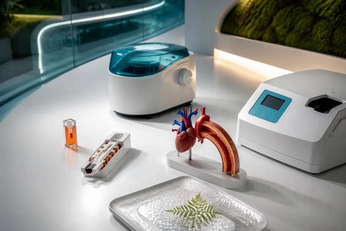
Кавасаки өвчин нь титэм судсанд нөлөөлж болзошгүй хүүхдийн васкулит юм. Америкийн Зүрхний Нийгэмлэгийн 2017 оны мэдэгдэлд эмчлээгүй тохиолдлын ойролцоогоор 25%-д титэм артерийн аневризм үүсдэг гэж тооцоолсон; энэ үзүүлэлт нь эмчлээгүй өвчнийг илэрхийлэхээс бус, удаан хугацаанд халуурсан эсвэл CRP-ийн хэмжээ өндөр гарсан хүүхэд бүрийг хэлээгүй (McCrindle et al., 2017). Өвчилсөн хүүхдүүдийн ихэнх нь 5-аас доош настай боловч түүнээс дээш насны хүүхдүүд ч өвчилж болно.
Үрэвслийн маркерын хэвийн бус үзүүлэлт нь дархлааны хариу урвалын нотолгоо болохоос өвчний нэр биш. CRP 80 mg/L байх нь Кавасаки өвчин, бактерийн халдвар эсвэл өөр үрэвсэлт эмгэгийн үед илэрч болох тул энэ үзүүлэлт дангаараа оношийг тогтоохгүй. Үүнтэй адил, ердөө 1 үзүүлэлтийг хэвийн бус гэж тэмдэглэсэн хариу нь сорьцыг эрт авсан эсвэл насанд хүрэгчдийн лавлах хэмжээгээр үнэлсэн бол эрсдэлийг дутуу үнэлж болзошгүй.
Kantesti нь Кавасаки өвчин оношлохгүйгээр CRP, CBC болон элэгний шинжилгээний нэр томьёог тайлбарлах хиймэл оюунд суурилсан цусны шинжилгээний анализатор юм. Манай хүүхдийн шинжилгээний хариуг тайлбарлах хамгаалалтын арга хэмжээ 3 настай хүүхдийн шинжилгээний хариуг насанд хүрэгчдийн таамаглалаар тайлбарлаж болохгүйг тайлбарлана. Мэдээлэл манай байгууллагын тухай энэхүү боловсролын үйлчилгээг хэн үзүүлдгийг тодорхойлдог; халууралт үргэлжилж байвал эмчид үзүүлэх шаардлагатай хэвээр.
Хүүхдийн үргэлжилсэн халууралтыг хэзээ үнэлүүлэх шаардлагатай вэ?
5 хоног үргэлжилсэн халууралттай хүүхдийг Кавасаки өвчнийг авч үзэх зэрэг эмнэлзүйн яаралтай үнэлгээнд хамруулах хэрэгтэй. Хүнд өвчин, сэтгэл түгшээсэн шинж тэмдэг илэрсэн эсвэл бага сартай нярай бол үүнээс өмнө эмнэлгийн тусламж аваарай; оношилгоонд халуурсан байх хугацааг заасан нь 5 дахь өдөр хүртэл гэртээ бай гэсэн үг биш.

3 сараас доош нярайд биеийн халуун 38°C буюу түүнээс дээш байвал яаралтай эмнэлгийн үнэлгээ хийлгэх шаардлагатай. Ямар ч насанд амьсгалахад хүндрэлтэй болох, ер бусын нойрмоглох, хариу үйлдэл муудах, дарахад арилахгүй тууралт гарах, хүнд шингэн алдалт эсвэл цусны эргэлт муудах шинж илэрвэл яаралтай тусламж авна. Эдгээр шинж нь Кавасаки өвөрмөц биш; сепсис болон цаг алдалгүй эмчлэх шаардлагатай бусад эмгэгийг мөн авч үзэх хэрэгтэй.
Парацетамол хэрэглэсний дараа халуун түр буурах нь Кавасаки өвчнийг үгүйсгэхгүй. Халууралт дахин нэмэгдэхийн хооронд хүүхдийн биеийн байдал сайжирсан мэт байж болох ч суурь өвчин үргэлжилж, шинж тэмдгүүд өөр өөр өдөр илэрч болно. Халуун анх хэзээ эхэлсэн, хэмжсэн бодит халууны заалт, хэрэглэсэн эм, шээсний гарц, нүд, уруул, гар, хөлд гарсан өөрчлөлтийг тэмдэглээрэй; термометр нэг удаа хэвийн заасан эсэхээр хязгаарлаж болохгүй.
6 сараас доош нярайд түгээмэл тууралт болон нүд улайх шинжгүйгээр удаан хугацаанд халуурч болно. Шалтгаан тодорхойгүй, дор хаяж 7 хоног үргэлжилсэн халууралт нь AHA-ийн алгоритмын дагуу дутуу илэрсэн өвчнийг үнэлэхэд харгалздаг шалгуур юм. Манай сорьц бэлтгэх заавар төлөвлөсөн шинжилгээнд хэрэгтэй боловч өвдсөн хүүхдийг мацаг барих, боломжтой цаг товлох эсвэл шинж тэмдгийн бүрэн жагсаалт бүрдэхийг хүлээлгэж огт болохгүй.
Эмнэлзүйн шинж тэмдгээр Кавасаки өвчнийг хэрхэн оношлох вэ?
Кавасаки өвчний сонгодог хэлбэрийг ихэвчлэн дор хаяж 5 хоног үргэлжилсэн халууралт болон эмнэлзүйн үндсэн 5 шинжийн дор хаяж 4 нь илэрсэн тохиолдолд оношилдог. Clinicians can diagnose some strongly characteristic cases earlier; neither the fever count nor the symptom count replaces clinical judgment or urgent evaluation.

The 5 principal features are bilateral non-purulent conjunctival redness, mouth changes, a polymorphous rash, extremity changes, and cervical lymphadenopathy. Mouth changes include red or cracked lips and a strawberry tongue; hand and foot changes include redness or swelling. The lymph-node criterion is usually a cervical node at least 1.5 cm across, often on one side.
The clinical features do not have to occur simultaneously. A photograph of a rash from illness day 3 may remain diagnostically useful when the skin looks different on day 6, and a previous episode of swollen hands should not disappear from the history because swelling has improved. Peeling around the fingertips often occurs during weeks 2–3, too late to use as a reason to delay assessment.
Certain findings point toward alternative explanations rather than strengthening the classic Kawasaki pattern. Purulent eye discharge, prominent exudative tonsillitis, or discrete mouth ulcers deserve separate consideration, although no single finding resolves every case. Our laboratory marker reference guide explains test terminology; it cannot substitute for examining the 5 clinical feature groups or arranging appropriate imaging.
Бүрэн бус Кавасаки өвчнийг лабораторийн шинжилгээ хэрхэн дэмждэг вэ?
Incomplete Kawasaki disease means a child has fewer classic features but enough clinical, laboratory, or echocardiographic evidence to warrant diagnosis and treatment. The AHA evaluation pathway considers children with fever for at least 5 days and 2–3 features, and certain infants with unexplained fever for at least 7 days.
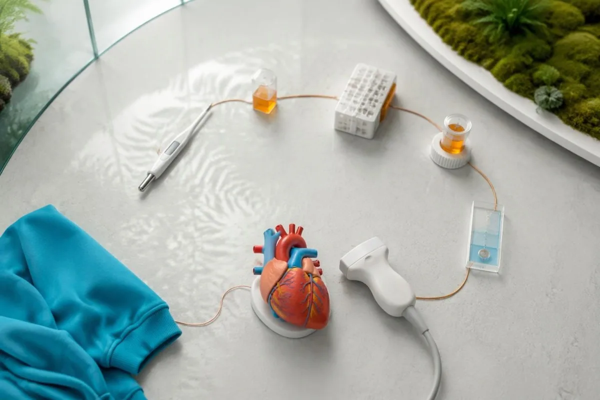
CRP ≥30 mg/L or ESR ≥40 mm/hour supports proceeding through the AHA incomplete-disease evaluation pathway. When inflammation reaches either threshold, clinicians assess echocardiography and supplementary laboratory criteria rather than waiting for another visible symptom. The thresholds are decision supports for a compatible illness—not proof that Kawasaki disease is the cause (McCrindle et al., 2017).
The 6 supplementary laboratory criteria are albumin ≤30 g/L, anemia for age, elevated ALT, WBC ≥15 × 10⁹/L, platelets ≥450 × 10⁹/L after day 7, and urine ≥10 white cells per high-power field. In the appropriate clinical setting, at least 3 supplementary criteria or a positive echocardiogram can support treatment through the pathway. These are not independent points to count without reviewing competing diagnoses.
Fewer than 3 supplementary criteria does not create a permanent rule-out result. Persistent fever may require repeated examination, repeat laboratory testing, and imaging, particularly if the first evaluation occurred early. Systemic juvenile idiopathic arthritis is one possible alternative; our childhood arthritis laboratory guide explains why shared inflammatory findings do not make these conditions interchangeable.
Кавасаки өвчний үед CRP болон ESR-ийн хариу ямар утгатай вэ?
CRP and ESR support Kawasaki evaluation by showing systemic inflammation, but neither identifies its cause. In the AHA incomplete-disease pathway, CRP ≥30 mg/L or ESR ≥40 mm/hour is a threshold for further assessment, not a severity scale or a stand-alone indication for treatment.
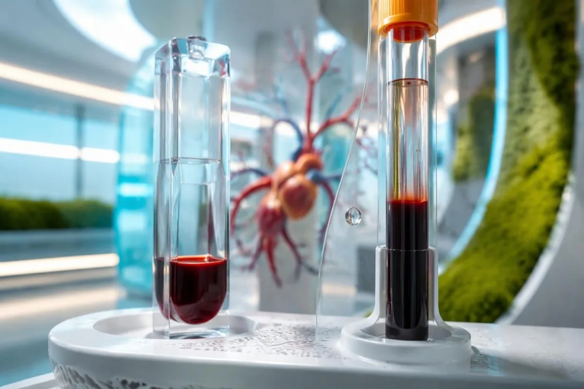
CRP of 3 mg/dL equals 30 mg/L. This 10-fold unit difference can make identical results appear very different when families compare reports from different laboratories, while common CRP reference limits are around 5 mg/L but vary by method. Our нь эдгээр тэмдэгнүүд яагаад зөрж болохыг тайлбарладаг. explains why a high CRP and a less impressive white-cell count can coexist.
ESR is influenced by factors beyond the immediate inflammatory response, including anemia and circulating proteins. A result of 45 mm/hour is not directly comparable to CRP of 45 mg/L; the tests measure different processes and have different kinetics. When both markers are below the pathway thresholds, Kawasaki disease becomes less likely, but an early sample or a concerning clinical course still requires judgment.
High-sensitivity CRP measures the same protein as conventional CRP, but adult cardiovascular-risk categories do not apply to a febrile child. A value of 30 mg/L during illness should not be interpreted using an adult 1–3 mg/L cardiovascular-risk framework. Our hs-CRP interpretation guide explains that distinction; for acute pediatric illness, the clinical question is inflammation and its cause.
| Түвшин | Хязгаар | Утга |
|---|---|---|
| CRP-ийн түгээмэл лавлах хязгаар | Often <5 mg/L; laboratory-specific | A reference interval, not a Kawasaki exclusion threshold; early disease may require reassessment. |
| Incomplete-disease CRP trigger | ≥30 mg/L, equivalent to ≥3 mg/dL | Supports further AHA pathway evaluation when fever and clinical history are compatible. |
| Common ESR reference interval | Often approximately 0–20 mm/hour; age- and method-dependent | Use the reporting laboratory's pediatric interval; ESR is affected by anemia and proteins. |
| Incomplete-disease ESR trigger | ≥40 mm/hour | Supports further evaluation; neither confirms Kawasaki disease nor defines an emergency by itself. |
CBC-ийн ямар үзүүлэлт Кавасаки өвчнийг дэмждэг вэ?
Kawasaki disease lab findings on a CBC can include neutrophil-predominant leukocytosis, anemia for age, and a later rise in platelets. WBC ≥15 × 10⁹/L and platelets ≥450 × 10⁹/L after day 7 are supplementary incomplete-disease criteria, but the illness day changes their meaning.
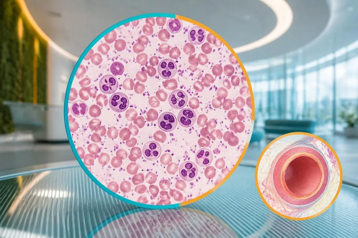
Platelets often rise during the second week rather than at the start of fever. A platelet count of 280 × 10⁹/L on illness day 4 therefore does not exclude Kawasaki disease, and waiting for thrombocytosis can delay care. Conversely, a count of 520 × 10⁹/L after day 7 supports the pattern but can also occur during recovery from other inflammatory illnesses.
Anemia in the incomplete-disease criteria is defined against the child's age, not a universal hemoglobin cutoff. For a hypothetical 3-year-old, hemoglobin of 9.8 g/dL would generally be low, but the cause could include inflammation, pre-existing iron deficiency, or both. WBC also needs age-specific context; our нейтрофилийн лавлах интервалын гарын авлага explains why pediatric differential counts differ from adult ones.
Low platelets can occur in severe Kawasaki presentations and should not be treated as reassuring. A febrile child with platelets of 90 × 10⁹/L, poor circulation, or worsening organ function needs urgent assessment for shock, sepsis, consumption, or macrophage activation. Platelet size indices and neutrophil-to-lymphocyte ratios remain contextual findings; they are not substitutes for the 6 established supplementary criteria.
Альбумин болон элэгний шинжилгээний үзүүлэлтүүд яагаад өөрчлөгддөг вэ?
Low albumin and elevated ALT can support incomplete Kawasaki disease because systemic inflammation affects protein distribution and liver function. Albumin ≤30 g/L is a defined supplementary criterion, while ALT is judged against the laboratory's pediatric upper reference limit rather than one universal Kawasaki cutoff.
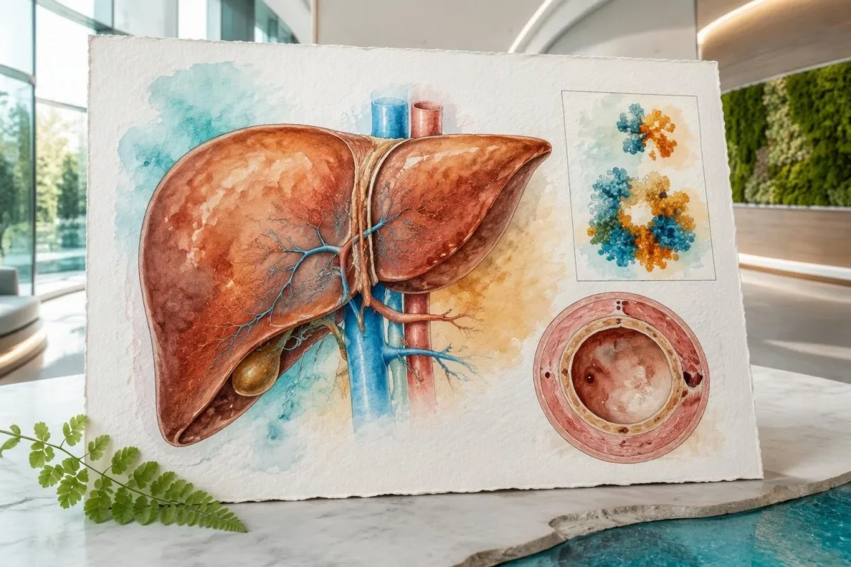
Albumin of 3.0 g/dL equals 30 g/L. During acute illness, a low result can reflect reduced synthesis, increased vascular permeability, dilution, or protein loss—not simply poor dietary protein intake. Our Цусны ийлдсийн уургийн тайлбарын гарын авлага explains these mechanisms; nutritional advice alone would miss the clinical significance of low albumin combined with persistent fever.
An ALT result above the pediatric upper limit counts as supporting evidence, but its magnitude does not diagnose Kawasaki disease. For example, a hypothetical child with albumin 28 g/L and ALT 72 IU/L may meet 2 supplementary criteria if that ALT exceeds the local limit. The same combination can arise from other illnesses or medication effects, as our ALT нэр томьёоны гарын авлага тайлбарладаг.
Kantesti is an AI blood test interpretation platform that can explain why albumin 28 g/L and ALT 72 IU/L require clinical context. Манай хиймэл оюун ухааны технологийн тайлбар describes the interpretation workflow, not a validated Kawasaki diagnostic test. Growing children can also have relatively high alkaline phosphatase from bone activity; that finding is not interchangeable with the ALT criterion.
Шээсний шинжилгээ болон натри үнэлгээнд тус болж чадах уу?
Urine white cells can support incomplete Kawasaki evaluation, while electrolyte changes help assess illness severity and alternative causes. Urine ≥10 white cells per high-power field is a supplementary criterion; low sodium is a possible associated finding but is not one of the 6 AHA supplementary laboratory criteria.
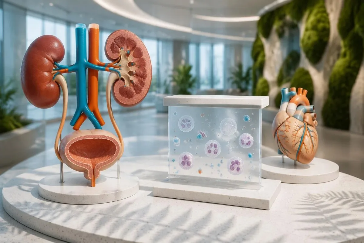
Sterile pyuria means urine white cells are present without a demonstrated bacterial cause on an appropriately collected culture. Kawasaki disease can produce this pattern, but a child with urine white cells still needs assessment for urinary infection. A urine dipstick alone cannot establish sterile pyuria; our шээсний тунадас тайлбарлах гарын авлагад distinguishes microscopy findings from dipstick screening results.
A report of 10 white cells per high-power field cannot be directly equated with an automated result of 10 cells/µL. Instruments, specimen preparation, and counting methods differ, so clinicians may need the laboratory's conversion or interpretive guidance. Collection quality also matters: contamination can introduce cells, whereas prior antibiotics can reduce culture yield without explaining the entire fever pattern.
Serum sodium below 135 mmol/L is generally classified as hyponatremia, but its cause must be assessed separately. Acute inflammation, fluid intake, gastrointestinal losses, and altered water regulation can all contribute; the number is not a Kawasaki confirmation test. Our электролитийн загвар гарын авлага explains why sodium 130 mmol/L with neurological symptoms is a different problem from an isolated mildly low value.
Зүрхний эхо шинжилгээ оношилгоонд юу нэмдэг вэ?
Echocardiography assesses coronary artery dimensions, heart function, and other cardiac findings that blood tests cannot show. A coronary artery Z-score ≥2.5 can support Kawasaki diagnosis in the incomplete-disease pathway, but a normal early echocardiogram does not exclude the illness or justify delaying indicated treatment.
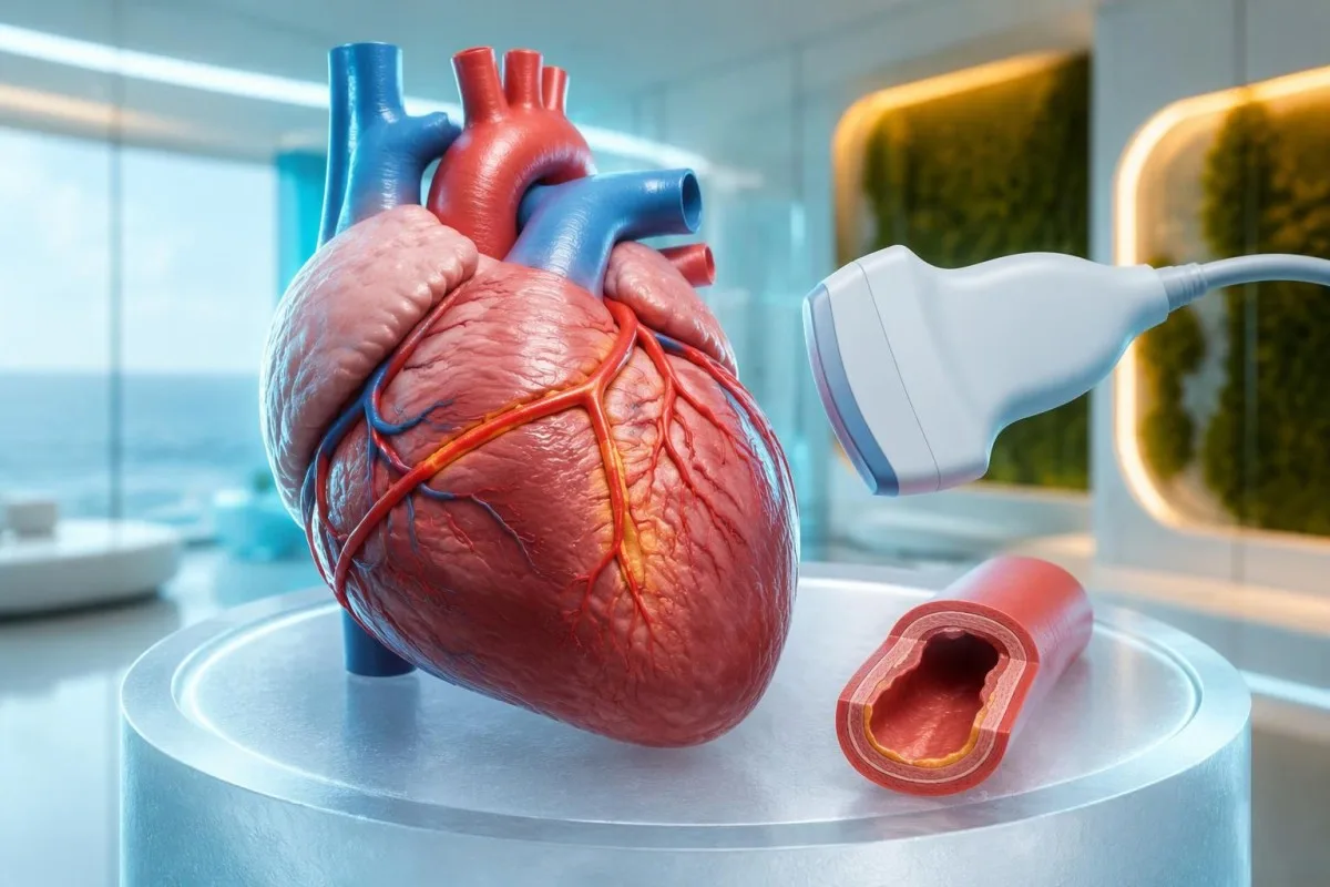
A coronary Z-score compares artery size with the expected size for a child's body surface area. A raw measurement such as 3 mm therefore has different implications in a small infant and a much larger child. The 2024 AHA statement emphasizes coronary assessment and risk-based management; the number should be interpreted with the measurement method and clinical setting (Jone et al., 2024).
A positive echocardiogram in the AHA incomplete-disease pathway can include a coronary aneurysm or a qualifying coronary Z-score. It can also involve at least 3 suggestive findings: decreased left-ventricular function, mitral regurgitation, pericardial effusion, or left anterior descending/right coronary Z-scores of 2 to less than 2.5. This is specialist image interpretation, not a checklist parents should apply to a brief scan summary.
Troponin and BNP or NT-proBNP may help evaluate cardiac involvement but do not replace echocardiography. A normal troponin on 1 sample cannot show that the coronary arteries are unaffected, and an elevated BNP can have several explanations. Our myocarditis laboratory guide explains the distinction between evidence of heart-muscle involvement and the separate question of coronary artery changes.
Эмч нар Кавасаки өвчнийг төстэй эмгэгүүдээс хэрхэн ялгадаг вэ?
Clinicians distinguish Kawasaki disease from other causes of fever by combining the illness timeline, examination, targeted infection testing, laboratory patterns, and heart imaging. MIS-C, bacterial infections, adenovirus, scarlet fever, and systemic inflammatory disorders can overlap; a CRP result of 80 mg/L cannot separate them reliably.
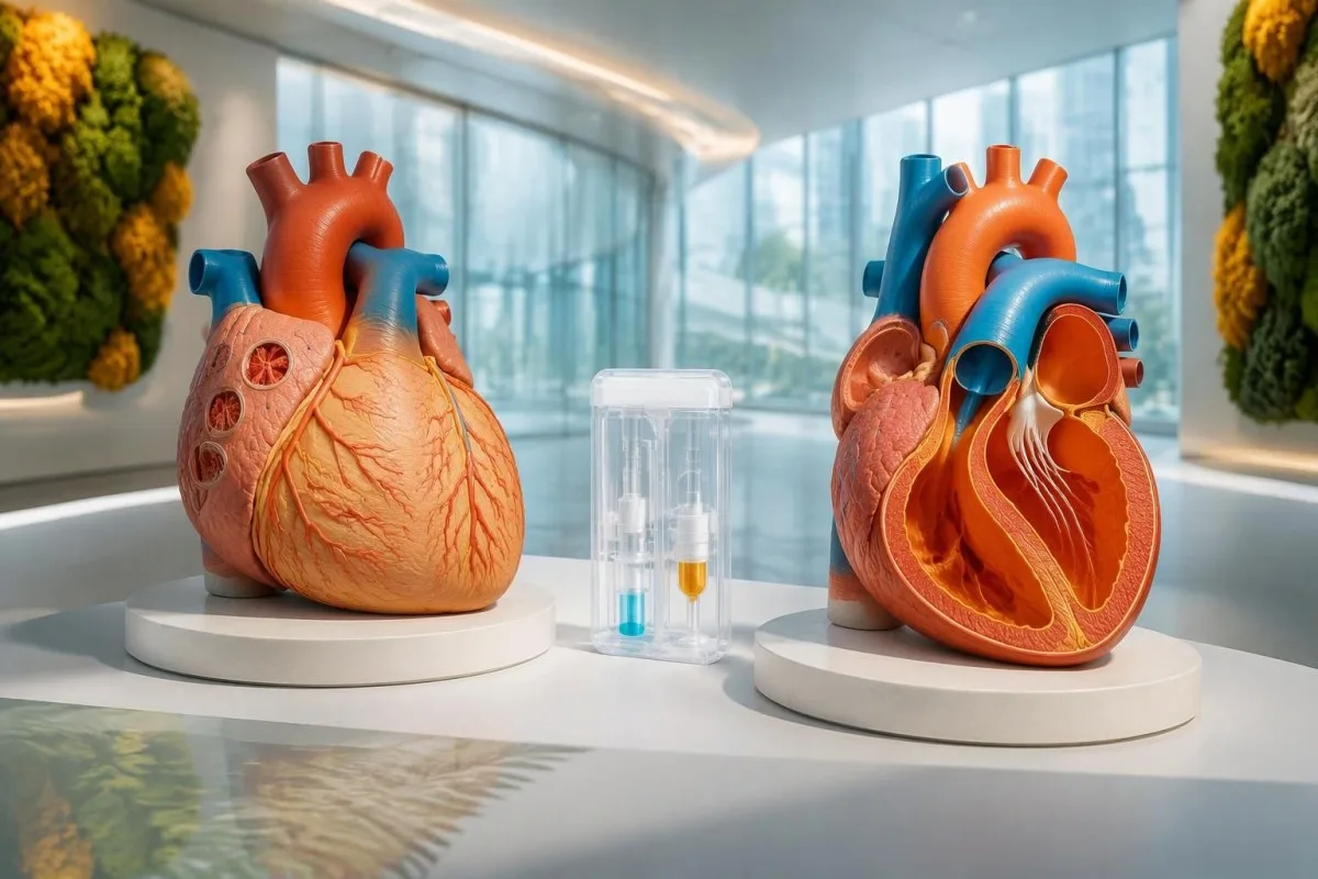
MIS-C often follows SARS-CoV-2 infection by approximately 2–6 weeks and frequently affects older children than classic Kawasaki disease. Prominent gastrointestinal symptoms, shock, lymphopenia, relatively low platelets, and ventricular dysfunction may favor MIS-C, but overlap is substantial. A positive SARS-CoV-2 antibody result alone does not settle the distinction because previous infection or vaccination can affect interpretation.
Procalcitonin can rise in Kawasaki disease and should not automatically be treated as proof of a bacterial cause. Clinicians assess cultures, examination, organ function, and the broader trajectory before deciding what an elevated result means. Our прокальцитониныг тайлбарлах гарын авлага explains why 1 biomarker cannot safely decide between sepsis, Kawasaki disease, or another inflammatory syndrome.
Finding a respiratory virus does not necessarily exclude coexisting Kawasaki disease. A hypothetical child with 6 days of fever, characteristic mouth changes, swollen feet, and an adenovirus-positive swab still needs a complete clinical assessment rather than automatic dismissal of the other findings. Travel, medication exposure, urinary symptoms, and recurrent fever patterns guide additional testing; there is no universal panel that rules out every mimic.
Өвчилсөн өдөр болон давтан шинжилгээ зураглалыг яагаад өөрчилдөг вэ?
Kawasaki laboratory findings evolve, so the illness day and treatment history are part of every result's meaning. A normal platelet count early in the first week can become elevated later, while intravenous immunoglobulin can raise ESR independently of ongoing inflammation and make post-treatment ESR misleading.
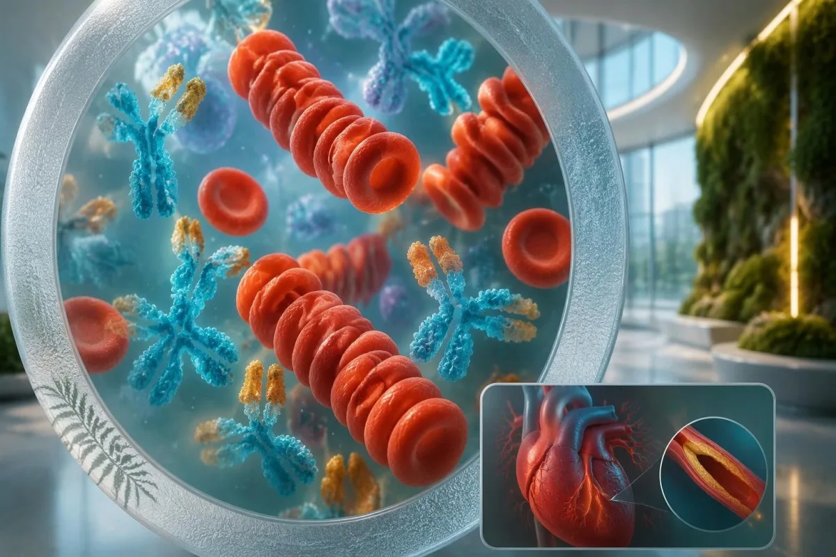
A changing CBC can strengthen suspicion without making the first sample wrong. For example, a hypothetical child might have WBC 13 × 10⁹/L and platelets 280 × 10⁹/L on day 4, then platelets 480 × 10⁹/L and albumin 29 g/L on day 7. Clinicians should not postpone a concerning assessment until those later findings appear; our лабораторийн цагийн хуваарийн гарын авлага explains evolving results.
ESR should not be used as the main measure of response after intravenous immunoglobulin. The infused proteins can increase sedimentation, so an ESR of 70 mm/hour after treatment does not necessarily mean treatment failure. Fever, clinical recovery, CRP trends, and cardiac assessment are more useful in context; the 2017 AHA statement specifically describes this post-infusion limitation (McCrindle et al., 2017).
Kantesti can help explain serial reports, but a trend graph cannot determine whether a child needs retreatment. Record the sample date, illness day, infusion completion time, and laboratory units alongside each value; comparing 2 results without that information can produce a false impression of deterioration. Immunoglobulin can also complicate some antibody tests through passive antibody transfer, so the treating team needs the infusion history.
Бүх шинж тэмдэг илрэхийг хүлээлгүй эмчлэх нь яагаад чухал вэ?
Treatment should begin promptly when Kawasaki disease is diagnosed, including incomplete disease, rather than waiting for every classic feature. Standard initial hospital treatment includes intravenous immunoglobulin at 2 g/kg, generally within the first 10 illness days; later treatment may still be indicated when fever or inflammation persists.
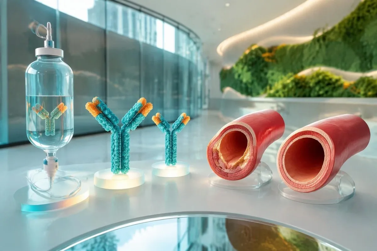
The 10-day treatment window is a target for timely care, not a reason to delay until day 10. The ACR/Vasculitis Foundation guideline recommends prompt treatment of incomplete Kawasaki disease once diagnosed, rather than waiting for the child to satisfy classic criteria (Gorelik et al., 2022). For a hypothetical 12-kg child, 2 g/kg represents 24 g of immunoglobulin administered under hospital supervision.
Aspirin treatment in Kawasaki disease requires a pediatric specialist's prescription and monitoring. Acute regimens vary internationally, including 30–50 or 80–100 mg/kg/day, followed in many cases by antiplatelet dosing of 3–5 mg/kg/day; families should not start, stop, or calculate treatment themselves. Our aspirin toxicity guidance explains why a prescribed Kawasaki regimen is not ordinary home fever treatment.
Approximately 10–20% of patients have persistent or recurrent fever after initial immunoglobulin treatment. Fever at least 36 hours after completion of the infusion is a commonly used threshold for IVIG resistance, after other causes are considered. Additional treatment may include corticosteroids or other specialist therapies; infants younger than 6 months and children with coronary Z-scores ≥2.5 can need particular attention, but risk scores do not perform equally across populations.
Цусны шинжилгээний үзүүлэлтүүд сайжирсан үед ямар хяналт шаардлагатай вэ?
Improving CRP and CBC results do not prove that coronary artery risk has resolved. Follow-up is based on clinical recovery and echocardiographic findings, including the largest coronary Z-score recorded; children with coronary aneurysms need a different surveillance plan from children whose coronary studies remain normal.
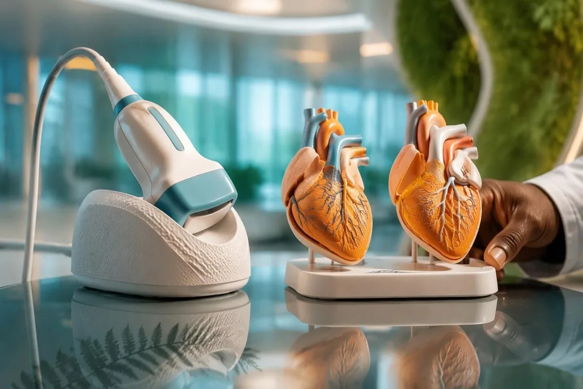
Uncomplicated follow-up often includes echocardiography around 1–2 weeks and 4–6 weeks after treatment, but the schedule is individualized. The 2024 AHA statement supports risk-based assessment, and normal early studies can affect whether further routine imaging adds value (Jone et al., 2024). Persistent coronary changes, poor visualization, recurrent symptoms, or an abnormal initial study can require a different timetable.
A platelet count that remains elevated during recovery is not, by itself, proof of treatment failure. A hypothetical count of 600 × 10⁹/L in week 2 may fit the expected illness phase even when fever has stopped and CRP is falling, but symptoms and cardiac findings still govern management. Our эмнэлгийн дараах лабораториын өөрчлөлтүүдийн нийтлэл guide explains why normalization is not simultaneous across all markers.
High-dose immunoglobulin can affect the timing of certain live vaccines. After IVIG 2 g/kg, measles- and varicella-containing vaccines are commonly deferred for 11 months under immunization guidance, with exceptions assessed by the child's team and local schedule. Ask for a written plan covering vaccines, medicines, activity, repeat imaging, and which symptoms require reassessment; coronary follow-up should not be replaced by an apparently reassuring laboratory report.
Үнэлгээний явцад хиймэл оюуны тайлбарыг хэрхэн аюулгүй ашиглах вэ?
AI explanations can help families understand laboratory language, but they cannot confirm Kawasaki disease, assess a child's circulation, or interpret coronary imaging independently. With fever lasting 5 days—or concerning symptoms sooner—clinical assessment takes priority over uploading reports, comparing reference ranges, or obtaining another automated interpretation.

Kantesti is an AI lab test interpretation service that can explain what CRP 30 mg/L or platelets 450 × 10⁹/L mean in a report. Those numbers are not sufficient to decide whether a child has Kawasaki disease, and an explanation cannot replace documenting illness day, examination findings, or an echocardiogram. Our хариуцлагатай AI-ийн зарчмууд describe why interpretation must remain separate from urgent pediatric decision-making.
A useful review packet combines at least 3 types of information: the fever timeline, complete laboratory reports, and the treating team's assessment. Preserve units, pediatric reference intervals, medication dates, and imaging conclusions rather than sharing only red flags or screenshots of isolated values. Our medical validation documentation describes evaluation standards and limitations; technical performance should not be read as condition-specific proof of diagnostic safety.
Kantesti's research publications listed below describe platform evaluation and aggregate report analysis, not a Kawasaki diagnostic assay. The Clinical Validation Framework v2.0 and Global Health Report 2026 are hosted on Zenodo, and their DOI identifiers establish persistent records rather than peer-review status. Neither publication should be interpreted as establishing sensitivity or specificity for incomplete Kawasaki disease; clinical recommendations here rely on the 2017 and 2024 AHA statements and the ACR/Vasculitis Foundation guideline.
Түгээмэл асуултууд
Кавасаки өвчнийг батлах цусны шинжилгээ байдаг уу?
Кавасаки өвчнийг батлах ганц цусны шинжилгээ байхгүй. Эмч нар халуурсан түүх, үзлэгийн олдвор, үрэвслийн маркер, нэмэлт лабораторийн шинжилгээний үр дүн болон эхокардиографийн шинжилгээг нэгтгэн үнэлдэг. AHA-ийн бүрэн бус өвчний алгоритмын дагуу CRP ≥30 мг/л эсвэл ESR ≥40 мм/цаг байх нь цаашдын үнэлгээ хийх үндэслэл болдог ч үрэвслийн шалтгааныг тодорхойлдоггүй. Ялангуяа өвчний эхэн үед тус тусдаа үзүүлэлт хэвийн гарах нь уг өвчнийг аюулгүйгээр үгүйсгэж чадахгүй.
Хүүхэд Кавасаки өвчний бүх шинж тэмдэг илрэхгүйгээр энэ өвчнөөр өвчилж болох уу?
Хүүхдэд Кавасаки өвчний үндсэн 5 эмнэлзүйн шинж бүгд илрээгүй байсан ч дутуу хэлбэрийн Кавасаки өвчин байж болно. AHA-ийн алгоритмд 5-аас доошгүй хоног халуурсан, 2–3 шинж тэмдэгтэй хүүхдүүд болон шалтгаан тодорхойгүй халууралт нь 7-оос доошгүй хоног үргэлжилсэн зарим нярай багтдаг. Лабораторийн шинжилгээний өөрчлөлт эсвэл титэм судасны шинж илэрвэл оношилж, эмчлэх үндэслэл болж болно. Арьс хуурах эсвэл өөр шинж тэмдэг илрэхийг хүлээлгүй, гэр бүлийнхэн яаралтай үнэлгээ хийлгэх хэрэгтэй.
Кавасаки өвчний үед цусны шинжилгээний ямар үзүүлэлтүүд түгээмэл илэрдэг вэ?
Кавасаки өвчний лабораторийн нийтлэг үзүүлэлтүүдэд CRP эсвэл ESR ихсэх, цагаан эсийн тоо нэмэгдэх, насандаа тохирсон хэмжээнээс цус багадалттай байх, альбумин буурах, ALT ихсэх болон хожуу үед ялтас эсийн тоо нэмэгдэх орно. Бүрэн бус хэлбэрийн өвчнийг дэмжих босго үзүүлэлтүүдэд альбумин ≤30 g/L, WBC ≥15 × 10⁹/L болон өвчний 7 дахь өдрөөс хойш ялтас эсийн тоо ≥450 × 10⁹/L байх багтана. Эдгээр үзүүлэлт Кавасаки өвчинд өвөрмөц биш тул биеийн үзлэг болон өвчний явцын хугацаатай уялдуулан тайлбарлах шаардлагатай. Шээсэн дэх цагаан эсүүд мөн үнэлгээнд дэмжлэг болж болох ч энэ нь цусны шинжилгээний үр дүн бус, шээсний үзүүлэлт юм.
Хэвийн ялтас эсвэл зүрхний эхокардиограмм Кавасаки өвчнийг үгүйсгэх үү?
Эхний долоо хоногт ялтас хэвийн байх, мөн эрт үеийн эхокардиограмм хэвийн гарах нь Кавасаки өвчнийг үгүйсгэхгүй. Ялтасны тоо хоёр дахь долоо хоногт ихэвчлэн нэмэгддэг бөгөөд титэм судасны өөрчлөлт эхний шинжилгээнд харагдахгүй байж болно. Иймээс өвчний эхэн үед ялтасны тоо 450 × 10⁹/L-ээс бага байх нь оношийг үгүйсгэхгүй. Халууралт үргэлжлэх эсвэл эмнэлзүйн шинж тэмдгийн төлөв байдал сэжигтэй байвал заалттай эмчилгээг хойшлуулахгүйгээр давтан үнэлгээ хийх шаардлагатай байж болно.
How long should a child's fever last before seeking help?
5 өдөр үргэлжилсэн халууралттай хүүхдийг эмнэлзүйн үзлэгт нэн даруй хамруулах хэрэгтэй боловч ноцтой шинж тэмдэг илэрвэл үүнээс ч эрт тусламж авах шаардлагатай. 3 сар хүрээгүй нярайд 38°C буюу түүнээс дээш халуурсан бол эмнэлгийн яаралтай үзлэгт хамруулах хэрэгтэй. Амьсгалахад хүндрэлтэй байх, хариу үйлдэл муутай байх, дарахад арилахгүй тууралт гарах, шингэн алдах эсвэл цусны эргэлт муудах тохиолдолд халуурсан хугацаанаас үл хамааран яаралтай тусламж авах шаардлагатай. Кавасаки өвчний 5 өдрийн шалгуур нь хүлээх заавар биш.
Кавасаки өвчний эмчилгээний дараа ESR яагаад өндөр хэвээр байна вэ?
Судсаар иммуноглобулин хийсний дараа сэлбэсэн уургууд тунадасжилтад нөлөөлдөг тул ESR өндөр хэвээр байх эсвэл нэмэгдэх боломжтой. Иймээс эмчилгээний дараах ESR 70 мм/цаг байх нь дангаараа эмчилгээ үр дүнгүй болсныг батлахгүй. Эмч нар дуслын дараах ESR-д найдахын оронд халууралт, эмнэлзүйн сайжрал, CRP-ийн явц болон зүрхний үзүүлэлтүүдийг үнэлдэг. Дусал хийж дууссанаас хойш дор хаяж 36 цагийн дараа халууралт үргэлжлэх эсвэл дахин илрэх тохиолдолд эмчлэгч баг дахин үнэлэх шаардлагатай.
Цусны шинжилгээний хариугаа тайлбарлуулаарай
Лабораторийн тайлангаа байршуулснаар Kantesti-ийн AI биомаркер бүрийг энгийн хэлээр хэдхэн секундэд тайлбарлана. Дэлхий даяар 2 сая гаруй хүн ашигладаг.
Лавлагаа
- Klein, T., Mitchell, S., & Weber, H. (2026). Эмнэлзүйн баталгаажуулалтын хүрээ v2.0 (Анагаахын баталгаажуулалтын хуудас). Kantesti AI-ийн анагаахын судалгаа. DOI
- Klein, T., Mitchell, S., & Weber, H. (2026). AI цусны шинжилгээний анализатор: 2.5M шинжилгээ шинжилсэн | Дэлхийн эрүүл мэндийн тайлан 2026. Kantesti AI-ийн анагаахын судалгаа. DOI
- McCrindle BW et al. (2017). Diagnosis, Treatment, and Long-Term Management of Kawasaki Disease: A Scientific Statement for Health Professionals From the American Heart Association. Circulation. DOI PubMed
- Jone PN et al. (2024). Update on Diagnosis and Management of Kawasaki Disease: A Scientific Statement From the American Heart Association. Circulation.
- Gorelik M et al. (2022). 2021 American College of Rheumatology/Vasculitis Foundation Guideline for the Management of Kawasaki Disease. Arthritis & Rheumatology.
- 2 сая+Шинжилсэн сорилууд
- 127+Улс орнууд
- 100+Хэлнүүд
Эмнэлгийн багийн тухай


E-E-A-T итгэлийн дохио
Туршлага
Эмчийн удирдлагатай лабораторийн тайлалын ажлын урсгалын клиник хяналт.
Мэргэшсэн байдал
Биомаркерууд клиникийн нөхцөлд хэрхэн ажиллаж/өөрчлөгдөж байгаад чиглэсэн лабораторийн анагаах ухаан.
Эрх мэдэл
Доктор Томас Кляйн эмнэлзүйн хувьд хянасан бөгөөд доктор Сара Митчелл, профессор доктор Ханс Вебер нар хувь нэмэр оруулсан.
Найдвартай байдал
Сэрэмжлүүлгийг бууруулахын тулд тодорхой дараагийн алхамтай, нотолгоонд суурилсан тайлал.



Эмнэлгийн хариуцлагаас татгалзах мэдэгдэл
Энэ нийтлэл зөвхөн боловсролын зорилготой бөгөөд эмнэлгийн зөвлөгөө биш болно. Оношилгоо, эмчилгээний шийдвэр гаргахдаа мэргэшсэн эрүүл мэндийн үйлчилгээ үзүүлэгчтэй заавал зөвлөлдөнө.
Хэвлэгдсэн · Редакцын багтай холбогдох
Kantesti Ltd компани Англи, Уэльст бүртгэлтэй, компанийн дугаар. 17090423, Лондон, Нэгдсэн Хаант Улс.
