
Արյան որևէ առանձին հետազոտություն չի հաստատում Կավասակիի հիվանդությունը։ Ախտորոշումը հիմնվում է տենդի պատմության, զննման, բորբոքային մարկերների, օժանդակ լաբորատոր արդյունքների և էխոկարդիոգրաֆիայի համադրության վրա․ երեխայի մոտ պահպանվող տենդը պահանջում է անհապաղ գնահատում, այլ ոչ թե սպասում մինչև բոլոր դասական ախտանշանների ի հայտ գալը։.

Հիմնական եզրակացությունները
- Ախտորոշում պահանջում է կլինիկական գնահատում․ Կավասակիի հիվանդության ոչ մի առանձին արյան հետազոտություն չի կարող հուսալիորեն հաստատել կամ բացառել այս վիճակը։.
- Պահպանվող տենդը 5 օր տևելու դեպքում պահանջում է Կավասակիի հիվանդության անհապաղ գնահատում, սակայն եթե երեխան շատ ծանր հիվանդ է թվում, նրան պետք է ավելի շուտ բուժօգնություն ցուցաբերել։.
- Բորբոքային մարկերներ CRP ≥30 mg/L կամ ESR ≥40 mm/hour ցուցանիշները օժանդակում են հետագա գնահատմանը՝ Ամերիկյան սրտի ասոցիացիայի ոչ լրիվ հիվանդության ուղեցույցի համաձայն։.
- Ոչ լրիվ Կավասակիի հիվանդությունը կարող է դրսևորվել դասական նշաններից միայն 2–3-ի առկայությամբ․ նորածինների և փոքր տարիքի երեխաների մոտ կարող է լինել երկարատև տենդ՝ շատ քիչ տեսանելի նշաններով։.
- Թրոմբոցիտներ ≥450 × 10⁹/L ցուցանիշը՝ 7-րդ օրվանից հետո, օժանդակ տվյալ է․ առաջին շաբաթվա ընթացքում թրոմբոցիտների նորմալ քանակը չի բացառում Կավասակիի հիվանդությունը։.
- Օժանդակ արդյունքները ներառում են ալբումին ≤30 g/L, տարիքին համապատասխան անեմիա, ALT-ի բարձրացում, WBC ≥15 × 10⁹/L և մեզում՝ տեսադաշտի մեծ խոշորացմամբ ≥10 լեյկոցիտ։.
- Ехокардіографія գնահատում է կորոնար զարկերակներն ու սրտի աշխատանքը․ նորմալ վաղ հետազոտությունը չի բացառում Կավասակիի հիվանդությունը։.
- Ժամանակի նշանակություն կարևոր է․ հիվանդանոցային բուժումը սովորաբար ներառում է ներերակային իմունոգլոբուլին, ցանկալի է՝ հիվանդության առաջին 10 օրվա ընթացքում՝ առանց բոլոր ախտանիշների ի հայտ գալուն սպասելու։.
Կարո՞ղ է Կավասակիի հիվանդության արյան հետազոտությունը հաստատել ախտորոշումը։
Արյան որևէ առանձին հետազոտությամբ հնարավոր չէ հաստատել Կավասակիի հիվանդության ախտորոշումը։. Արյան հետազոտությունները չափում են բորբոքումը և օրգանների արձագանքները, մինչդեռ բժիշկները հիվանդությունը ճանաչում են ջերմության, զննման արդյունքների և սրտի պատկերային հետազոտության հիման վրա․ 5 օր ջերմություն ունեցող երեխայի վիճակը պետք է անհապաղ գնահատվի, նույնիսկ եթե ախտանիշների ամբողջական համակցությունը բացակայում է։.
Կավասակիի հիվանդությունը մանկական վասկուլիտ է, որը կարող է ախտահարել կորոնար զարկերակները։. Ամերիկյան սրտաբանական ասոցիացիայի 2017 թվականի հայտարարության համաձայն՝ չբուժված դեպքերի մոտավորապես 25%-ում առաջանում են կորոնար զարկերակների անևրիզմաներ․ այդ թիվը վերաբերում է չբուժված հիվանդությանը, ոչ թե երկարատև ջերմություն կամ CRP-ի բարձր ցուցանիշ ունեցող յուրաքանչյուր երեխայի (McCrindle et al., 2017)։ Հիվանդացած երեխաների մեծ մասը 5 տարեկանից փոքր է, թեև հիվանդությունը կարող է զարգանալ նաև ավելի մեծ երեխաների մոտ։.
Բորբոքման ցուցանիշի շեղումը վկայում է իմունային պատասխանի մասին, այլ ոչ թե ցույց տալիս կոնկրետ հիվանդությունը։. 80 mg/L CRP-ի ցուցանիշ կարող է լինել Կավասակիի հիվանդության, բակտերիալ հիվանդության կամ մեկ այլ բորբոքային վիճակի դեպքում, ուստի միայն այդ արդյունքով հնարավոր չէ հաստատել ախտորոշումը։ Նույն կերպ, ընդամենը 1 շեղված ցուցանիշով պատասխանը կարող է թերագնահատել մտահոգության հիմքերը, եթե նմուշը վերցվել է վաղ փուլում կամ արդյունքները գնահատվել են մեծահասակների նորմերի համեմատ։.
Kantesti-ն արյան հետազոտությունների վերլուծության AI գործիք է, որը կարող է բացատրել CRP-ի, CBC-ի և լյարդի հետազոտությունների տերմինաբանությունը՝ առանց Կավասակիի հիվանդությունը ախտորոշելու։. Մեր մանկական տարիքի արդյունքների մեկնաբանման երաշխիքներ բացատրել, թե ինչու 3-ամյա երեխայի արդյունքները չի կարելի մեկնաբանել մեծահասակների համար նախատեսված չափանիշներով։ Տեղեկություններ մեր կազմակերպության մասին ներկայացնում է, թե ով է մատուցում այս կրթական ծառայությունը․ շարունակվող ջերմությունը դեռևս պահանջում է բժշկի գնահատում։.
Ե՞րբ պետք է գնահատել երեխայի մոտ պահպանվող տենդը։
5 օր շարունակվող ջերմություն ունեցող երեխան պետք է անհապաղ անցնի կլինիկական գնահատում՝ այդ թվում՝ Կավասակիի հիվանդության դիտարկմամբ։. Լուրջ հիվանդության, մտահոգիչ ախտանիշների կամ փոքր տարիքի դեպքում դիմեք բժշկի ավելի շուտ․ ախտորոշման համար սահմանված ջերմության տևողությունը չի նշանակում, որ մինչև 5-րդ օրը պետք է մնալ տանը։.

3 ամսականից փոքր մանկան 38°C կամ ավելի բարձր ջերմությունը պահանջում է անհետաձգելի բժշկական գնահատում։. Ցանկացած տարիքում շնչառության դժվարացումը, արտասովոր քնկոտությունը, թույլ արձագանքելը, սեղմելիս չգունատվող ցանը, արտահայտված ջրազրկումը կամ արյան շրջանառության խանգարման նշանները պահանջում են շտապ օգնություն։ Այս նշանները բնորոշ չեն միայն Կավասակիի հիվանդությանը․ պետք է հաշվի առնել նաև սեպսիսը և ժամանակին միջամտություն պահանջող այլ վիճակներ։.
Պարացետամոլից հետո ջերմության ժամանակավոր նվազումը չի բացառում Կավասակիի հիվանդությունը։. Երեխան կարող է ավելի լավ զգալ ջերմության բարձրացումների միջև, մինչդեռ հիմքում ընկած հիվանդությունը շարունակվում է, իսկ ախտանիշները կարող են ի հայտ գալ տարբեր օրերի։ Գրանցեք, թե երբ է առաջին անգամ սկսվել ջերմությունը, ջերմաչափի իրական ցուցումները, դեղերը, մեզի քանակը և աչքերի, շուրթերի, ձեռքերի կամ ոտքերի փոփոխությունները, ոչ թե միայն այն, թե արդյոք ջերմաչափի ցուցումն ընդամենը մեկ անգամ նորմալ է եղել։.
6 ամսականից փոքր մանկան մոտ կարող է երկարատև ջերմություն լինել՝ առանց բնորոշ ցանի և աչքերի կարմրության։. Առնվազն 7 օր տևող՝ անհայտ պատճառի ջերմությունը ԱՀԱ-ի ուղեցույցում ճանաչված ազդակ է ոչ ամբողջական ձևի գնահատման համար։ Մեր նմուշի պատրաստման ուղեցույցը օգտակար է նախապես պլանավորված հետազոտությունների համար, սակայն հիվանդ երեխային երբեք չպետք է սպասեցնել՝ մինչև քաղցած մնալը, հարմար ժամադրությունը կամ ախտանիշների ամբողջական ցանկի կազմումը։.
Ինչպե՞ս է Կավասակիի հիվանդությունն ախտորոշվում կլինիկական նշաններով։
Կավասակիի հիվանդության դասական ձևը սովորաբար ախտորոշվում է առնվազն 5 օր տևող ջերմության և 5 հիմնական կլինիկական հատկանիշներից առնվազն 4-ի առկայության հիման վրա։. Clinicians can diagnose some strongly characteristic cases earlier; neither the fever count nor the symptom count replaces clinical judgment or urgent evaluation.

The 5 principal features are bilateral non-purulent conjunctival redness, mouth changes, a polymorphous rash, extremity changes, and cervical lymphadenopathy. Mouth changes include red or cracked lips and a strawberry tongue; hand and foot changes include redness or swelling. The lymph-node criterion is usually a cervical node at least 1.5 cm across, often on one side.
The clinical features do not have to occur simultaneously. A photograph of a rash from illness day 3 may remain diagnostically useful when the skin looks different on day 6, and a previous episode of swollen hands should not disappear from the history because swelling has improved. Peeling around the fingertips often occurs during weeks 2–3, too late to use as a reason to delay assessment.
Certain findings point toward alternative explanations rather than strengthening the classic Kawasaki pattern. Purulent eye discharge, prominent exudative tonsillitis, or discrete mouth ulcers deserve separate consideration, although no single finding resolves every case. Our laboratory marker reference guide explains test terminology; it cannot substitute for examining the 5 clinical feature groups or arranging appropriate imaging.
Ինչպե՞ս են լաբորատոր հետազոտություններն օժանդակում ոչ լրիվ Կավասակիի հիվանդության ախտորոշմանը։
Incomplete Kawasaki disease means a child has fewer classic features but enough clinical, laboratory, or echocardiographic evidence to warrant diagnosis and treatment. The AHA evaluation pathway considers children with fever for at least 5 days and 2–3 features, and certain infants with unexplained fever for at least 7 days.
CRP ≥30 mg/L or ESR ≥40 mm/hour supports proceeding through the AHA incomplete-disease evaluation pathway. When inflammation reaches either threshold, clinicians assess echocardiography and supplementary laboratory criteria rather than waiting for another visible symptom. The thresholds are decision supports for a compatible illness—not proof that Kawasaki disease is the cause (McCrindle et al., 2017).
The 6 supplementary laboratory criteria are albumin ≤30 g/L, anemia for age, elevated ALT, WBC ≥15 × 10⁹/L, platelets ≥450 × 10⁹/L after day 7, and urine ≥10 white cells per high-power field. In the appropriate clinical setting, at least 3 supplementary criteria or a positive echocardiogram can support treatment through the pathway. These are not independent points to count without reviewing competing diagnoses.
Fewer than 3 supplementary criteria does not create a permanent rule-out result. Persistent fever may require repeated examination, repeat laboratory testing, and imaging, particularly if the first evaluation occurred early. Systemic juvenile idiopathic arthritis is one possible alternative; our childhood arthritis laboratory guide explains why shared inflammatory findings do not make these conditions interchangeable.
Ի՞նչ են նշանակում CRP-ի և ESR-ի արդյունքները Կավասակիի հիվանդության դեպքում։
CRP and ESR support Kawasaki evaluation by showing systemic inflammation, but neither identifies its cause. In the AHA incomplete-disease pathway, CRP ≥30 mg/L or ESR ≥40 mm/hour is a threshold for further assessment, not a severity scale or a stand-alone indication for treatment.
CRP of 3 mg/dL equals 30 mg/L. This 10-fold unit difference can make identical results appear very different when families compare reports from different laboratories, while common CRP reference limits are around 5 mg/L but vary by method. Our WBC և CRP համեմատությունը explains why a high CRP and a less impressive white-cell count can coexist.
ESR is influenced by factors beyond the immediate inflammatory response, including anemia and circulating proteins. A result of 45 mm/hour is not directly comparable to CRP of 45 mg/L; the tests measure different processes and have different kinetics. When both markers are below the pathway thresholds, Kawasaki disease becomes less likely, but an early sample or a concerning clinical course still requires judgment.
High-sensitivity CRP measures the same protein as conventional CRP, but adult cardiovascular-risk categories do not apply to a febrile child. A value of 30 mg/L during illness should not be interpreted using an adult 1–3 mg/L cardiovascular-risk framework. Our hs-CRP interpretation guide explains that distinction; for acute pediatric illness, the clinical question is inflammation and its cause.
| Մակարդակ | Միջակայք | Ինչ է նշանակում |
|---|---|---|
| CRP-ի տարածված սահմանային արժեքը | Often <5 mg/L; laboratory-specific | A reference interval, not a Kawasaki exclusion threshold; early disease may require reassessment. |
| Incomplete-disease CRP trigger | ≥30 mg/L, equivalent to ≥3 mg/dL | Supports further AHA pathway evaluation when fever and clinical history are compatible. |
| Common ESR reference interval | Often approximately 0–20 mm/hour; age- and method-dependent | Use the reporting laboratory's pediatric interval; ESR is affected by anemia and proteins. |
| Incomplete-disease ESR trigger | ≥40 mm/hour | Supports further evaluation; neither confirms Kawasaki disease nor defines an emergency by itself. |
CBC-ի ո՞ր ցուցանիշներն են օժանդակում Կավասակիի հիվանդության ախտորոշմանը։
Kawasaki disease lab findings on a CBC can include neutrophil-predominant leukocytosis, anemia for age, and a later rise in platelets. WBC ≥15 × 10⁹/L and platelets ≥450 × 10⁹/L after day 7 are supplementary incomplete-disease criteria, but the illness day changes their meaning.
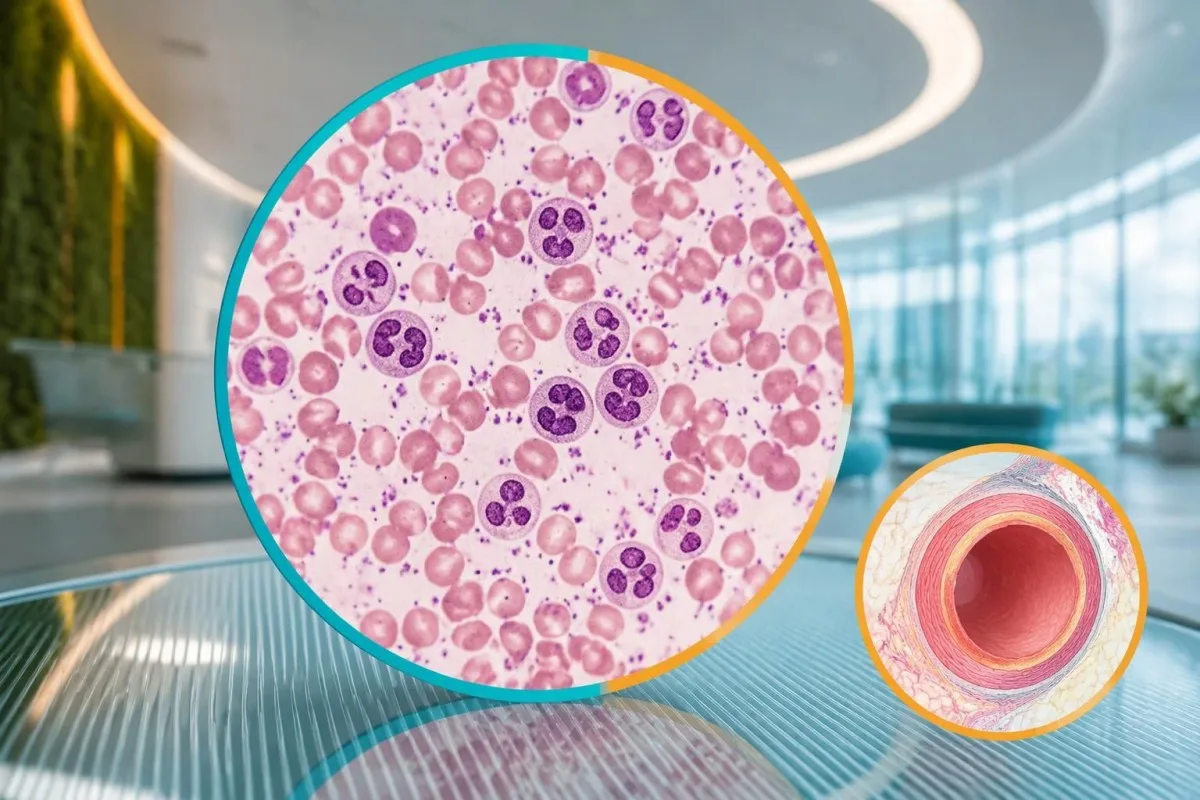
Platelets often rise during the second week rather than at the start of fever. A platelet count of 280 × 10⁹/L on illness day 4 therefore does not exclude Kawasaki disease, and waiting for thrombocytosis can delay care. Conversely, a count of 520 × 10⁹/L after day 7 supports the pattern but can also occur during recovery from other inflammatory illnesses.
Anemia in the incomplete-disease criteria is defined against the child's age, not a universal hemoglobin cutoff. For a hypothetical 3-year-old, hemoglobin of 9.8 g/dL would generally be low, but the cause could include inflammation, pre-existing iron deficiency, or both. WBC also needs age-specific context; our նեյտրոֆիլների հղումային միջակայքի ուղեցույցը explains why pediatric differential counts differ from adult ones.
Low platelets can occur in severe Kawasaki presentations and should not be treated as reassuring. A febrile child with platelets of 90 × 10⁹/L, poor circulation, or worsening organ function needs urgent assessment for shock, sepsis, consumption, or macrophage activation. Platelet size indices and neutrophil-to-lymphocyte ratios remain contextual findings; they are not substitutes for the 6 established supplementary criteria.
Ինչո՞ւ են ալբումինի և լյարդի հետազոտությունների ցուցանիշները փոխվում։
Low albumin and elevated ALT can support incomplete Kawasaki disease because systemic inflammation affects protein distribution and liver function. Albumin ≤30 g/L is a defined supplementary criterion, while ALT is judged against the laboratory's pediatric upper reference limit rather than one universal Kawasaki cutoff.
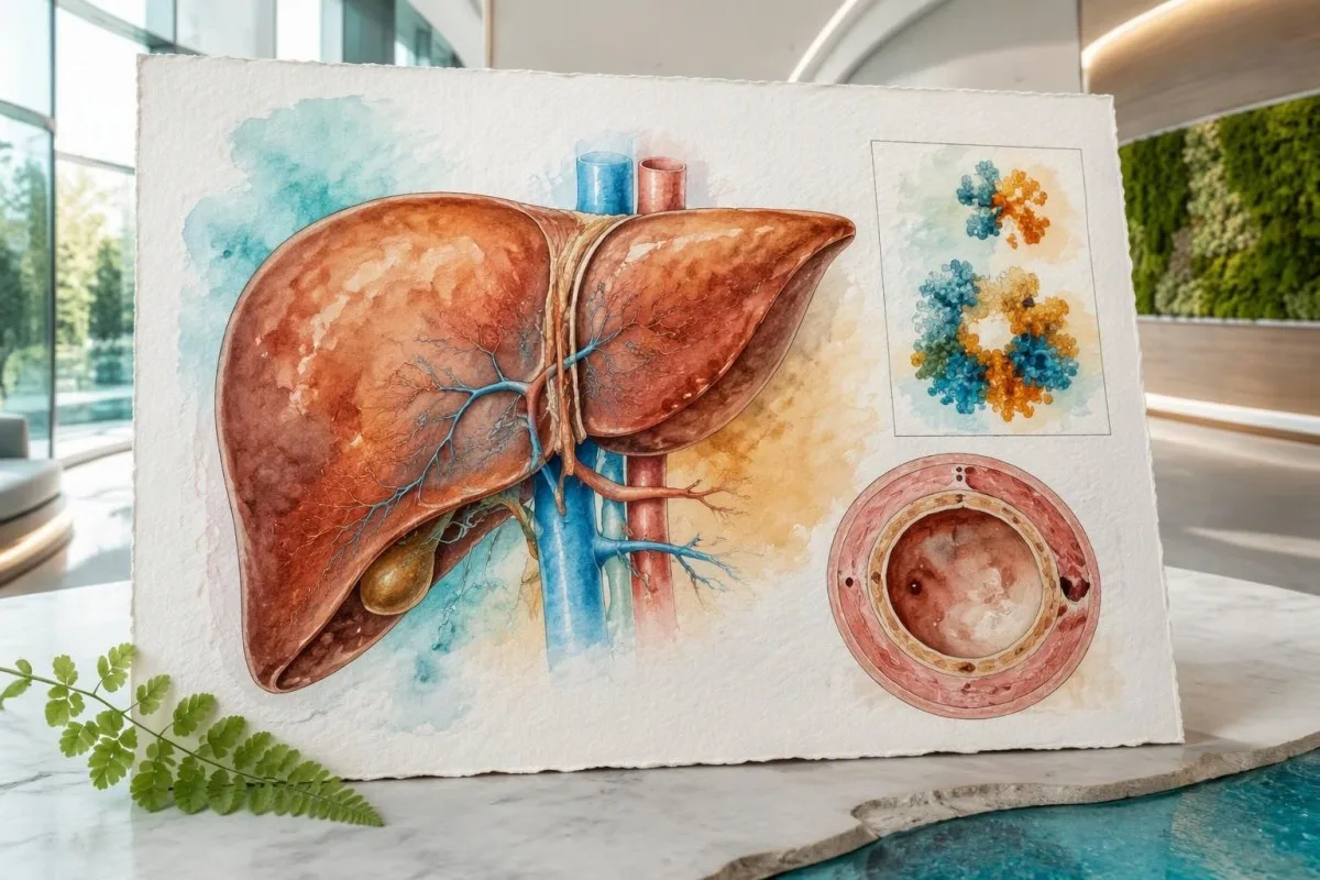
Albumin of 3.0 g/dL equals 30 g/L. During acute illness, a low result can reflect reduced synthesis, increased vascular permeability, dilution, or protein loss—not simply poor dietary protein intake. Our շիճուկային սպիտակուցների մեկնաբանման ուղեցույց explains these mechanisms; nutritional advice alone would miss the clinical significance of low albumin combined with persistent fever.
An ALT result above the pediatric upper limit counts as supporting evidence, but its magnitude does not diagnose Kawasaki disease. For example, a hypothetical child with albumin 28 g/L and ALT 72 IU/L may meet 2 supplementary criteria if that ALT exceeds the local limit. The same combination can arise from other illnesses or medication effects, as our ALT-ի տերմինաբանական ուղեցույցը է բացատրում։.
Kantesti is an AI blood test interpretation platform that can explain why albumin 28 g/L and ALT 72 IU/L require clinical context. Մեր AI տեխնոլոգիայի բացատրություն describes the interpretation workflow, not a validated Kawasaki diagnostic test. Growing children can also have relatively high alkaline phosphatase from bone activity; that finding is not interchangeable with the ALT criterion.
Կարո՞ղ են մեզի հետազոտությունները և նատրիումի մակարդակը օժանդակել գնահատմանը։
Urine white cells can support incomplete Kawasaki evaluation, while electrolyte changes help assess illness severity and alternative causes. Urine ≥10 white cells per high-power field is a supplementary criterion; low sodium is a possible associated finding but is not one of the 6 AHA supplementary laboratory criteria.
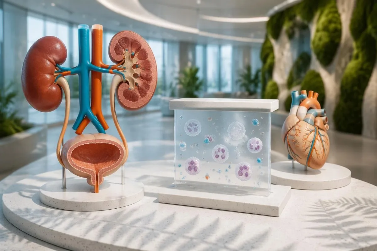
Sterile pyuria means urine white cells are present without a demonstrated bacterial cause on an appropriately collected culture. Kawasaki disease can produce this pattern, but a child with urine white cells still needs assessment for urinary infection. A urine dipstick alone cannot establish sterile pyuria; our մեզի նստվածքի մեկնաբանման ուղեցույցում distinguishes microscopy findings from dipstick screening results.
A report of 10 white cells per high-power field cannot be directly equated with an automated result of 10 cells/µL. Instruments, specimen preparation, and counting methods differ, so clinicians may need the laboratory's conversion or interpretive guidance. Collection quality also matters: contamination can introduce cells, whereas prior antibiotics can reduce culture yield without explaining the entire fever pattern.
Serum sodium below 135 mmol/L is generally classified as hyponatremia, but its cause must be assessed separately. Acute inflammation, fluid intake, gastrointestinal losses, and altered water regulation can all contribute; the number is not a Kawasaki confirmation test. Our էլեկտրոլիտային պատկերի ուղեցույց explains why sodium 130 mmol/L with neurological symptoms is a different problem from an isolated mildly low value.
Ի՞նչ լրացուցիչ տվյալ է տալիս էխոկարդիոգրամը ախտորոշման համար։
Echocardiography assesses coronary artery dimensions, heart function, and other cardiac findings that blood tests cannot show. A coronary artery Z-score ≥2.5 can support Kawasaki diagnosis in the incomplete-disease pathway, but a normal early echocardiogram does not exclude the illness or justify delaying indicated treatment.
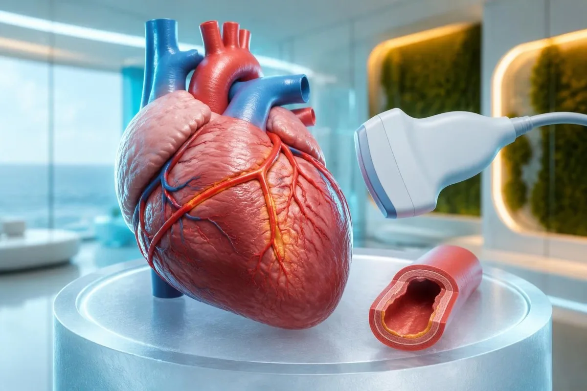
A coronary Z-score compares artery size with the expected size for a child's body surface area. A raw measurement such as 3 mm therefore has different implications in a small infant and a much larger child. The 2024 AHA statement emphasizes coronary assessment and risk-based management; the number should be interpreted with the measurement method and clinical setting (Jone et al., 2024).
A positive echocardiogram in the AHA incomplete-disease pathway can include a coronary aneurysm or a qualifying coronary Z-score. It can also involve at least 3 suggestive findings: decreased left-ventricular function, mitral regurgitation, pericardial effusion, or left anterior descending/right coronary Z-scores of 2 to less than 2.5. This is specialist image interpretation, not a checklist parents should apply to a brief scan summary.
Troponin and BNP or NT-proBNP may help evaluate cardiac involvement but do not replace echocardiography. A normal troponin on 1 sample cannot show that the coronary arteries are unaffected, and an elevated BNP can have several explanations. Our myocarditis laboratory guide explains the distinction between evidence of heart-muscle involvement and the separate question of coronary artery changes.
Ինչպե՞ս են բժիշկները տարբերակում Կավասակիի հիվանդությունը նմանակող այլ հիվանդություններից։
Clinicians distinguish Kawasaki disease from other causes of fever by combining the illness timeline, examination, targeted infection testing, laboratory patterns, and heart imaging. MIS-C, bacterial infections, adenovirus, scarlet fever, and systemic inflammatory disorders can overlap; a CRP result of 80 mg/L cannot separate them reliably.
MIS-C often follows SARS-CoV-2 infection by approximately 2–6 weeks and frequently affects older children than classic Kawasaki disease. Prominent gastrointestinal symptoms, shock, lymphopenia, relatively low platelets, and ventricular dysfunction may favor MIS-C, but overlap is substantial. A positive SARS-CoV-2 antibody result alone does not settle the distinction because previous infection or vaccination can affect interpretation.
Procalcitonin can rise in Kawasaki disease and should not automatically be treated as proof of a bacterial cause. Clinicians assess cultures, examination, organ function, and the broader trajectory before deciding what an elevated result means. Our պրոկալցիտոնինի մեկնաբանման ուղեցույցը explains why 1 biomarker cannot safely decide between sepsis, Kawasaki disease, or another inflammatory syndrome.
Finding a respiratory virus does not necessarily exclude coexisting Kawasaki disease. A hypothetical child with 6 days of fever, characteristic mouth changes, swollen feet, and an adenovirus-positive swab still needs a complete clinical assessment rather than automatic dismissal of the other findings. Travel, medication exposure, urinary symptoms, and recurrent fever patterns guide additional testing; there is no universal panel that rules out every mimic.
Ինչո՞ւ են հիվանդության օրը և կրկնակի հետազոտությունները փոխում պատկերը։
Kawasaki laboratory findings evolve, so the illness day and treatment history are part of every result's meaning. A normal platelet count early in the first week can become elevated later, while intravenous immunoglobulin can raise ESR independently of ongoing inflammation and make post-treatment ESR misleading.
A changing CBC can strengthen suspicion without making the first sample wrong. For example, a hypothetical child might have WBC 13 × 10⁹/L and platelets 280 × 10⁹/L on day 4, then platelets 480 × 10⁹/L and albumin 29 g/L on day 7. Clinicians should not postpone a concerning assessment until those later findings appear; our լաբորատոր ժամկետային ուղեցույցը explains evolving results.
ESR should not be used as the main measure of response after intravenous immunoglobulin. The infused proteins can increase sedimentation, so an ESR of 70 mm/hour after treatment does not necessarily mean treatment failure. Fever, clinical recovery, CRP trends, and cardiac assessment are more useful in context; the 2017 AHA statement specifically describes this post-infusion limitation (McCrindle et al., 2017).
Kantesti can help explain serial reports, but a trend graph cannot determine whether a child needs retreatment. Record the sample date, illness day, infusion completion time, and laboratory units alongside each value; comparing 2 results without that information can produce a false impression of deterioration. Immunoglobulin can also complicate some antibody tests through passive antibody transfer, so the treating team needs the infusion history.
Ինչո՞ւ չպետք է բուժումը հետաձգել մինչև բոլոր ախտանշանների ի հայտ գալը։
Treatment should begin promptly when Kawasaki disease is diagnosed, including incomplete disease, rather than waiting for every classic feature. Standard initial hospital treatment includes intravenous immunoglobulin at 2 g/kg, generally within the first 10 illness days; later treatment may still be indicated when fever or inflammation persists.
The 10-day treatment window is a target for timely care, not a reason to delay until day 10. The ACR/Vasculitis Foundation guideline recommends prompt treatment of incomplete Kawasaki disease once diagnosed, rather than waiting for the child to satisfy classic criteria (Gorelik et al., 2022). For a hypothetical 12-kg child, 2 g/kg represents 24 g of immunoglobulin administered under hospital supervision.
Aspirin treatment in Kawasaki disease requires a pediatric specialist's prescription and monitoring. Acute regimens vary internationally, including 30–50 or 80–100 mg/kg/day, followed in many cases by antiplatelet dosing of 3–5 mg/kg/day; families should not start, stop, or calculate treatment themselves. Our aspirin toxicity guidance explains why a prescribed Kawasaki regimen is not ordinary home fever treatment.
Approximately 10–20% of patients have persistent or recurrent fever after initial immunoglobulin treatment. Fever at least 36 hours after completion of the infusion is a commonly used threshold for IVIG resistance, after other causes are considered. Additional treatment may include corticosteroids or other specialist therapies; infants younger than 6 months and children with coronary Z-scores ≥2.5 can need particular attention, but risk scores do not perform equally across populations.
Ի՞նչ հետագա հսկողություն է անհրաժեշտ, երբ արյան հետազոտությունների արդյունքները բարելավվում են։
Improving CRP and CBC results do not prove that coronary artery risk has resolved. Follow-up is based on clinical recovery and echocardiographic findings, including the largest coronary Z-score recorded; children with coronary aneurysms need a different surveillance plan from children whose coronary studies remain normal.
Uncomplicated follow-up often includes echocardiography around 1–2 weeks and 4–6 weeks after treatment, but the schedule is individualized. The 2024 AHA statement supports risk-based assessment, and normal early studies can affect whether further routine imaging adds value (Jone et al., 2024). Persistent coronary changes, poor visualization, recurrent symptoms, or an abnormal initial study can require a different timetable.
A platelet count that remains elevated during recovery is not, by itself, proof of treatment failure. A hypothetical count of 600 × 10⁹/L in week 2 may fit the expected illness phase even when fever has stopped and CRP is falling, but symptoms and cardiac findings still govern management. Our պոստ‑հիվանդանոցային լաբորատորիայի փոփոխությունների մասին guide explains why normalization is not simultaneous across all markers.
High-dose immunoglobulin can affect the timing of certain live vaccines. After IVIG 2 g/kg, measles- and varicella-containing vaccines are commonly deferred for 11 months under immunization guidance, with exceptions assessed by the child's team and local schedule. Ask for a written plan covering vaccines, medicines, activity, repeat imaging, and which symptoms require reassessment; coronary follow-up should not be replaced by an apparently reassuring laboratory report.
Ինչպե՞ս կարելի է գնահատման ընթացքում անվտանգ օգտագործել արհեստական բանականության բացատրությունները։
AI explanations can help families understand laboratory language, but they cannot confirm Kawasaki disease, assess a child's circulation, or interpret coronary imaging independently. With fever lasting 5 days—or concerning symptoms sooner—clinical assessment takes priority over uploading reports, comparing reference ranges, or obtaining another automated interpretation.

Kantesti is an AI lab test interpretation service that can explain what CRP 30 mg/L or platelets 450 × 10⁹/L mean in a report. Those numbers are not sufficient to decide whether a child has Kawasaki disease, and an explanation cannot replace documenting illness day, examination findings, or an echocardiogram. Our արհեստական բանականության պատասխանատու օգտագործման սկզբունքները describe why interpretation must remain separate from urgent pediatric decision-making.
A useful review packet combines at least 3 types of information: the fever timeline, complete laboratory reports, and the treating team's assessment. Preserve units, pediatric reference intervals, medication dates, and imaging conclusions rather than sharing only red flags or screenshots of isolated values. Our medical validation documentation describes evaluation standards and limitations; technical performance should not be read as condition-specific proof of diagnostic safety.
Kantesti's research publications listed below describe platform evaluation and aggregate report analysis, not a Kawasaki diagnostic assay. The Clinical Validation Framework v2.0 and Global Health Report 2026 are hosted on Zenodo, and their DOI identifiers establish persistent records rather than peer-review status. Neither publication should be interpreted as establishing sensitivity or specificity for incomplete Kawasaki disease; clinical recommendations here rely on the 2017 and 2024 AHA statements and the ACR/Vasculitis Foundation guideline.
Հաճախ տրվող հարցեր
Կա՞ արդյոք արյան անալիզ, որը հաստատում է Կավասակիի հիվանդությունը։
Կավասակիի հիվանդությունը հաստատող առանձին արյան անալիզ չկա։ Բժիշկները համադրում են տենդի պատմությունը, զննման արդյունքները, բորբոքային մարկերները, լրացուցիչ լաբորատոր հետազոտությունների արդյունքները և էխոկարդիոգրաֆիան։ CRP ≥30 mg/L կամ ESR ≥40 mm/hour ցուցանիշները հիմք են տալիս հետագա գնահատման համար AHA-ի ոչ լրիվ հիվանդության ուղեցույցի շրջանակում, սակայն չեն բացահայտում բորբոքման պատճառը։ Առանձին նորմալ արդյունքը, հատկապես հիվանդության վաղ փուլում, չի կարող անվտանգ կերպով բացառել այս վիճակը։.
Կարո՞ղ է երեխան ունենալ Կավասակիի հիվանդություն՝ առանց բոլոր ախտանշանների։
Երեխայի մոտ կարող է լինել Կավասակիի ոչ լրիվ հիվանդություն՝ առանց բոլոր 5 հիմնական կլինիկական նշանների։ AHA-ի ուղեցույցը ներառում է առնվազն 5 օր տևող ջերմությամբ և 2–3 նշանով երեխաներին, ինչպես նաև որոշ նորածինների, որոնց մոտ առնվազն 7 օր տևողությամբ անհասկանալի ջերմություն կա։ Ախտորոշումն ու բուժումը կարող են հիմնավորվել այն աջակցող լաբորատոր շեղումներով կամ կորոնար զարկերակների ախտահարման նշաններով։ Ընտանիքները պետք է անհապաղ դիմեն գնահատման, այլ ոչ թե սպասեն, մինչև մաշկը սկսի թեփոտվել կամ ի հայտ գա մեկ այլ ախտանիշ։.
Արյան ո՞ր ցուցանիշների շեղումներն են հաճախ հանդիպում Կավասակիի հիվանդության դեպքում:
Կավասակիի հիվանդության լաբորատոր տարածված հայտնաբերումները ներառում են CRP-ի կամ ESR-ի բարձրացում, լեյկոցիտների քանակի ավելացում, տարիքին համապատասխան անեմիա, ալբումինի ցածր մակարդակ, ALT-ի բարձրացում և ավելի ուշ՝ թրոմբոցիտների քանակի աճ։ Ոչ լրիվ հիվանդության լրացուցիչ շեմերը ներառում են ալբումին ≤30 g/L, WBC ≥15 × 10⁹/L և թրոմբոցիտներ ≥450 × 10⁹/L՝ 7-րդ օրվանից հետո։ Այս հայտնաբերումները հատուկ չեն Կավասակիի հիվանդությանը և պետք է մեկնաբանվեն զննման արդյունքների ու հիվանդության ընթացքի ժամանակագրության հետ համատեղ։ Մեզում լեյկոցիտների առկայությունը նույնպես կարող է օգնել գնահատմանը, թեև դա մեզի հայտնաբերում է, ոչ թե արյան հետազոտության արդյունք։.
Նորմալ թրոմբոցիտները կամ նորմալ էխոկարդիոգրաման բացառո՞ւմ են Կավասակիի հիվանդությունը։
Առաջին շաբաթվա ընթացքում թրոմբոցիտների նորմալ քանակը և վաղ շրջանում կատարված էխոկարդիոգրաֆիայի նորմալ արդյունքը չեն բացառում Կավասակիի հիվանդությունը։ Թրոմբոցիտների քանակը հաճախ բարձրանում է երկրորդ շաբաթվա ընթացքում, իսկ կորոնար զարկերակների փոփոխությունները կարող են տեսանելի չլինել նախնական հետազոտության ժամանակ։ Հետևաբար, հիվանդության վաղ շրջանում թրոմբոցիտների քանակը 450 × 10⁹/L-ից ցածր լինելը չի բացառում ախտորոշումը։ Շարունակվող տենդը կամ մտահոգիչ կլինիկական պատկերը կարող են պահանջել կրկնակի գնահատում՝ առանց ցուցված բուժումը հետաձգելու։.
How long should a child's fever last before seeking help?
5 օր տևող ջերմություն ունեցող երեխան պետք է անհապաղ կլինիկական գնահատում անցնի, սակայն լուրջ ախտանիշների դեպքում օգնություն պետք է ստանալ ավելի շուտ։ 3 ամսականից փոքր նորածնի 38°C կամ ավելի բարձր ջերմությունը պահանջում է հրատապ բժշկական գնահատում։ Շնչառության դժվարացումը, թույլ արձագանքը, սեղմելիս չգունատվող ցանը, ջրազրկումը կամ արյան վատ շրջանառությունը պահանջում են հրատապ կամ շտապ օգնություն՝ անկախ ջերմության տևողությունից։ Կավասակիի հիվանդության 5-օրյա չափանիշը սպասելու հրահանգ չէ։.
Ինչո՞ւ է Կավասակի հիվանդության բուժումից հետո ԷՆԱ-ն դեռ բարձր մնում?
ESR-ը կարող է մնալ բարձր կամ բարձրանալ ներերակային իմունոգլոբուլինից հետո, քանի որ ներարկված սպիտակուցներն ազդում են նստեցման վրա։ Հետևաբար, բուժումից հետո 70 մմ/ժամ ESR-ն ինքնին չի հաստատում բուժման ձախողումը։ Բժիշկները գնահատում են ջերմությունը, կլինիկական ապաքինումը, CRP-ի միտումները և սրտի հետազոտության արդյունքները՝ հետներարկումային ESR-ի վրա հենվելու փոխարեն։ Ներարկման ավարտից առնվազն 36 ժամ անց պահպանվող կամ կրկնվող ջերմությունը պահանջում է բուժող թիմի կողմից վերագնահատում։.
Ստացեք ձեր արյան անալիզի արդյունքների բացատրությունը
Վերբեռնեք ձեր լաբորատոր հաշվետվությունը, և Kantesti-ի AI-ն վայրկյանների ընթացքում պարզ լեզվով կբացատրի յուրաքանչյուր կենսամարկեր։ Օգտագործում է ավելի քան 2 միլիոն մարդ ամբողջ աշխարհում։.
Հղումներ
- Klein, T., Mitchell, S., & Weber, H. (2026). Կլինիկական վավերացման շրջանակ v2.0 (Բժշկական վավերացման էջ)։. Kantesti AI բժշկական հետազոտություն։. ԴՕԻ
- Klein, T., Mitchell, S., & Weber, H. (2026). AI արյան անալիզի անալիզատոր. վերլուծված 2.5M թեստ | Համաշխարհային առողջապահական զեկույց 2026։. Kantesti AI բժշկական հետազոտություն։. ԴՕԻ
- McCrindle BW et al. (2017). Diagnosis, Treatment, and Long-Term Management of Kawasaki Disease: A Scientific Statement for Health Professionals From the American Heart Association. Circulation։. ԴՕԻ PubMed
- Jone PN et al. (2024). Update on Diagnosis and Management of Kawasaki Disease: A Scientific Statement From the American Heart Association. Circulation։.
- Gorelik M et al. (2022). 2021 American College of Rheumatology/Vasculitis Foundation Guideline for the Management of Kawasaki Disease. Arthritis & Rheumatology.
- 2 միլիոն+Վերլուծված թեստեր
- 127+Երկրներ
- 100+Լեզուներ
Բժշկական թիմի մասին


E-E-A-T վստահության ազդանշաններ
Փորձառություն
Բժշկի կողմից ղեկավարվող կլինիկական վերանայում՝ լաբորատոր մեկնաբանության աշխատանքային հոսքերի համար։.
Մասնագիտություն
Լաբորատոր բժշկության կենտրոնացում՝ այն բանի վրա, թե ինչպես են բիոմարկերները դրսևորվում կլինիկական համատեքստում։.
Հեղինակություն
Բժշկական գրախոսությունը՝ դոկտոր Թոմաս Քլայնի կողմից, դոկտոր Սառա Միտչելի և պրոֆ. դոկտոր Հանս Վեբերի մասնակցությամբ։.
Հուսալիություն
Ապացույցների վրա հիմնված մեկնաբանություն՝ հստակ հետագա քայլերի ուղիներով՝ ահազանգերը նվազեցնելու համար։.




Բժշկական հրաժարում պատասխանատվությունից
Այս հոդվածը միայն կրթական նպատակների համար է և չի հանդիսանում բժշկական խորհրդատվություն։ Ախտորոշման և բուժման որոշումների համար միշտ խորհրդակցեք որակավորված բուժաշխատողի հետ։.
Հրապարակված · Կապ հաստատել խմբագրական թիմի հետ
Հրատարակված է Kantesti Ltd-ի կողմից, գրանցված Անգլիայում և Ուելսում, ընկերության համար. 17090423, Լոնդոն, Միացյալ Թագավորություն։.
