
Gjin inkele bloedtest befêstiget Kawasaki-sykte. De diagnoaze kombinearret de koartsskiednis, ûndersyk, ûntstekkingsmarkers, stypjende laboratoariumfynsten en echokardiografy—oanhâldende koarts by in bern freget om prompt ûndersyk, net om te wachtsjen oant alle klassike symptomen der binne.

Wichtichste punten
- Diagnoaze fereasket klinyske beoardieling; gjin inkele bloedtest foar Kawasaki-sykte kin de oandwaning betrouber befêstigje of útslute.
- Oanhâldende koarts dy't 5 dagen duorret, freget om prompt ûndersyk nei Kawasaki-sykte, mar in bern dat slim siik liket, hat earder soarch nedich.
- Ynflammatoryske markers CRP ≥30 mg/L of ESR ≥40 mm/hour stypje fierder ûndersyk neffens it paad foar ûnfolsleine sykte fan de American Heart Association.
- Unfolsleine Kawasaki-sykte kin foarkomme mei mar 2–3 klassike skaaimerken; berntsjes kinne lang koarts hawwe mei mar hiel wat sichtbere tekens.
- Bloedplaatjes ≥450 × 10⁹/L nei dei 7 is in stypjende fynst; normale bloedplaatjes yn de earste wike slute Kawasaki-sykte net út.
- Stypjende resultaten omfetsje albumine ≤30 g/L, bloedearmoede foar de leeftyd, ferhege ALT, WBC ≥15 × 10⁹/L, en urine mei ≥10 wite bloedsellen per mikroskoopfjild mei hege fergrutting.
- Echocardiography beoardielet kransslagieren en de hertfunksje; in normale iere scan slút de sykte fan Kawasaki net út.
- Behannelingstiidstip is wichtich: sikehûsbehanneling omfettet meastal yntraveneuze immunoglobuline, leafst binnen de earste 10 syktedagen, sûnder op alle symptomen te wachtsjen.
Kin in bloedtest foar Kawasaki-sykte de diagnoaze befêstigje?
Gjin inkele bloedtest foar de sykte fan Kawasaki befêstiget de diagnoaze. Bloedtests mjitte ûntstekking en reaksjes fan organen, wylst kliïnten de sykte identifisearje op basis fan koarts, befiningen by ûndersyk en byldfoarming fan it hert; in bern mei 5 dagen koarts moat prompt beoardiele wurde, ek as it folsleine symptoompatroan ûntbrekt.
De sykte fan Kawasaki is in bernefaskulitis dy't de kransslagieren oantaaste kin. De ferklearring fan 'e American Heart Association út 2017 skat dat kransslaggeraneurysma's foarkomme by likernôch 25% fan 'e net behannele gefallen; dat sifer beskriuwt de net behannele sykte, net elk bern mei lang oanhâldende koarts of in ferhege CRP-resultaat (McCrindle et al., 2017). De measte troffen bern binne jonger as 5 jier, hoewol't âldere bern it ek krije kinne.
In abnormale ûntstekingsmarker is bewiis foar in ymmúnreaksje, net foar in spesifike sykte. In CRP fan 80 mg/L kin foarkomme by de sykte fan Kawasaki, in baktearjele sykte of in oare ûntstekkingstastân, dus it resultaat kin op himsels de diagnoaze net fêststelle. Likegoed kin in rapport mei mar 1 markearre wearde de soarch ûnderskatte as it stekproef betiid ôfnommen is of beoardiele is oan 'e hân fan referinsjewearden foar folwoeksenen.
Kantesti is in AI-bloedtestanalysator dy't CRP, CBC en terminology fan leverûndersiken útlizze kin sûnder de sykte fan Kawasaki te diagnostisearjen. Ús feiligensmaatregels foar pediatryske ynterpretaasje útlizze wêrom't de resultaten fan in 3-jierrige net beoardiele wurde kinne op basis fan oannamen foar folwoeksenen. Ynformaasje side oer ús organisaasje jout oan wa't dizze edukative tsjinst leveret; oanhâldende koarts moat noch altyd troch in kliïnt beoardiele wurde.
Wannear moat oanhâldende koarts by in bern ûndersocht wurde?
In bern mei koarts dy't 5 dagen oanhâldt, moat prompt klinysk beoardiele wurde, mei omtinken foar de sykte fan Kawasaki. Sykje earder help by earnstige sykte, soarchlike symptomen of in jonge poppe; de diagnostyske doer fan 'e koarts is gjin ynstruksje om oant dei 5 thús te bliuwen.

In temperatuer fan 38°C of heger by in poppe jonger as 3 moannen fereasket driuwende medyske beoardieling. Op elke leeftyd freegje sykheljen dy't dreech giet, ûngewoane slûgens, min reagearjen, in útslach dy't net ferdwynt by druk, slimme útdroeging of tekens fan minne bloedsirkulaasje om needsoarch. Dizze befiningen binne net spesifyk foar de sykte fan Kawasaki; sepsis en oare tiidkrityske tastannen moatte ek yn omtinken nommen wurde.
Koarts dy't tydlik reagearret op paracetamol slút de sykte fan Kawasaki net út. In bern kin him tusken temperatuerpiken nofliker fiele, wylst de ûnderlizzende sykte trochgiet, en symptomen kinne op ferskillende dagen ferskine. Hâld by wannear't de koarts foar it earst begûn, de feitlike temperatuerwearden, medisinen, urineproduksje en feroaringen oan eagen, lippen, hannen of fuotten—net allinnich oft de termometer ien kear normaal wie.
Poppen jonger as 6 moannen kinne lang oanhâldende koarts hawwe sûnder it bekende patroan fan útslach en reade eagen. Unferklearre koarts dy't op syn minst 7 dagen oanhâldt, is in erkende oanlieding foar evaluaasje fan in ûnfolsleine sykte yn it AHA-protokol. Us rjochtlinen foar it tarieden fan stekproeven binne nuttich foar plande tests, mar in siik bern moat nea wachtsje op fêstjen, in handich ôfspraakmomint of in folsleine checklist fan symptomen.
Hoe wurdt Kawasaki-sykte diagnostisearre op basis fan klinyske tekens?
Klassike sykte fan Kawasaki wurdt meastal diagnostisearre op basis fan koarts dy't op syn minst 5 dagen oanhâldt, plus op syn minst 4 fan 'e 5 wichtichste klinyske skaaimerken. Clinicians can diagnose some strongly characteristic cases earlier; neither the fever count nor the symptom count replaces clinical judgment or urgent evaluation.

The 5 principal features are bilateral non-purulent conjunctival redness, mouth changes, a polymorphous rash, extremity changes, and cervical lymphadenopathy. Mouth changes include red or cracked lips and a strawberry tongue; hand and foot changes include redness or swelling. The lymph-node criterion is usually a cervical node at least 1.5 cm across, often on one side.
The clinical features do not have to occur simultaneously. A photograph of a rash from illness day 3 may remain diagnostically useful when the skin looks different on day 6, and a previous episode of swollen hands should not disappear from the history because swelling has improved. Peeling around the fingertips often occurs during weeks 2–3, too late to use as a reason to delay assessment.
Certain findings point toward alternative explanations rather than strengthening the classic Kawasaki pattern. Purulent eye discharge, prominent exudative tonsillitis, or discrete mouth ulcers deserve separate consideration, although no single finding resolves every case. Our laboratory marker reference guide explains test terminology; it cannot substitute for examining the 5 clinical feature groups or arranging appropriate imaging.
Hoe stypje laboratoariumtests de diagnoaze fan ûnfolsleine Kawasaki-sykte?
Incomplete Kawasaki disease means a child has fewer classic features but enough clinical, laboratory, or echocardiographic evidence to warrant diagnosis and treatment. The AHA evaluation pathway considers children with fever for at least 5 days and 2–3 features, and certain infants with unexplained fever for at least 7 days.
CRP ≥30 mg/L or ESR ≥40 mm/hour supports proceeding through the AHA incomplete-disease evaluation pathway. When inflammation reaches either threshold, clinicians assess echocardiography and supplementary laboratory criteria rather than waiting for another visible symptom. The thresholds are decision supports for a compatible illness—not proof that Kawasaki disease is the cause (McCrindle et al., 2017).
The 6 supplementary laboratory criteria are albumin ≤30 g/L, anemia for age, elevated ALT, WBC ≥15 × 10⁹/L, platelets ≥450 × 10⁹/L after day 7, and urine ≥10 white cells per high-power field. In the appropriate clinical setting, at least 3 supplementary criteria or a positive echocardiogram can support treatment through the pathway. These are not independent points to count without reviewing competing diagnoses.
Fewer than 3 supplementary criteria does not create a permanent rule-out result. Persistent fever may require repeated examination, repeat laboratory testing, and imaging, particularly if the first evaluation occurred early. Systemic juvenile idiopathic arthritis is one possible alternative; our childhood arthritis laboratory guide explains why shared inflammatory findings do not make these conditions interchangeable.
Wat betsjutte CRP- en ESR-resultaten by Kawasaki-sykte?
CRP and ESR support Kawasaki evaluation by showing systemic inflammation, but neither identifies its cause. In the AHA incomplete-disease pathway, CRP ≥30 mg/L or ESR ≥40 mm/hour is a threshold for further assessment, not a severity scale or a stand-alone indication for treatment.
CRP of 3 mg/dL equals 30 mg/L. This 10-fold unit difference can make identical results appear very different when families compare reports from different laboratories, while common CRP reference limits are around 5 mg/L but vary by method. Our WBC en CRP fergeliking explains why a high CRP and a less impressive white-cell count can coexist.
ESR is influenced by factors beyond the immediate inflammatory response, including anemia and circulating proteins. A result of 45 mm/hour is not directly comparable to CRP of 45 mg/L; the tests measure different processes and have different kinetics. When both markers are below the pathway thresholds, Kawasaki disease becomes less likely, but an early sample or a concerning clinical course still requires judgment.
High-sensitivity CRP measures the same protein as conventional CRP, but adult cardiovascular-risk categories do not apply to a febrile child. A value of 30 mg/L during illness should not be interpreted using an adult 1–3 mg/L cardiovascular-risk framework. Our hs-CRP interpretation guide explains that distinction; for acute pediatric illness, the clinical question is inflammation and its cause.
| Nivo | Berik | Wat it betsjut |
|---|---|---|
| Gewoane CRP-referinsjegrins | Often <5 mg/L; laboratory-specific | A reference interval, not a Kawasaki exclusion threshold; early disease may require reassessment. |
| Incomplete-disease CRP trigger | ≥30 mg/L, equivalent to ≥3 mg/dL | Supports further AHA pathway evaluation when fever and clinical history are compatible. |
| Common ESR reference interval | Often approximately 0–20 mm/hour; age- and method-dependent | Use the reporting laboratory's pediatric interval; ESR is affected by anemia and proteins. |
| Incomplete-disease ESR trigger | ≥40 mm/hour | Supports further evaluation; neither confirms Kawasaki disease nor defines an emergency by itself. |
Hokker CBC-fynsten stypje de diagnoaze fan Kawasaki-sykte?
Kawasaki disease lab findings on a CBC can include neutrophil-predominant leukocytosis, anemia for age, and a later rise in platelets. WBC ≥15 × 10⁹/L and platelets ≥450 × 10⁹/L after day 7 are supplementary incomplete-disease criteria, but the illness day changes their meaning.
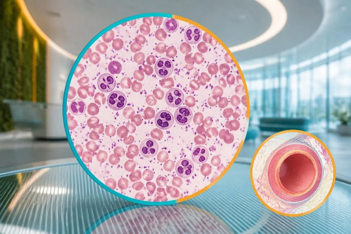
Platelets often rise during the second week rather than at the start of fever. A platelet count of 280 × 10⁹/L on illness day 4 therefore does not exclude Kawasaki disease, and waiting for thrombocytosis can delay care. Conversely, a count of 520 × 10⁹/L after day 7 supports the pattern but can also occur during recovery from other inflammatory illnesses.
Anemia in the incomplete-disease criteria is defined against the child's age, not a universal hemoglobin cutoff. For a hypothetical 3-year-old, hemoglobin of 9.8 g/dL would generally be low, but the cause could include inflammation, pre-existing iron deficiency, or both. WBC also needs age-specific context; our gids foar it referinsjeberik fan neutrofielen explains why pediatric differential counts differ from adult ones.
Low platelets can occur in severe Kawasaki presentations and should not be treated as reassuring. A febrile child with platelets of 90 × 10⁹/L, poor circulation, or worsening organ function needs urgent assessment for shock, sepsis, consumption, or macrophage activation. Platelet size indices and neutrophil-to-lymphocyte ratios remain contextual findings; they are not substitutes for the 6 established supplementary criteria.
Wêrom feroarje albumine- en levertestwearden?
Low albumin and elevated ALT can support incomplete Kawasaki disease because systemic inflammation affects protein distribution and liver function. Albumin ≤30 g/L is a defined supplementary criterion, while ALT is judged against the laboratory's pediatric upper reference limit rather than one universal Kawasaki cutoff.
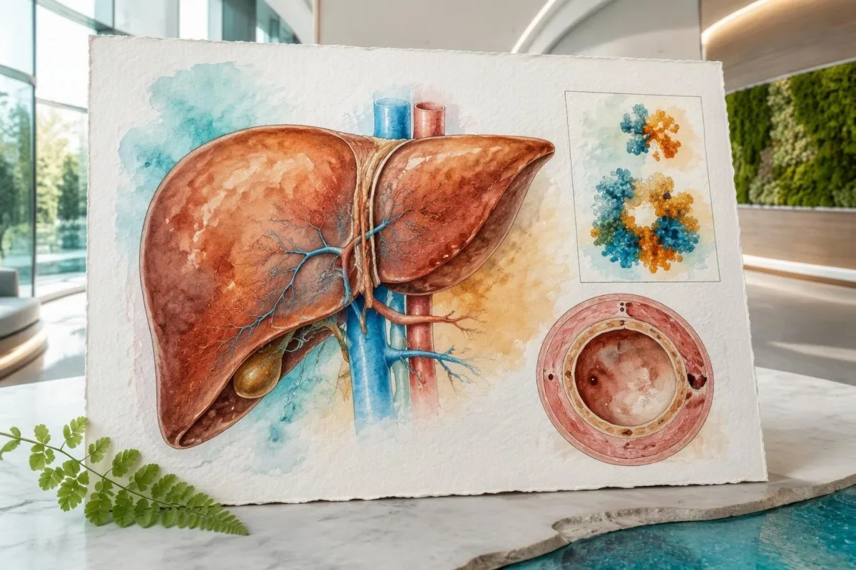
Albumin of 3.0 g/dL equals 30 g/L. During acute illness, a low result can reflect reduced synthesis, increased vascular permeability, dilution, or protein loss—not simply poor dietary protein intake. Our gids foar it ynterpretearen fan serumeiwiten explains these mechanisms; nutritional advice alone would miss the clinical significance of low albumin combined with persistent fever.
An ALT result above the pediatric upper limit counts as supporting evidence, but its magnitude does not diagnose Kawasaki disease. For example, a hypothetical child with albumin 28 g/L and ALT 72 IU/L may meet 2 supplementary criteria if that ALT exceeds the local limit. The same combination can arise from other illnesses or medication effects, as our hantlieding foar ALT-terminology útljocht.
Kantesti is an AI blood test interpretation platform that can explain why albumin 28 g/L and ALT 72 IU/L require clinical context. Ús útlis fan AI-technology describes the interpretation workflow, not a validated Kawasaki diagnostic test. Growing children can also have relatively high alkaline phosphatase from bone activity; that finding is not interchangeable with the ALT criterion.
Kinne urineûndersiken en natrium helpe by de evaluaasje?
Urine white cells can support incomplete Kawasaki evaluation, while electrolyte changes help assess illness severity and alternative causes. Urine ≥10 white cells per high-power field is a supplementary criterion; low sodium is a possible associated finding but is not one of the 6 AHA supplementary laboratory criteria.
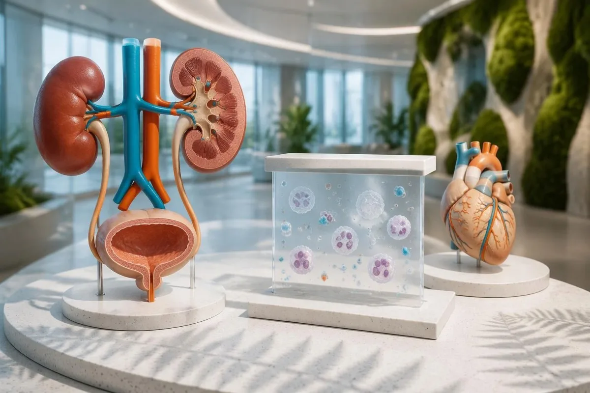
Sterile pyuria means urine white cells are present without a demonstrated bacterial cause on an appropriately collected culture. Kawasaki disease can produce this pattern, but a child with urine white cells still needs assessment for urinary infection. A urine dipstick alone cannot establish sterile pyuria; our hantlieding foar ynterpretaasje fan urinesedimint distinguishes microscopy findings from dipstick screening results.
A report of 10 white cells per high-power field cannot be directly equated with an automated result of 10 cells/µL. Instruments, specimen preparation, and counting methods differ, so clinicians may need the laboratory's conversion or interpretive guidance. Collection quality also matters: contamination can introduce cells, whereas prior antibiotics can reduce culture yield without explaining the entire fever pattern.
Serum sodium below 135 mmol/L is generally classified as hyponatremia, but its cause must be assessed separately. Acute inflammation, fluid intake, gastrointestinal losses, and altered water regulation can all contribute; the number is not a Kawasaki confirmation test. Our elektrolyt patroan gids explains why sodium 130 mmol/L with neurological symptoms is a different problem from an isolated mildly low value.
Wat foeget in echokardiogram ta oan de diagnoaze?
Echocardiography assesses coronary artery dimensions, heart function, and other cardiac findings that blood tests cannot show. A coronary artery Z-score ≥2.5 can support Kawasaki diagnosis in the incomplete-disease pathway, but a normal early echocardiogram does not exclude the illness or justify delaying indicated treatment.
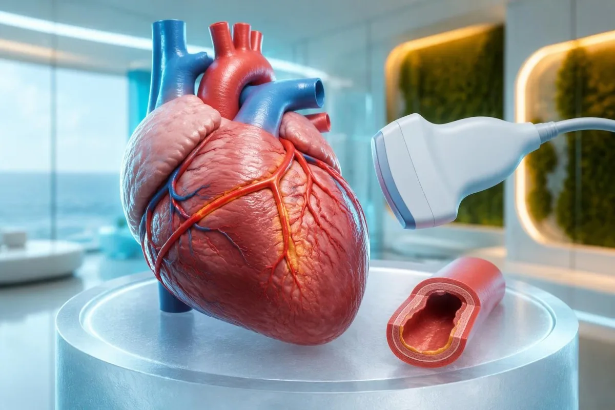
A coronary Z-score compares artery size with the expected size for a child's body surface area. A raw measurement such as 3 mm therefore has different implications in a small infant and a much larger child. The 2024 AHA statement emphasizes coronary assessment and risk-based management; the number should be interpreted with the measurement method and clinical setting (Jone et al., 2024).
A positive echocardiogram in the AHA incomplete-disease pathway can include a coronary aneurysm or a qualifying coronary Z-score. It can also involve at least 3 suggestive findings: decreased left-ventricular function, mitral regurgitation, pericardial effusion, or left anterior descending/right coronary Z-scores of 2 to less than 2.5. This is specialist image interpretation, not a checklist parents should apply to a brief scan summary.
Troponin and BNP or NT-proBNP may help evaluate cardiac involvement but do not replace echocardiography. A normal troponin on 1 sample cannot show that the coronary arteries are unaffected, and an elevated BNP can have several explanations. Our myocarditis laboratory guide explains the distinction between evidence of heart-muscle involvement and the separate question of coronary artery changes.
Hoe ûnderskiede klinisy Kawasaki-sykte fan sykten mei ferlykbere symptomen?
Clinicians distinguish Kawasaki disease from other causes of fever by combining the illness timeline, examination, targeted infection testing, laboratory patterns, and heart imaging. MIS-C, bacterial infections, adenovirus, scarlet fever, and systemic inflammatory disorders can overlap; a CRP result of 80 mg/L cannot separate them reliably.
MIS-C often follows SARS-CoV-2 infection by approximately 2–6 weeks and frequently affects older children than classic Kawasaki disease. Prominent gastrointestinal symptoms, shock, lymphopenia, relatively low platelets, and ventricular dysfunction may favor MIS-C, but overlap is substantial. A positive SARS-CoV-2 antibody result alone does not settle the distinction because previous infection or vaccination can affect interpretation.
Procalcitonin can rise in Kawasaki disease and should not automatically be treated as proof of a bacterial cause. Clinicians assess cultures, examination, organ function, and the broader trajectory before deciding what an elevated result means. Our gids foar de ynterpretaasje fan procalcitonine explains why 1 biomarker cannot safely decide between sepsis, Kawasaki disease, or another inflammatory syndrome.
Finding a respiratory virus does not necessarily exclude coexisting Kawasaki disease. A hypothetical child with 6 days of fever, characteristic mouth changes, swollen feet, and an adenovirus-positive swab still needs a complete clinical assessment rather than automatic dismissal of the other findings. Travel, medication exposure, urinary symptoms, and recurrent fever patterns guide additional testing; there is no universal panel that rules out every mimic.
Wêrom feroarje de syktedei en werhelle testen it byld?
Kawasaki laboratory findings evolve, so the illness day and treatment history are part of every result's meaning. A normal platelet count early in the first week can become elevated later, while intravenous immunoglobulin can raise ESR independently of ongoing inflammation and make post-treatment ESR misleading.
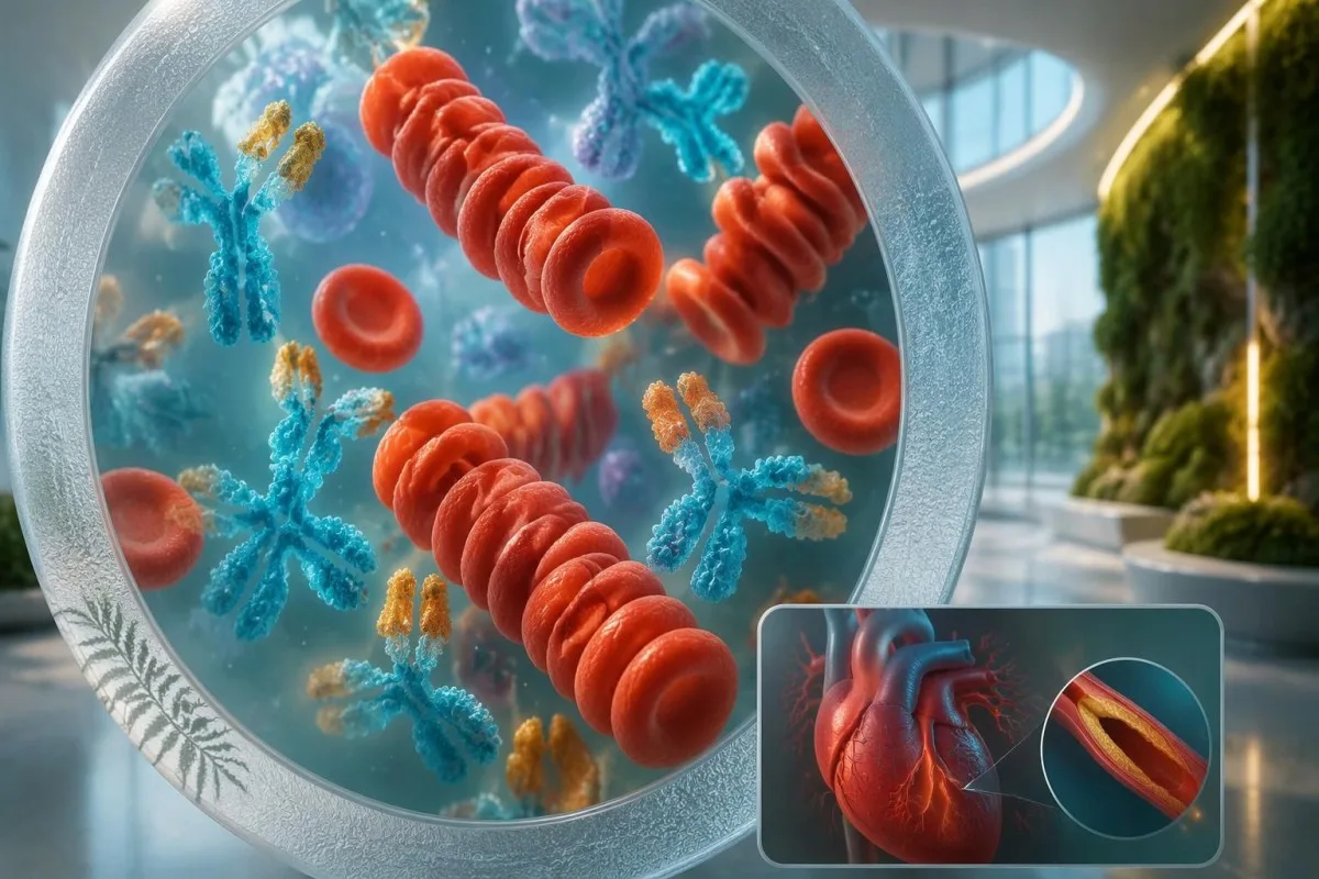
A changing CBC can strengthen suspicion without making the first sample wrong. For example, a hypothetical child might have WBC 13 × 10⁹/L and platelets 280 × 10⁹/L on day 4, then platelets 480 × 10⁹/L and albumin 29 g/L on day 7. Clinicians should not postpone a concerning assessment until those later findings appear; our laboratoriumtiidlinegids explains evolving results.
ESR should not be used as the main measure of response after intravenous immunoglobulin. The infused proteins can increase sedimentation, so an ESR of 70 mm/hour after treatment does not necessarily mean treatment failure. Fever, clinical recovery, CRP trends, and cardiac assessment are more useful in context; the 2017 AHA statement specifically describes this post-infusion limitation (McCrindle et al., 2017).
Kantesti can help explain serial reports, but a trend graph cannot determine whether a child needs retreatment. Record the sample date, illness day, infusion completion time, and laboratory units alongside each value; comparing 2 results without that information can produce a false impression of deterioration. Immunoglobulin can also complicate some antibody tests through passive antibody transfer, so the treating team needs the infusion history.
Wêrom moat behanneling net wachtsje oant alle symptomen der binne?
Treatment should begin promptly when Kawasaki disease is diagnosed, including incomplete disease, rather than waiting for every classic feature. Standard initial hospital treatment includes intravenous immunoglobulin at 2 g/kg, generally within the first 10 illness days; later treatment may still be indicated when fever or inflammation persists.
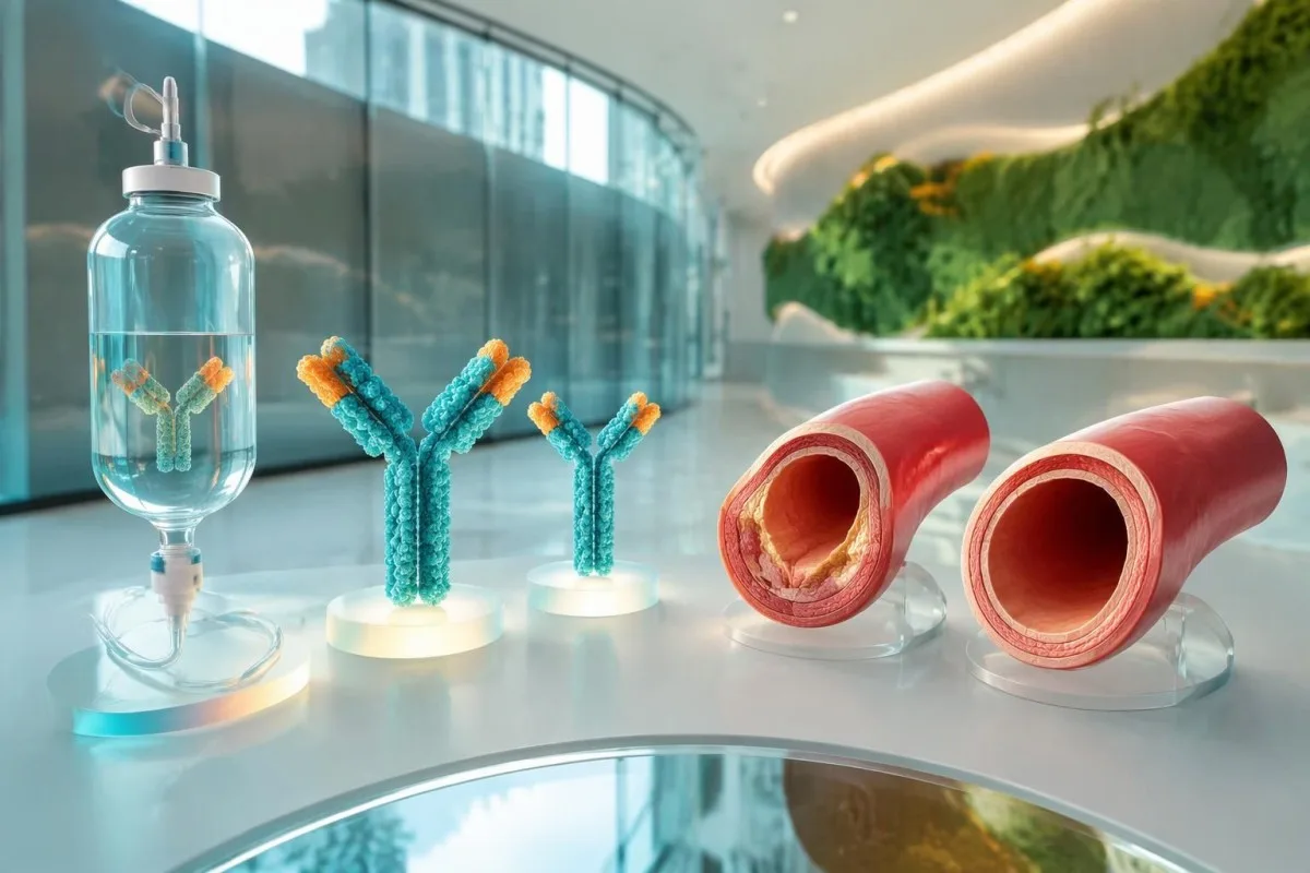
The 10-day treatment window is a target for timely care, not a reason to delay until day 10. The ACR/Vasculitis Foundation guideline recommends prompt treatment of incomplete Kawasaki disease once diagnosed, rather than waiting for the child to satisfy classic criteria (Gorelik et al., 2022). For a hypothetical 12-kg child, 2 g/kg represents 24 g of immunoglobulin administered under hospital supervision.
Aspirin treatment in Kawasaki disease requires a pediatric specialist's prescription and monitoring. Acute regimens vary internationally, including 30–50 or 80–100 mg/kg/day, followed in many cases by antiplatelet dosing of 3–5 mg/kg/day; families should not start, stop, or calculate treatment themselves. Our aspirin toxicity guidance explains why a prescribed Kawasaki regimen is not ordinary home fever treatment.
Approximately 10–20% of patients have persistent or recurrent fever after initial immunoglobulin treatment. Fever at least 36 hours after completion of the infusion is a commonly used threshold for IVIG resistance, after other causes are considered. Additional treatment may include corticosteroids or other specialist therapies; infants younger than 6 months and children with coronary Z-scores ≥2.5 can need particular attention, but risk scores do not perform equally across populations.
Hokker follow-up is nedich as bloedtestwearden ferbetterje?
Improving CRP and CBC results do not prove that coronary artery risk has resolved. Follow-up is based on clinical recovery and echocardiographic findings, including the largest coronary Z-score recorded; children with coronary aneurysms need a different surveillance plan from children whose coronary studies remain normal.
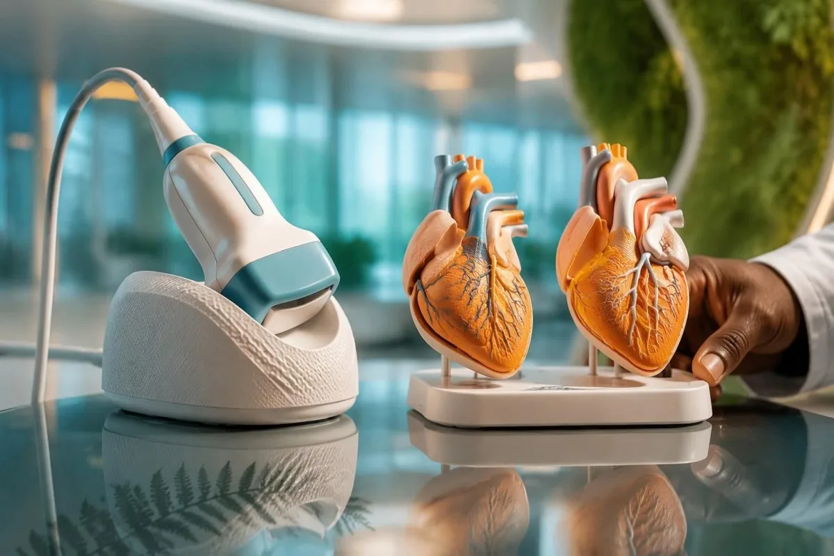
Uncomplicated follow-up often includes echocardiography around 1–2 weeks and 4–6 weeks after treatment, but the schedule is individualized. The 2024 AHA statement supports risk-based assessment, and normal early studies can affect whether further routine imaging adds value (Jone et al., 2024). Persistent coronary changes, poor visualization, recurrent symptoms, or an abnormal initial study can require a different timetable.
A platelet count that remains elevated during recovery is not, by itself, proof of treatment failure. A hypothetical count of 600 × 10⁹/L in week 2 may fit the expected illness phase even when fever has stopped and CRP is falling, but symptoms and cardiac findings still govern management. Our post‑hospital laboratorium‑feroarings guide explains why normalization is not simultaneous across all markers.
High-dose immunoglobulin can affect the timing of certain live vaccines. After IVIG 2 g/kg, measles- and varicella-containing vaccines are commonly deferred for 11 months under immunization guidance, with exceptions assessed by the child's team and local schedule. Ask for a written plan covering vaccines, medicines, activity, repeat imaging, and which symptoms require reassessment; coronary follow-up should not be replaced by an apparently reassuring laboratory report.
Hoe kinne AI-útlisjes feilich brûkt wurde by de evaluaasje?
AI explanations can help families understand laboratory language, but they cannot confirm Kawasaki disease, assess a child's circulation, or interpret coronary imaging independently. With fever lasting 5 days—or concerning symptoms sooner—clinical assessment takes priority over uploading reports, comparing reference ranges, or obtaining another automated interpretation.

Kantesti is an AI lab test interpretation service that can explain what CRP 30 mg/L or platelets 450 × 10⁹/L mean in a report. Those numbers are not sufficient to decide whether a child has Kawasaki disease, and an explanation cannot replace documenting illness day, examination findings, or an echocardiogram. Our prinsipes foar ferantwurde AI describe why interpretation must remain separate from urgent pediatric decision-making.
A useful review packet combines at least 3 types of information: the fever timeline, complete laboratory reports, and the treating team's assessment. Preserve units, pediatric reference intervals, medication dates, and imaging conclusions rather than sharing only red flags or screenshots of isolated values. Our medical validation documentation describes evaluation standards and limitations; technical performance should not be read as condition-specific proof of diagnostic safety.
Kantesti's research publications listed below describe platform evaluation and aggregate report analysis, not a Kawasaki diagnostic assay. The Clinical Validation Framework v2.0 and Global Health Report 2026 are hosted on Zenodo, and their DOI identifiers establish persistent records rather than peer-review status. Neither publication should be interpreted as establishing sensitivity or specificity for incomplete Kawasaki disease; clinical recommendations here rely on the 2017 and 2024 AHA statements and the ACR/Vasculitis Foundation guideline.
Faak stelde fragen
Is der in bloedtest dy't de sykte fan Kawasaki befêstiget?
Gjin inkele bloedtest befêstiget de sykte fan Kawasaki. Klinisy kombinearje de skiednis fan 'e koarts, befiningen by ûndersyk, ûntstekkingsmarkers, oanfoljende laboratoariumresultaten en echokardiografy. CRP ≥30 mg/L of ESR ≥40 mm/hour stipet fierdere beoardieling yn it AHA-trajekt foar ûnfolsleine sykte, mar identifisearret de oarsaak fan 'e ûntstekking net. In normale yndividuele útslach, benammen betiid yn 'e sykte, kin de oandwaning net feilich útslute.
Kin in bern Kawasaki-sykte hawwe sûnder alle symptomen?
In bern kin incomplete Kawasaki-sykte hawwe sûnder alle 5 wichtichste klinyske skaaimerken. It AHA-protokol omfettet bern mei op syn minst 5 dagen koarts en 2–3 skaaimerken, en ek bepaalde poppen mei op syn minst 7 dagen ûnferklearre koarts. Stypjende laboratoariumôfwikingen of koronêre befiningen kinne in diagnoaze en behanneling rjochtfeardigje. Famyljes moatte gau in beoardieling sykje en net wachtsje oant de hûd begjint te skiljen of der in oar symptoom ferskynt.
Hokker bloedresultaten komme faak foar by de sykte fan Kawasaki?
Algemiene laboratoariumfynsten by de sykte fan Kawasaki binne in ferhege CRP of ESR, in ferhege tal wite bloedsellen, bloedearmoed foar de leeftyd, leech albumine, in ferhege ALT en in lettere taname fan it tal bloedplaatjes. Oanfoljende drompelwearden foar de ûnfolsleine foarm fan de sykte binne albumine ≤30 g/L, WBC ≥15 × 10⁹/L en bloedplaatjes ≥450 × 10⁹/L nei dei 7. Dizze fynsten binne net spesifyk foar de sykte fan Kawasaki en moatte ynterpretearre wurde yn kombinaasje mei it ûndersyk en it ferrin fan de sykte. Wite bloedsellen yn de urine kinne ek helpe by de evaluaasje, al giet it om in urinefynst en net om in bloedresultaat.
Slute normale bloedplaatjes of in normale echokardiogram de sykte fan Kawasaki út?
Normale bloedplaatsjes yn 'e earste wike en in normaal ier echokardiogram slute de sykte fan Kawasaki net út. It tal bloedplaatsjes nimt yn 'e twadde wike faak ta, en feroarings oan 'e kransslachieren binne mooglik net sichtber op 'e earste scan. In tal bloedplaatsjes ûnder 450 × 10⁹/L ier yn 'e sykte slút de diagnoaze dêrom net út. Oanhâldende koarts of in soarchlik klinysk patroan kin in werhelle beoardieling fereaskje sûnder in oanjûne behanneling út te stellen.
How long should a child's fever last before seeking help?
0 In bern mei koarts dy't 5 dagen duorret, moat gau klinysk beoardiele wurde, mar by serieuze symptomen is earder soarch nedich. In temperatuer fan 38°C of heger by in poppe jonger as 3 moannen freget om driuwende medyske beoardieling. Swierrichheden mei sykheljen, min reagearjen, in útslach dy't net bleek wurdt by druk, útdroeging of minne bloedsirkulaasje freegje om driuwende of needsoarch, ûnôfhinklik fan hoe lang de koarts duorret. It Kawasaki-kritearium fan 5 dagen is gjin ynstruksje om te wachtsjen.
Wêrom is de ESR noch altyd heech nei de behanneling fan Kawasaki?
ESR kin nei intravenous immunoglobulin heech bliuwe of tanimme, om't de ynfúzjearre aaiwiten ynfloed hawwe op de sedimintaasje. In ESR fan 70 mm/oere nei behanneling wiist dêrom op himsels net op mislearjen fan de behanneling. Klinisy beoardielje de koarts, it klinyske herstel, de trends yn CRP en de hertbefiningen, ynstee fan ôf te gean op ESR nei de ynfúzje. Oanhâldende of weromkommende koarts op syn minst 36 oeren nei ôfrin fan de ynfúzje fereasket in opnij beoardieling troch it behanneljende team.
Lit jo bloedtest resultaten útlizze
Laad jo laboratoariumrapport op en de AI fan Kantesti leit elke biomarker binnen sekonden út yn gewoane taal. Brûkt troch mear as 2 miljoen minsken wrâldwiid.
Referinsjes
- Klein, T., Mitchell, S., & Weber, H. (2026). Klinysk Falidaasjekader v2.0 (Medyske Falidaasjeside). Kantesti AI Medysk Undersyk. DOI
- Klein, T., Mitchell, S., & Weber, H. (2026). AI Bloedtest Analyzer: 2.5M Tests Analiseerd | Global Health Report 2026. Kantesti AI Medysk Undersyk. DOI
- McCrindle BW et al. (2017). Diagnosis, Treatment, and Long-Term Management of Kawasaki Disease: A Scientific Statement for Health Professionals From the American Heart Association. Circulation. DOI PubMed
- Jone PN et al. (2024). Update on Diagnosis and Management of Kawasaki Disease: A Scientific Statement From the American Heart Association. Circulation.
- Gorelik M et al. (2022). 2021 American College of Rheumatology/Vasculitis Foundation Guideline for the Management of Kawasaki Disease. Arthritis & Rheumatology.
- 2M+Analysearre tests
- 127+Lannen
- 100+Talen
Oer it medyske team


E-E-A-T fertrouwensignalen
Ûnderfining
Dokter-oandreaune klinyske resinsje fan lab-ynterpretaasje-wurkprosessen.
Ekspertize
Fokus fan laboratoariummedisyne op hoe’t biomarkers har gedrage yn in klinyske kontekst.
Autoriteit
Medysk beoardiele troch Dr. Thomas Klein, mei bydragen fan Dr. Sarah Mitchell en prof. dr. Hans Weber.
Betrouberens
Bewiis-basearre ynterpretaasje mei dúdlike ferfolchpaadkes om alarm te ferminderjen.




Medyske disclaimer
Dit artikel is allinnich foar ûnderwiisdoelen en foarmet gjin medysk advys. Rieplachtsje altyd in kwalifisearre soarchferliener foar besluten oer diagnoaze en behanneling.
Publisearre · Nim kontakt op mei it redaksjeteam
Publisearre troch Kantesti Ltd, registrearre yn Ingelân en Wales, bedriuwsnûmer. 17090423, Londen, Feriene Keninkryk.
